

Capsular attachment on the anterosuperior femoral head-neck junction: A hypothesis about femoroacetabular impingement
- PMID: 38590168
- PMCID: PMC11259747
- DOI: 10.1111/joa.14046
Capsular attachment on the anterosuperior femoral head-neck junction: A hypothesis about femoroacetabular impingement
Abstract
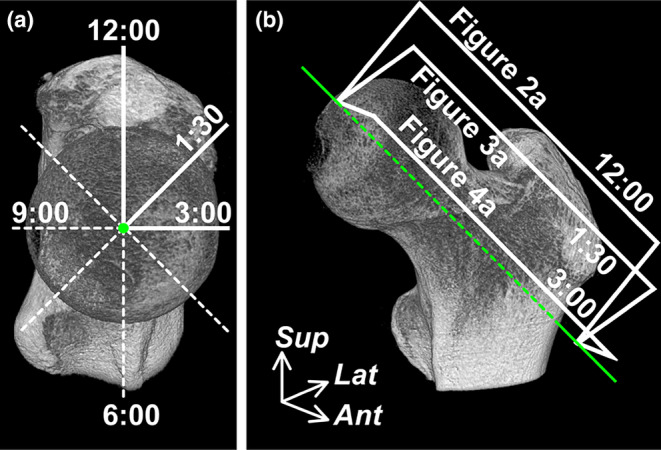
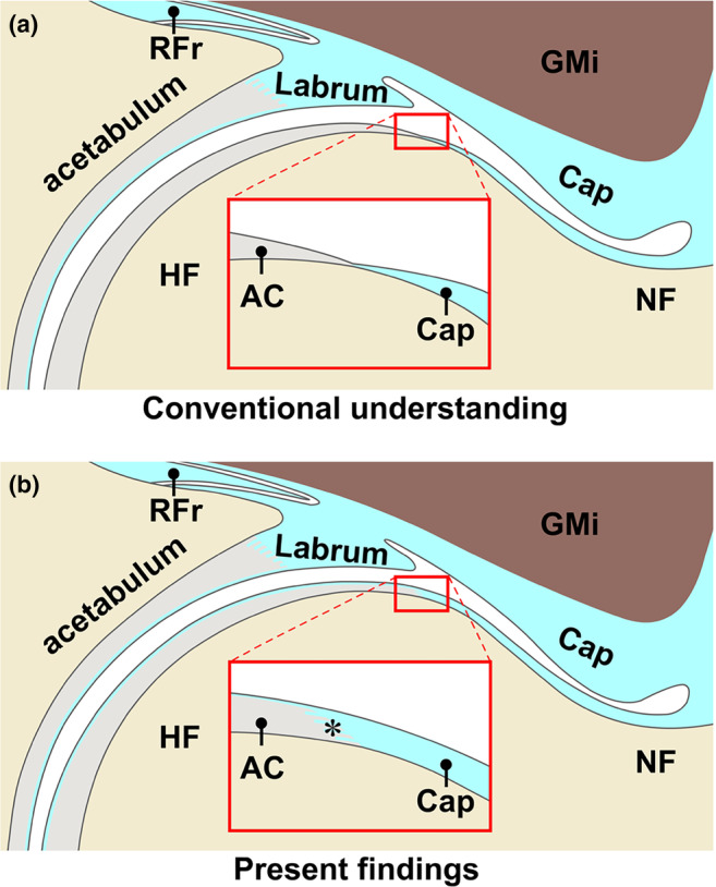
Femoroacetabular impingement (FAI), characterized by a pathological contact between the proximal femur and acetabulum, is a common precursor of hip osteoarthritis. Cam morphology is a bony prominence that causes FAI and frequently forms on the anterosuperior femoral head-neck junction. Despite anatomical consensus regarding the femoral head-neck junction as a boundary area covered by the articular cartilage and joint capsule, it remains unclear whether the joint capsule is continuous with the anterosuperior articular cartilage. For the anatomical consideration of cam morphology formation, this study aimed to investigate the histological characteristics of the capsular attachment on the anterosuperior femoral head-neck junction, particularly focusing on the presence or absence of continuity of the joint capsule to the articular cartilage. A total of 21 anterosuperior regions (seven hips each for the 12:00, 1:30, and 3:00 positions) from seven hips (three males and four females; mean age at death, 68.7 years) were histologically analyzed in this study for quantitative evaluation of the capsular thickness using histological sections stained with Masson's trichrome, as well as qualitative evaluation of the capsular attachment. The present study showed that the joint capsule, which folded proximally to the femoral head-neck junction from the recess, exhibited a blend of the fibrous and synovial regions. Notably, it not only continued with the superficial layer of the articular cartilage, but also attached to the articular cartilage via the fibrocartilage. This continuous region was relatively fibrous with dense connective tissue running in the longitudinal direction. The capsular thickness at the recess point (mean, 1.7 ± 0.9 mm) and those at the distal end of the articular cartilage (0.35 ± 0.16 mm) were significantly greater than the control value for the most superficial layer thickness of the articular cartilage (0.019 ± 0.003 mm) (Dunnett's T3, both p-value <0.001). Based on the fibrous continuity between the joint capsule and articular cartilage and its thickness, this study suggests the anatomical possibility that some mechanical stress can be transmitted from the joint capsule to the articular cartilage at the frequent sites of cam morphology.
Keywords: articular cartilage; cam morphology; femoral head–neck junction; hip joint capsule; histology.
© 2024 The Authors. Journal of Anatomy published by John Wiley & Sons Ltd on behalf of Anatomical Society.
Conflict of interest statement
None.
Figures


Similar articles
-
The Pattern of Acetabular Cartilage Wear Is Hip Morphology-dependent and Patient Demographic-dependent.Clin Orthop Relat Res. 2019 May;477(5):1021-1033. doi: 10.1097/CORR.0000000000000649. Clin Orthop Relat Res. 2019. PMID: 30998630 Free PMC article.
-
The Otto Aufranc Award. On the etiology of the cam deformity: a cross-sectional pediatric MRI study.Clin Orthop Relat Res. 2014 Feb;472(2):430-6. doi: 10.1007/s11999-013-2990-y. Clin Orthop Relat Res. 2014. PMID: 23604603 Free PMC article.
-
[Femoroacetabular impingement: a new direction in the diagnosis and treatment of the hip joint].Harefuah. 2011 Feb;150(2):148-52, 205. Harefuah. 2011. PMID: 22164944 Review. Hebrew.
-
Hip capsular thickness correlates with range of motion limitations in femoroacetabular impingement.Knee Surg Sports Traumatol Arthrosc. 2018 Oct;26(10):3178-3187. doi: 10.1007/s00167-018-4915-5. Epub 2018 Mar 24. Knee Surg Sports Traumatol Arthrosc. 2018. PMID: 29574547
-
Arthroscopic femoral osteoplasty/chielectomy for cam-type femoroacetabular impingement in the athlete.Sports Med Arthrosc Rev. 2010 Jun;18(2):90-9. doi: 10.1097/JSA.0b013e3181dfce63. Sports Med Arthrosc Rev. 2010. PMID: 20473127 Review.
Cited by
-
Femur osteoid osteoma in children: are there location-dependent differences in MRI findings?Pediatr Radiol. 2025 Mar;55(3):520-529. doi: 10.1007/s00247-024-06149-3. Epub 2025 Jan 20. Pediatr Radiol. 2025. PMID: 39831979
References
-
- Adams, M.A. (2021) Functional anatomy of the musculoskeltal system. In: Standring, S. (Ed.) Gray's anatomy: the anatomical basis of clinical practice. New York: Elsevier Health Sciences, pp. 85–126.
-
- Agricola, R. , Bessems, J.H. , Ginai, A.Z. , Heijboer, M.P. , van der Heijden, R. , Verhaar, J.A. et al. (2012) The development of cam‐type deformity in adolescent and young male soccer players. American Journal of Sports Medicine, 40(5), 1099–1106. - PubMed
-
- Agricola, R. , Heijboer, M.P. , Ginai, A.Z. , Roels, P. , Zadpoor, A.A. , Verhaar, J.A.N. et al. (2014) A cam deformity is gradually acquired during skeletal maturation in adolescent and young male soccer players: a prospective study with minimum 2‐year follow‐up. American Journal of Sports Medicine, 42(4), 798–806. - PubMed
-
- Casartelli, N.C. , Maffiuletti, N.A. , Valenzuela, P.L. , Grassi, A. , Ferrari, E. , van Buuren, M.M.A. et al. (2021) Is hip morphology a risk factor for developing hip osteoarthritis? A systematic review with meta‐analysis. Osteoarthritis and Cartilage, 29(9), 1252–1264. - PubMed
Publication types
MeSH terms
Grants and funding
LinkOut - more resources
Full Text Sources
