

Vanillic acid protects mortality and toxicity induced by N-ethyl-N-nitrosourea in mice; in vivo model of chronic lymphocytic leukemia
- PMID: 38590344
- PMCID: PMC10999465
- DOI: 10.1016/j.toxrep.2024.03.013
Vanillic acid protects mortality and toxicity induced by N-ethyl-N-nitrosourea in mice; in vivo model of chronic lymphocytic leukemia
Abstract
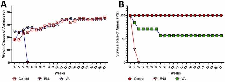
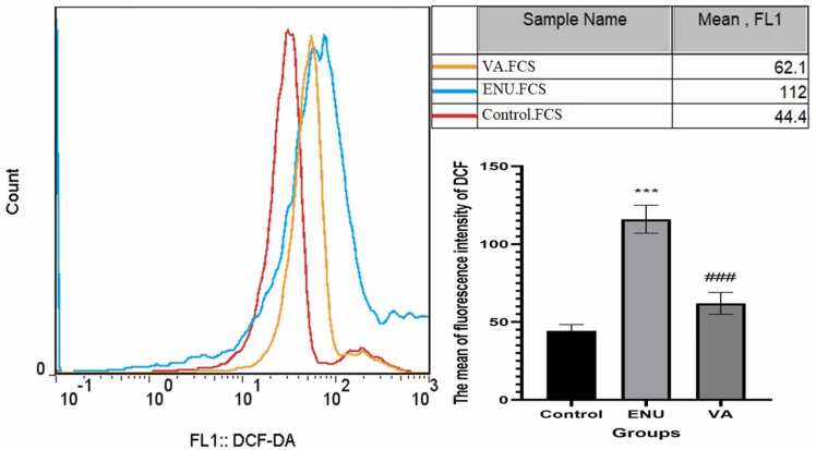
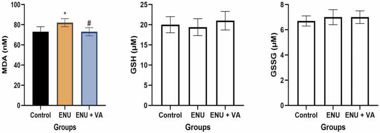
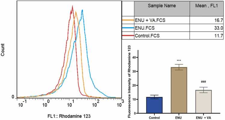
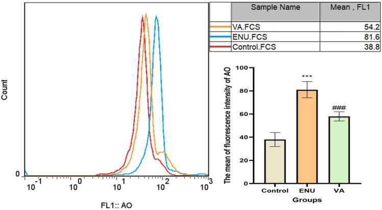
Alkylating agents such as N-Ethyl-N-Nitrosourea (ENU) are ubiquitous within living cells and in the environment. This study designed to evaluate the chemopreventive activity of vanillic acid on ENU-induced toxicity and carcinogenesis in mice as an animal model of chronic lymphocytic leukemia (CLL). The female, Swiss albino mice were divided into three groups each with 7 mice, group I received normal saline, group II, mice received ENU at a dose of 80 mg/kg body weight i.p. to induce CLL on the 31th day of the study, and group III, the mice pretreated with vanillic acid at a dose of 20 mg/kg body weight/day, i.p. up to 30 days and received ENU. The animals were monitored for weight changes and mortality during 120 days, and then were sacrificed for isolation of lymphocytes, as target cells in CLL. Cellular parameters like reactive oxygen species (ROS) formation, malondialdehyde (MDA) production, depletion of glutathione (GSH), mitochondrial membrane potential (MMP) and lysosomal membrane integrity were studied. We found that pretreatment with vanillic acid significantly increased the survival of mice up to 57%, delay in death time (30%) and prevented weight changes after exposure to ENU. In addition, it was found that vanillic acid protected ROS formation, lipid peroxidation mitochondrial dysfunction, and lysosomal membrane destabilization in isolated lymphocytes. These data suggest that vanillic acid exhibited significant protection against ENU-induced toxicity and carcinogenicity, which might be related to the protection of the mitochondria and lysosomes and the reduction of ROS formation and oxidative stress.
Keywords: Cancer chemoprevention; Carcinogenesis; Dietary; Leukemia; Phytochemicals.
© 2024 The Authors.
Conflict of interest statement
The authors declare that they have no known competing financial interests or personal relationships that could have appeared to influence the work reported in this paper.
Figures

Similar articles
-
Preparation of gallic acid-loaded chitosan nanoparticles and their chemoprotective effects on N-ethyl-N-nitrosourea-induced hepatotoxicity and mortality in rats.J Mol Histol. 2024 Nov 25;56(1):1. doi: 10.1007/s10735-024-10280-8. J Mol Histol. 2024. PMID: 39585491
-
Hesperidin as a bioactive compound in citrus fruits reduces N-ethyl-N-nitrosourea-induced mortality and toxicity in mice: as a model for chronic lymphocytic leukemia.Naunyn Schmiedebergs Arch Pharmacol. 2025 Apr;398(4):4009-4018. doi: 10.1007/s00210-024-03531-8. Epub 2024 Oct 14. Naunyn Schmiedebergs Arch Pharmacol. 2025. PMID: 39400715
-
Sinapic Acid Protects Mortality and Toxicity Induced by N-Ethyl-N-Nitrosourea, a Full Carcinogen Agent, in Mice.Drug Res (Stuttg). 2025 Sep 8. doi: 10.1055/a-2687-0870. Online ahead of print. Drug Res (Stuttg). 2025. PMID: 40921182
-
Iranian Mesobuthus Eupeus Crude Venom Induces Selective Toxicity in Chronic Lymphocytic Leukemia B-Lymphocytes Through Lysosomal/Mitochondrial Dysfunction and Reactive Oxygen Species Formation.Asian Pac J Cancer Prev. 2022 Jul 1;23(7):2309-2316. doi: 10.31557/APJCP.2022.23.7.2309. Asian Pac J Cancer Prev. 2022. PMID: 35901336 Free PMC article.
-
A review of the mutagenic potential of N-ethyl-N-nitrosourea (ENU) to induce hematological malignancies.J Biochem Mol Toxicol. 2022 Jul;36(7):e23067. doi: 10.1002/jbt.23067. Epub 2022 Apr 8. J Biochem Mol Toxicol. 2022. PMID: 35393684 Review.
Cited by
-
Regulating Sirtuin 3-mediated mitochondrial dynamics through vanillic acid improves muscle atrophy in cancer-induced cachexia.Commun Biol. 2025 Apr 9;8(1):585. doi: 10.1038/s42003-025-07770-0. Commun Biol. 2025. PMID: 40204937 Free PMC article.
-
Preparation of gallic acid-loaded chitosan nanoparticles and their chemoprotective effects on N-ethyl-N-nitrosourea-induced hepatotoxicity and mortality in rats.J Mol Histol. 2024 Nov 25;56(1):1. doi: 10.1007/s10735-024-10280-8. J Mol Histol. 2024. PMID: 39585491
-
Therapeutic potential of traditional herbal plants and their polyphenols in alleviation of mercury toxicity.Naunyn Schmiedebergs Arch Pharmacol. 2025 Jul;398(7):7737-7763. doi: 10.1007/s00210-025-03807-7. Epub 2025 Feb 6. Naunyn Schmiedebergs Arch Pharmacol. 2025. PMID: 39912903 Review.
References
-
- Jenkins G., Doak S., Johnson G., Quick E., Waters E., Parry J. Do dose response thresholds exist for genotoxic alkylating agents? Mutagenesis. 2005;20(6):389–398. - PubMed
-
- Swenberg J., Lai Y., Yu R., Sharma V., Moeller B.C., Hartwell H., Gibson J., Nakamura J. Thresholds of Genotoxic Carcinogens. Elsevier; 2016. The role of endogenous versus exogenous DNA damage in risk assessment; pp. 83–102.
-
- Bartsch H., Ohshima H., Shuker D.E., Pignatelli B., Calmels S. Exposure of humans to endogenous N-nitroso compounds: implications in cancer etiology. Mutat. Res./Rev. Genet. Toxicol. 1990;238(3):255–267. - PubMed
LinkOut - more resources
Full Text Sources
