. 2024 Mar 15;14(3):1087-1100.
doi: 10.62347/WMQV6643. eCollection 2024.

Duloxetine enhances the sensitivity of non-small cell lung cancer cells to EGFR inhibitors by REDD1-induced mTORC1/S6K1 suppression

Affiliations

Duloxetine enhances the sensitivity of non-small cell lung cancer cells to EGFR inhibitors by REDD1-induced mTORC1/S6K1 suppression

Se-Kyeong Jang et al. Am J Cancer Res. .

Abstract

Although epidermal growth factor receptor-tyrosine kinase inhibitors (EGFR-TKIs) have been effective targeted therapies for non-small cell lung cancer (NSCLC), most advanced NSCLC inevitably develop resistance to these therapies. Combination therapies emerge as valuable approach to preventing, delaying, or overcoming disease progression. Duloxetine, an antidepressant known as a serotonin-noradrenaline reuptake inhibitor, is commonly prescribed for the treatment of chemotherapy-induced peripheral neuropathy. In the present study, we investigated the combined effects of duloxetine and EGFR-TKIs and their possible mechanism in NSCLC cells. Compared with either monotherapy, the combination of duloxetine and EGFR-TKIs leads to synergistic cell death. Mechanistically, duloxetine suppresses 70-kDa ribosomal protein S6 kinase 1 (p70S6K1) activity through mechanistic target of rapamycin complex 1 (mTORC1), and this effect is associated with the synergistic induction of cell death of duloxetine combined with EGFR-TKIs. More importantly, activating transcription factor 4 (ATF4)-induced regulated in development and DNA damage response 1 (REDD1) is responsible for the suppression of mTORC1/S6K1 activation. Additionally, we found that the combination effect was significantly attenuated in REDD1 knockout NSCLC cells. Taken together, our findings reveal that the ATF4/REDD1/mTORC1/S6K1 signaling axis, as a novel mechanism, is responsible for the synergistic therapeutic effect of duloxetine with EGFR-TKIs. These results suggest that combining EGFR-TKIs with duloxetine appears to be a promising way to improve EGFR-TKI efficacy against NSCLC.

Keywords: ATF4; Duloxetine; EGFR-TKI; REDD1; S6K1; mTORC1; non-small cell lung cancer.


Conflict of interest statement

None.

Figures

Figure 1
Figure 1
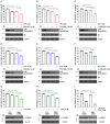
Duloxetine, a serotonin-norepinephrine reuptake inhibitor, sensitizes non-small cell lung cancer (NSCLC) cells to EGFR inhibitors. A. H1299, H460, and A549 cells were treated with 5 μM lapatinib and/or 10 μM duloxetine for 48 h. B. H1299, H460, and A549 cells were treated with 5 μM gefitinib and/or 10 μM duloxetine for 48 h. C. H1299, H460, and A549 cells were treated with 5 μM erlotinib and/or 10 μM duloxetine for 48 h. A-C. Cell viability was measured by MTT assay. The data are presented as the mean percentage of control ± SD (n=3; ***P<0.001). DLX: duloxetine, ERL: erlotinib, GEF: gefitinib, LPT: lapatinib.
Figure 2
Figure 2
Duloxetine, but not milnacipran, venlafaxine, or desvenlafaxine, sensitizes NSCLC cells to lapatinib. A-C. H1299, H460, and A549 cells were treated with 5 μM lapatinib and the indicated concentrations of duloxetine, milnacipran, venlafaxine or desvenlafaxine for 48 h. D. H1299, H460, and A549 cells were treated with 5 μM lapatinib and/or 10 μM duloxetine for 48 h. A-C. Cell viability was measured by MTT assay. The data are presented as the mean percentage of control ± SD (n=3). D. Cell death was detected as the percentage of Annexin V and/or Propidium Iodide (PI) positive cells. DLX: duloxetine, LPT: lapatinib, PI: Propidium Iodide.
Figure 3
Figure 3
Duloxetine and/or lapatinib reduces 70-kDa ribosomal protein S6 kinase 1 (p70S6K1)/mechanistic target of rapamycin complex 1 (mTORC1) activity. A. H1299 cells were treated with the indicated concentrations of lapatinib for 18 h. B. H1299 cells were treated with the indicated concentrations of duloxetine for 18 h. C. H1299 cells were treated with the indicated concentrations of duloxetine, milnacipran, venlafaxine and des venlafaxine for 18 h. D. H1299 cells were treated with 5 μM lapatinib and/or 10 μM duloxetine for 18 h. A-D. The indicated protein levels were estimated by western blot analysis. Data are representative of three independent experiments. DLX: duloxetine, LPT: lapatinib.
Figure 4
Figure 4
Duloxetine enhanced lapatinib-induced cell sensitivity by inhibiting mTORC1/S6K1. A. H1299 cells were treated with 5 μM lapatinib and/or 10 nM RAD-001 for 48 h. B. H1299 cells were treated with 5 μM lapatinib and/or 10 μM PF-4708671 for 48 h. C. H1299 cells were treated with 5 μM lapatinib and/or 3 μM LY2584702 for 48 h. D. H1299 cells were treated with 10 μM duloxetine and/or 10 nM RAD-001 for 48 h. E. H1299 cells were treated with 10 μM duloxetine and/or 10 μM PF-4708671 for 48 h. F. H1299 cells were treated with 10 μM duloxetine and/or 3 μM LY2584702 for 48 h. G. H1299 cells were transfected with CTL siRNA or S6K1 siRNA and then treated with 5 μM lapatinib. H. H1299 cells were transfected with CTL siRNA or S6K1 siRNA and then treated with 10 μM duloxetine. I. H1299 cells were transfected with CTL siRNA or S6K1 siRNA and then treated with 10 μM duloxetine and 5 μM lapatinib. A-I. Cell viability was measured by MTT assay. The indicated protein levels were estimated by western blot analysis. The data are presented as the mean percentage of control ± SD (n=3; ***P<0.001). CTL: control, DLX: duloxetine, LPT: lapatinib.
Figure 5
Figure 5
Duloxetine enhanced the lapatinib-induced inhibition of p70S6K1 phosphorylation via induction of regulated in development and DNA damage response 1 (REDD1). A. H1299 cells were treated with the indicated concentrations of lapatinib for 18 h. PC: H1299 cells were transiently transfected with control siRNA for 18 h and subsequently incubated in lysine-free medium for 6 h. NC: H1299 cells were transiently transfected with REDD1 siRNA for 18 h and subsequently incubated in lysine-free medium for 6 h. B. H1299 cells were treated with the indicated concentrations of duloxetine for 18 h. H1299 cells were deprived with lysine as a positive control. C. H1299 cells were treated with 10 μM duloxetine and/or 5 μM lapatinib for 18 h. D. H1299 cells or REDD1 k/o H1299 cells were treated with 5 μM lapatinib for 18 h. E. H1299 cells or REDD1 k/o H1299 cells were treated with 10 μM duloxetine for 18 h. F, G. H1299 cells or REDD1 k/o H1299 cells were treated with 10 μM duloxetine and 5 μM lapatinib for 18 h (F) or 48 h (G). H, I. H1299 cells that stably overexpressed vector and REDD1 were treated with 10 μM duloxetine and 5 μM lapatinib for 12 h (H) or 24 h (I). A-C. The indicated mRNA levels were estimated by real-time PCR analysis. (A-F, H) The indicated protein levels were estimated by western blot analysis. G-I. Cell viability was measured by MTT assay. The data are presented as the mean percentage of control ± SD (n=3; *P<0.05, ***P<0.001, ns: not significantly different). DLX: duloxetine, LPT: lapatinib, PC: positive control, NC: negative control.
Figure 6
Figure 6
Duloxetine enhanced the lapatinib-induced inhibition of p70S6K1 phosphorylation via induction of REDD1 in an activating transcription factor 4 (ATF4)-dependent manner. A. H1299 cells were treated with the indicated concentrations of lapatinib for 18 h. PC: H1299 cells were transiently transfected with control siRNA for 18 h and subsequently incubated in lysine-free medium for 6 h. NC: H1299 cells were transiently transfected with REDD1 siRNA for 18 h and subsequently incubated in lysine-free medium for 6 h. B. H1299 cells were treated with the indicated concentrations of duloxetine for 18 h. C. H1299 cells were treated with 10 μM duloxetine and/or 5 μM lapatinib for 18 h. D. H1299 cells were transfected with control or ATF4 siRNA for 12 h followed by treatment with 5 μM lapatinib. E. H1299 cells were transfected with control or ATF4 siRNA for 12 h followed by treatment with 10 μM duloxetine for 18 h. F, G. H1299 cells were transfected with control or ATF4 siRNA for 12 h followed by treatment with 10 μM duloxetine and 5 μM lapatinib for 18 h (F) or 48 h (G). The data are presented as the mean percentage of control ± SD (n=3; ***P<0.001, ns: not significantly different). CTL: control, DLX: duloxetine, LPT: lapatinib, PC: positive control, NC: negative control.
Figure 7
Figure 7
Duloxetine, a serotonin-norepinephrine reuptake inhibitor, sensitizes EGFR inhibitors in the EGFR-TKI-resistant lung cancer cell line H1975 (L858R/T790M-mutant EGFR). A. H1975 cells were treated with 5 μM lapatinib, 5 μM gefitinib or 5 μM erlotinib with duloxetine for 48 h. B, C. H1975 cells were treated with lapatinib and duloxetine, milnacipran, venlafaxine or desvenlafaxine for 48 h. A, B. Cell viability was measured by MTT assay. The data are presented as the mean percentage of control ± SD (n=3). C. Cell death was detected as the percentage of Annexin V and/or PI positive cells. The data are presented as the mean percentage of control ± SD (n=3; *P<0.05, ***P<0.001, ns: not significantly different). DLX: duloxetine, ERL: erlotinib, GEF: gefitinib, LPT: lapatinib, PI: Propidium Iodide.
Figure 8
Figure 8
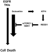
Proposed mechanism by which duloxetine enhances cell death to EGFR-TKIs in NSCLC cells by downregulating mTORC1/S6K1 through upregulation of REDD1 induced by ATF4 activation.

References

    1. Zappa C, Mousa SA. Non-small cell lung cancer: current treatment and future advances. Transl Lung Cancer Res. 2016;5:288–300. - PMC - PubMed
    1. Sher T, Dy GK, Adjei AA. Small cell lung cancer. Mayo Clin Proc. 2008;83:355–367. - PubMed
    1. Lynch TJ, Bell DW, Sordella R, Gurubhagavatula S, Okimoto RA, Brannigan BW, Harris PL, Haserlat SM, Supko JG, Haluska FG, Louis DN, Christiani DC, Settleman J, Haber DA. Activating mutations in the epidermal growth factor receptor underlying responsiveness of non-small-cell lung cancer to gefitinib. N Engl J Med. 2004;350:2129–2139. - PubMed
    1. Paez JG, Janne PA, Lee JC, Tracy S, Greulich H, Gabriel S, Herman P, Kaye FJ, Lindeman N, Boggon TJ, Naoki K, Sasaki H, Fujii Y, Eck MJ, Sellers WR, Johnson BE, Meyerson M. EGFR mutations in lung cancer: correlation with clinical response to gefitinib therapy. Science. 2004;304:1497–1500. - PubMed
    1. Huang L, Fu L. Mechanisms of resistance to EGFR tyrosine kinase inhibitors. Acta Pharm Sin B. 2015;5:390–401. - PMC - PubMed

LinkOut - more resources