

Adult-onset Sacral Meningocele Causing a Specific Headache Triggered by Compression or Adoption of a Sitting or Supine Posture
- PMID: 38590924
- PMCID: PMC10999460
- DOI: 10.2176/jns-nmc.2023-0272
Adult-onset Sacral Meningocele Causing a Specific Headache Triggered by Compression or Adoption of a Sitting or Supine Posture
Abstract
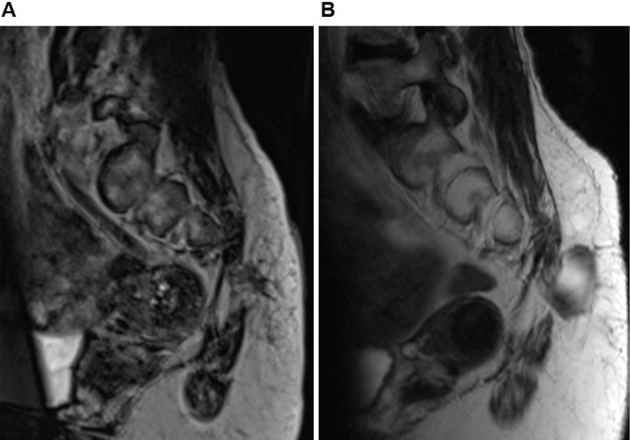
We report a rare case of adult-onset sacral meningocele where compression triggered a specific headache. A 46-year-old woman presented with a headache, which worsened when she was in a sitting or supine position. A subcutaneous mass was observed on her left buttock, the compression of which also induced headache. No neurological deficits were evident. Lumbar and sacral magnetic resonance imaging demonstrated a meningocele in the left dorsal buttock, connecting to the sacral cerebrospinal fluid (CSF) space, and spinal computed tomography revealed sacral dysplasia. Initial meningocele resection improved the patient's headache, but the cyst recurred 2 years later. Following repeated surgery to reinforce the meningocele orifice, the headache was relieved and has been absent for more than 6 years. The headache was due to intracranial pressure fluctuations due to CSF influx into and drainage from the meningocele. Meningocele development in adulthood can be owing to a spinal bone defect and pressure load on the spinal dura. Surgical resection can improve symptoms resulting from meningocele, and reinforcement of the orifice using an artificial surgical membrane effectively prevents recurrence.
Keywords: adult-onset; headache; meningocele; supine posture.
© 2024 The Japan Neurosurgical Society.
Conflict of interest statement
All authors have no conflicts of interest to declare.
Figures

Similar articles
-
Posterior Surgical Ligation and Cyst Decompression -via Needle Puncture- of a Large Anterior Sacral Pelvic Meningocele Through Posterior Sacral Laminectomy.Acta Neurochir Suppl. 2023;135:447-451. doi: 10.1007/978-3-031-36084-8_68. Acta Neurochir Suppl. 2023. PMID: 38153507
-
Anterior sacral pseudomeningocele following minimal trauma: case report.J Neurosurg Spine. 2013 Sep;19(3):384-8. doi: 10.3171/2013.6.SPINE12956. Epub 2013 Jul 5. J Neurosurg Spine. 2013. PMID: 23829288
-
Giant anterior sacral meningocele presenting as bacterial meningitis in a previously healthy adult.Orthopedics. 2008 Feb;31(2):182. doi: 10.3928/01477447-20080201-21. Orthopedics. 2008. PMID: 19292186 Review.
-
Anterior endoscopic treatment of a huge anterior sacral meningocele: technical case report.Neurosurgery. 2003 May;52(5):1231-3; discussion 1233-4. doi: 10.1093/neurosurgery/52.5.1231. Neurosurgery. 2003. PMID: 12699572
-
Holocord syringomyelia secondary to tethered spinal cord associated with anterior sacral meningocele and tailgut cyst: case report and review of literature.Childs Nerv Syst. 2014 Jun;30(6):1141-6. doi: 10.1007/s00381-014-2379-6. Epub 2014 Feb 22. Childs Nerv Syst. 2014. PMID: 24562417 Review.
References
-
- Pang D, Wilberger JE Jr: Tethered cord syndrome in adults. J Neurosurg 57: 32-47, 1982 - PubMed
-
- Fidas A, MacDonald HL, Elton RA, Wild SR, Chisholm GD, Scott R: Prevalence and patterns of spina bifida occulta in 2707 normal adults. Clin Radiol 38: 537-542, 1987 - PubMed
-
- Miyati T, Mase M, Kasai H, et al. : Noninvasive MRI assessment of intracranial compliance in idiopathic normal pressure hydrocephalus. J Magn Reson Imaging 26: 274-278, 2007 - PubMed