

Papillary renal neoplasm with reverse polarity: an observational study of histology, immunophenotypes, and molecular variation
- PMID: 38590969
- PMCID: PMC10999029
- DOI: 10.21037/tau-23-518
Papillary renal neoplasm with reverse polarity: an observational study of histology, immunophenotypes, and molecular variation
Abstract
Background: Papillary renal neoplasm with reverse polarity (PRNRP) is a novel entity with unique clinicopathological characteristics, and only a small number of patients with PRNRP have been described.
Methods: We retrospectively analyzed the data for nine patients with PRNRP and evaluated differences in the clinical, histomorphological, immunohistochemical, and molecular features; prognosis; and differential diagnosis of PRNRP from other renal tumors with papillary structure.
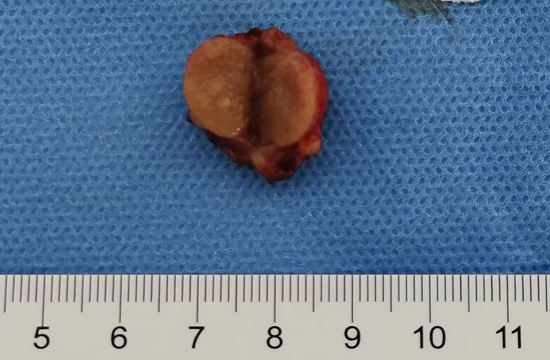
Results: There were six males and three females aged 36 to 74 years (mean: 62.33 years; median: 68 years). All the tumors were solitary and ranged from 1 to 3.7 cm (mean: 2.17 cm; median: 2 cm), with three and six tumors arose in the left and right renal tract, respectively. Pathologically, PRNRP is a small, well-circumscribed neoplasm with predominant papillary formations. The lining epithelium is composed of a monolayer of cuboidal to low-columnar cells with low-grade nuclei arranged against the apical pole of the tumor cells. Edema, mucinous degeneration, and hyaline degeneration are found in the fibrovascular cores. Foamy macrophages, psammoma bodies, hemosiderin deposition, and infiltrative tumor boundaries were present in some patients. Immunohistochemically, all tumors showed diffuse positive staining for GATA3. Sanger sequencing confirmed the presence of KRAS mutation in seven patients. All patients had a good prognosis after surgery and were relapse free. Positive staining for GATA3 and negative staining for vimentin were the most significant markers for differentiating PRNRP from other renal tumors with analogous structure.
Conclusions: These findings suggested that PRNRP is a distinctive subtype of renal tumor with specific pathological features and indolent behaviors that should be distinguished from other renal tumors, especially papillary renal cell carcinoma. A monolayer of tumor cells with an inverted nuclear pattern, positive staining for GATA3, and KRAS mutation are essential for pathological diagnosis. Owing to its satisfactory prognosis, the surveillance and follow-up of patients with PRNRP should be additionally formulated.
Keywords: GATA3; KRAS mutation; Papillary renal cell carcinoma (PRCC); renal tumor.
2024 Translational Andrology and Urology. All rights reserved.
Conflict of interest statement
Conflicts of Interest: All authors have completed the ICMJE uniform disclosure form (available at https://tau.amegroups.com/article/view/10.21037/tau-23-518/coif). The authors have no conflicts of interest to declare.
Figures


References
-
- Delahunt B, Eble JN. Papillary renal cell carcinoma: a clinicopathologic and immunohistochemical study of 105 tumors. Mod Pathol 1997;10:537-44. - PubMed
-
- Moch H, Humphrey PA, Ulbright TM, et al. WHO classification of tumours of the urinary system and male genital organs. 4th edition. Lyon: IARC Press; 2016. - PubMed
-
- WHO classification of tumours editorial board. WHO classification of tumours of the urinary and male genital organs. 5th edition. Lyon: IARC Press; 2022.
LinkOut - more resources
Full Text Sources
Miscellaneous