Review
. 2024 Mar 24:26:101028.
doi: 10.1016/j.mtbio.2024.101028. eCollection 2024 Jun.

Semiconducting polymer dots for multifunctional integrated nanomedicine carriers

Affiliations
Review

Semiconducting polymer dots for multifunctional integrated nanomedicine carriers

Ze Zhang et al. Mater Today Bio. .

Abstract

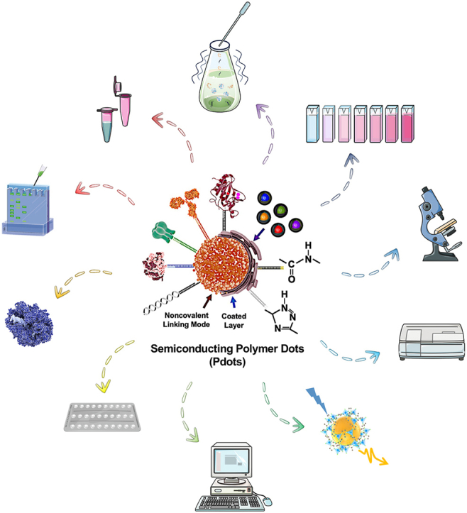
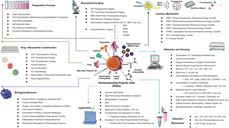
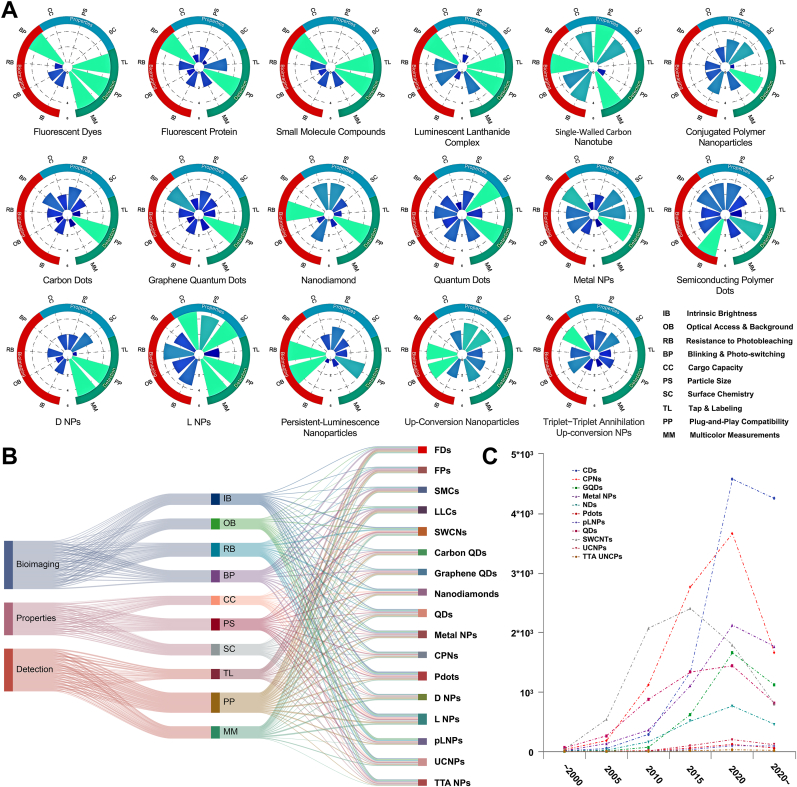
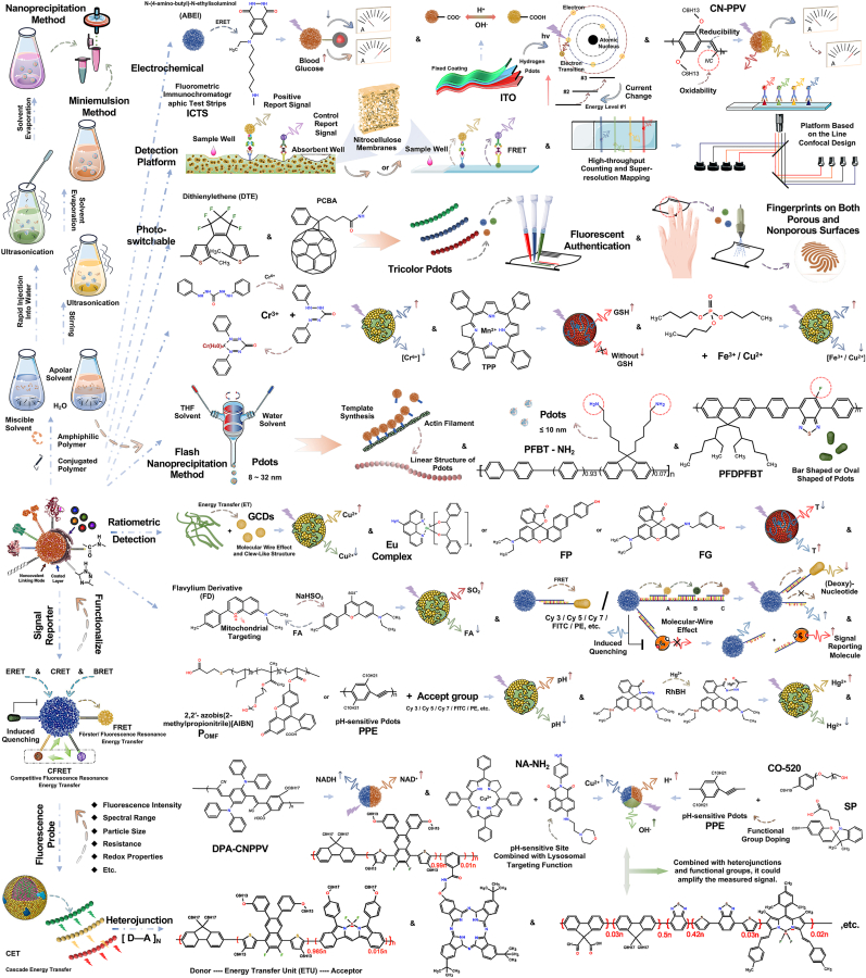
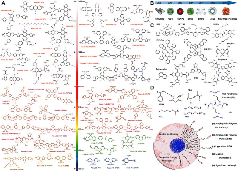
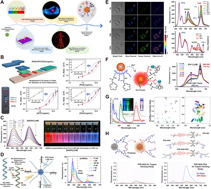
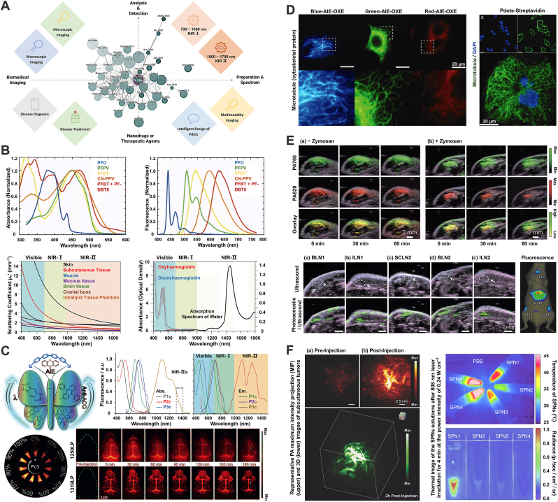
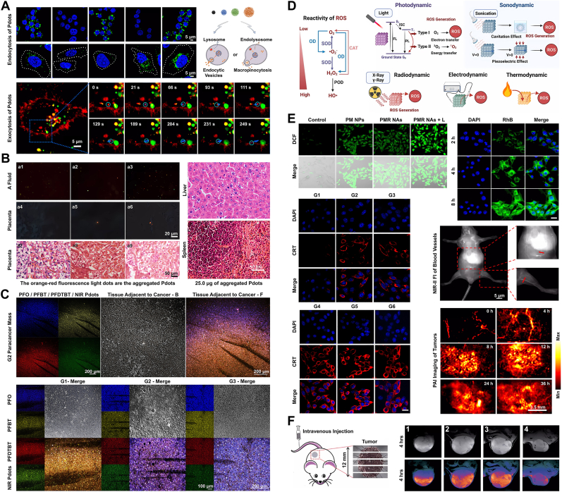
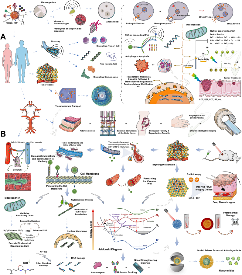
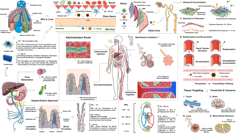
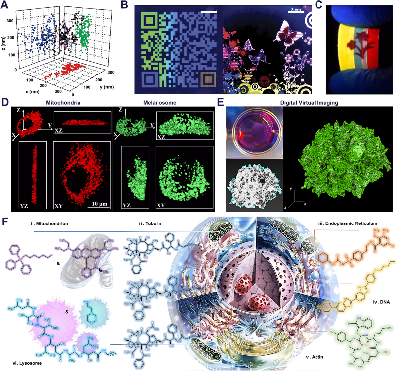
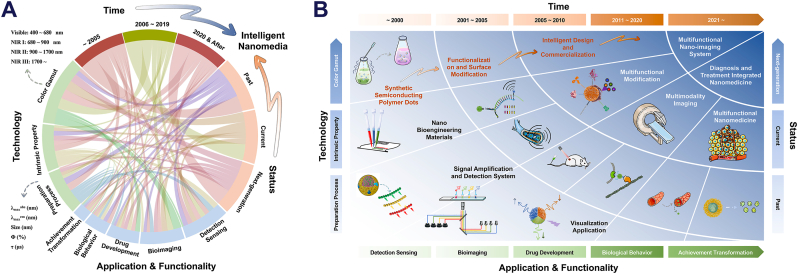
The expansion applications of semiconducting polymer dots (Pdots) among optical nanomaterial field have long posed a challenge for researchers, promoting their intelligent application in multifunctional nano-imaging systems and integrated nanomedicine carriers for diagnosis and treatment. Despite notable progress, several inadequacies still persist in the field of Pdots, including the development of simplified near-infrared (NIR) optical nanoprobes, elucidation of their inherent biological behavior, and integration of information processing and nanotechnology into biomedical applications. This review aims to comprehensively elucidate the current status of Pdots as a classical nanophotonic material by discussing its advantages and limitations in terms of biocompatibility, adaptability to microenvironments in vivo, etc. Multifunctional integration and surface chemistry play crucial roles in realizing the intelligent application of Pdots. Information visualization based on their optical and physicochemical properties is pivotal for achieving detection, sensing, and labeling probes. Therefore, we have refined the underlying mechanisms and constructed multiple comprehensive original mechanism summaries to establish a benchmark. Additionally, we have explored the cross-linking interactions between Pdots and nanomedicine, potential yet complete biological metabolic pathways, future research directions, and innovative solutions for integrating diagnosis and treatment strategies. This review presents the possible expectations and valuable insights for advancing Pdots, specifically from chemical, medical, and photophysical practitioners' standpoints.

Keywords: Bioimaging; Biosensor; Diagnosis; Nanomedicine; Semiconducting polymer dots; Translational medicine; Treatment.


Conflict of interest statement

All authors disclosed no relevant relationships.

Figures

Image 1
We summarize the situation of semiconducting polymer dots (Pdots) in the field of classical nanophotonic materials, discuss the development and application progress of functionalized designs based on substrate detection purposes, and the development of diagnosis and treatment integrated nanotechnology platforms for biomedical applications. This review aims to provide new ideas for designing and exploring the next generation of fluorescent probes and integrated nanomedical materials.
Fig. 1
Fig. 1
Newsflash Chart of Semiconducting Polymer Dots. Progression and future of semiconducting polymer dots within and beyond the optical properties and toward the expanded applications in analytical testing and biomedical imaging. Starting from the preparation process, the wide application of visual signals in inspection, sensing, labeling probes, drug carriers, and other fields is elaborated, and the advantages and limitations of Pdots in optical properties, versatility, biocompatibility, and microenvironment adaptability are explained. Progression and future of Pdots within and beyond the optical properties and toward the expanded applications in analytical testing and biomedical imaging.
Fig. 2
Fig. 2
The research focuses on luminescent molecular nanomaterials. and the quantity trend of published literature. A.) Qualitative scores for luminescent molecular nanomaterials (LMNs) in various functional categories are shown as radar plots. These qualitative scores might reflect variations in exploring potential, the difficulty of preparation, Spectral coverage range, medical-biological imaging, detection analysis, and achievement transformation, as discussed in the text. The higher the assignment scores, the more in-depth the research progress. Doped and labeled NPs are listed as D&L NPs. Drawing inspiration and reprinted with permission from Ref. [2]. Copyright 2020 American Chemical Society. B). Sankey diagram based on the score of each LMNs in Fig. 3A. Among the three areas, researchers pay more attention to the application of optical materials in analysis, monitoring, and biological imaging, while less energy is devoted to the properties of materials. C). Approximate numbers of indexed publications per year of various optical nanomaterials. Abbreviations: SWCNTs, single-walled carbon nanotubes; CDs, carbon dots; CPNs, conjugated polymer nanoparticles; UCNPs, lanthanide-doped upconversion nanoparticles; metal NCs, metal nanoclusters; Pdots, semiconducting polymer dots; pLNPs, persistent-luminescence nanoparticles; TTA-NPs, triplet−triplet annihilation upconversion nanoparticles. Source: PubMed (https://pubmed.ncbi.nlm.nih.gov).
Fig. 3
Fig. 3
The preparation process of semiconducting polymer dots (Pdots), the mechanisms used in analysis and detection, and representative applications. Upper Left). Schematic diagrams of two common preparation processes. Left Lower). The inherent mechanism applied to substrate detection and analysis. Upper Right). Schematic diagram of electrochemical detection, ICTS and anti-counterfeiting stamp. Middle Part). Special preparation process. Right Lower). Schematic diagram of substrate detection based on energy transfer and heterojunction. Abbreviations: AIE, Aggregation-Induced Emission; AIQ, Aggregation-Induced Quenching; BRET, bioluminescence resonance energy transfer; CET, cascade energy transfer; CRET, chemiluminescence resonance energy transfer; CFRET, competitive fluorescence resonance energy transfer; CV, cyclic voltammetry; D–A: donor-(single or multiple) energy transfer unit (ETU)-accepter; DPV, differential pulsed voltammetry; ECL, electrochemiluminescence; ERET, electrochemiluminescence resonance energy transfer; FRET, Förster/fluorescence resonance energy transfer; ET, energy transfer; ICTS, fluorometric immunochromatographic test strips; NRC, nanoreactors; PEC, photoelectrochemical; TCL, thermo-chemiluminescence.
Fig. 4
Fig. 4
Molecular Design Strategies and Surface Modification. A). Chemical structures of the published Pdots. The chemical structural formula's color represents the emission peak's spectral position. Rx represents the side chain chemical structural formula. Drawing inspiration and reprinted with permission from Ref. [65]. Copyright 2021 American Chemical Society. B). Hotspot optical nanomaterials at different time nodes. Abbreviations: SWCNTs, single-walled carbon nanotubes; QDs, quantum dots; RENPs, rare-earth-doped nanoprobes; SPNs, supramolecular polymer networks; SMDs, small-molecule dyes; AIEs, aggregation-induced emission luminogens. Drawing inspiration and reprinted with permission from Ref. [92]. Copyright 2020 American Chemical Society. C). The common doped and combined fluorescent dyes (donor-acceptor or donor-bridge-acceptor) inside or outside the π-conjugated chain. Abbreviations: BTE, (1,2-bis(2,4-dimethyl-5-phenyl-3-thienyl)-3,3,4,4,5,5-hexafluoro-1-cyclopentene; CPPO, Bis{3,4,6-trichloro-2-[(pentyloxy)carbonyl]phenyl} oxalate; BODIPY, 10-(4-(2,5-dioxo-2,5-dihydro-1H-pyrrol-1-yl)phenyl)-5,5; DBS, dithienyl-benzo selenadiazole; PtBzPor, (meso-tetraphenyl tetrabenzoporphyrin platinum (II), etc. D). Common amphiphilic polymers or compounds used for surface chemical modification. Drawing inspiration and reprinted with permission from Ref. [2]. Copyright 2020 American Chemical Society. Abbreviations: PSMA, poly (styrene-co-maleic anhydride); PDA, polydiacetylene; PEG, polyethylene glycol; PCL, Poly (ε-caprolactone), etc. Exposed chemical groups that can be used for functionalization after modification, which could form a single or multiple layer surface modification coating. Or include processes such as electrostatic bonding and self-forming coating.
Fig. 5
Fig. 5
Multifunctional Pdots through the design of π-conjugated chains and surface functionalized modification to reflect unique sensor architecture for special functions categories of use. A). Five representative nodes of time for Pdots with optical sensing performance, including (i) the fabrication of high-brightness Pdots, (ii) the concept of Pdots in the first region of the NIR-I, (iii) in vivo applications of biosensors based on Pdots, (iv) the concept of Pdots in the second region of the NIR-II, and (v) proposal of the concept of intelligent nano platform. Reprinted with permission from Refs. [3,248]. Copyright 2013 John Wiley and Sons Ltd. & Reprinted with permission from Refs. [249,114]. Copyright 2016 and 2023 American Chemical Society. B). The structure and principle of multiplexed ICTS based on PFO/PFCN/PFTC6FQ Pdots for detecting [CEA/AFP/PSA]. The graph in the lower corner represents the detection results of multiplexed CTMs concentration (0/0/5, 0/5/0, 5/0/0, 5/5/5 ng/mL). C). Emission spectra of DPA-CN PPV Pdots with 0∼2 mM [NADH] (left). DPA-CN PPV Pdots' color gradient law at 0–2 mM [NADH] with excitation at 365 nm under UV light (right). Reprinted with permission from Ref. [101]. Copyright 2021 John Wiley and Sons Ltd. D). The structure and principle of multiplexed Pdots-anti miRNAs based on PFO/PFBT/PFDTBT/CN PPV Pdots for detecting the pathological grade specific miRNAs (left). High sensitivity tests spectral results based on energy transfer models (right). E). Fluorescence imaging of intracellular 1O2 detection using SOSG-Pdots with 10 μg/mL [Ce6+] after different irradiation times at 0/30/60/120 s (LEFT). Spectral changes of the SOSG-Pdots in the presence of 1O2 (right, [Ce6+] contain oxidizing properties). F). Schematic diagram of color changes with increasing concentrations of Pb2+ and the fluorescence spectra of PFBT-DBT Pdots with different concentrations of Pb2+. G). Fluorescence spectra of the three Pdots formed from PFO Pdots and Pdots doped with BODIPY 520 & BODIPY 680 (left). The 2D distribution of 20 Pdots spectral-intensity barcodes plotted against the green-to-blue (x-axis) and red-to-blue (y-axis) fluorescence intensity ratios. The excitation wavelength was 405 nm, and blue, green, and red fluorescence were collected through the band-pass of 450/50 nm, 525/50 nm, and 670/30 nm, respectively, by flow cytometry (right). H). The structure and principle of protease sensor based on Pdots (upper). Protease-triggered “Switch on or off” leads to a change in fluorescence spectra. Fluorescence spectra of shell cross-linked Pdots before (red) and after (blue) incubation with trypsin (lower). Reprinted with permission from Refs. [180,[140], [142], [145], [250]], and [251]. Figure B and D ∼ H Copyright 2012, 2015, 2017, 2018, and 2022 American Chemical Society.
Fig. 6
Fig. 6
Multiple imaging methods and classic cases for Pdots of the π-conjugation chain. A). Pdots-related developments and applications are typically implemented on a nano- or microscale, including (i) preparation and spectrum, (ii) analysis and detection, (iii) biomedical imaging, and (iv) nanodrugs or therapeutic agents. The squares in the figure represent the main influencing factors in these practical applications. Source: Connected Papers (https://www.connectedpapers.com/). B). Typical representative of Pdots whose emission spectra lie in the visible range of light. And the challenges to be faced in the development of NIR Pdots nanoprobes. Absorption spectra - upper left, emission spectra - upper right, scattering coefficients of different biological tissues & intralipid - lower left, absorption spectra of oxyhaemoglobin (red), deoxyhaemoglobin (blue) and water (black) through a 1-mm-long path in human blood - lower right. Reprinted with permission from Refs. [297,298]. Copyright 2008 and 2015 American Chemical Society. C). Method for NIR NPs synthesis of visible light fluorescent materials - aggregation-induced emission (AIE). Absorption and fluorescence spectra of P1c, P2c, and P3c polymers in water & THF - upper right. Fluorescence intensity changes of P3a, P3b, and P3c in THF/water mixtures containing different water fractions (v/v % = THF/water %). In vivo NIR-II fluorescence imaging of mouse brain at designated time points after intravenous injection of P3c Pdots – lower right. Reprinted with permission from Ref. [51]. Copyright 2020 John Wiley and Sons Ltd. D). Applications of Pdots in the field of super-resolution imaging. Fluorescence images of microtubules in HeLa cells labeled with the AIE Pdots - left. And the same cells were incubated with both biotinylated anti-α-tubulin and PFBT–NH–PIMA Pdots-streptavidin - right. Reprinted with permission from Refs. [35,299]. Copyright 2012 and 2017 John Wiley and Sons Ltd. E). In vivo photoacoustic imaging of tumor (liver), bronchial lymph nodes (BLN), inguinal lymph nodes (ILN), supraclavicular lymph nodes (SLN) and lymph nodes (LN) (lower). Reprinted with permission from Ref. [300]. Copyright 2013 Springer Nature. & F). In vivo PA imaging and 3D images of tumor incubated with Pdots (SPN4) - left. Thermal image of the SPNs solutions after 808 nm laser irradiation for 4 min. Reprinted with permission from Ref. [301]. Copyright 2015 John Wiley and Sons Ltd.
Fig. 7
Fig. 7
Multiple disease diagnosis & treatment methods and classic cases for Pdots of the π-conjugation chain. A). Pdots with small particle size and spherical physical morphology largely depend on the clathrin-mediated endocytic pathway for transmembrane transport, and Pdots with large particle size or surface functionalized modification mainly rely on the macropinocytosis pathway for transmembrane transport. Reprinted with permission from Ref. [401]. Copyright 2017 American Chemical Society. B). Reproductive toxicity of Pdots. By tail vein injection, Pdots accumulated mainly in the liver and spleen, with no significant effect on maternal body weight or organ coefficient (note: A Fluid: amniotic fluid). Reprinted with permission from Ref. [400]. Copyright 2017 Ivyspring International Publishers. C). The staining ability of pathological grading with multi-Pdots-anti miRNAs. As the cell source gradually changed from normal tissue to malignant hepatocellular carcinoma, the color palette, after calculation and stacking, showed a gradual transition from bright orange for approximately normal tissue to violet-blue for cancerous tissue (upper part). The lower part shows bright orange-G1 (cell morphology is like normal tissue), violet-G2 (highly differentiated tumor cells), and dark blue or blue−yellow-G3 (poorly differentiated tumor cell) channel images to show pathology grading. Reprinted with permission from Ref. [180]. Copyright 2022 American Chemical Society. D). Schematic representation of photodynamic therapy (PDT). Photosensitizers generate ROS via light-induced electron transfer or energy transfer. Other effects, such as energy production and internal irradiation, are also included. Reprinted with permission from Ref. [406]. Copyright 2023 John Wiley and Sons Ltd. E).To improve the efficacy of immunotherapy, using Mn2+ ions as a coordination node, self-transferring NIR-II phototherapeutic nano adjuvants (PMR-NAs) were successfully prepared through the coordination self-assembly of ultrasmall NIR-II Pdots with toll-like receptor agonist resiquimod (R848).PMR-NAs can achieve TME-responsive drug release and tumor-targeted delivery of NIR-II fluorescence/photoacoustic/magnetic resonance imaging-mediated drugs and complete photothermal synergistic chemotherapy, stimulating effective anti-tumor immune responses through ICD effects. Reprinted with permission from Ref. [407]. Copyright 2023 Elsevier. F). The pH/hypoxia programmable triggered cancer photo-chemotherapy based on a Pdots hybridized mesoporous silica framework. Reprinted with permission from Ref. [408]. Copyright 2018 Royal Society of Chemistry.
Fig. 8
Fig. 8
Strategies for the application of semiconducting polymer dots (Pdots) in biomedical fields, the mechanisms used in disease diagnosis & treatment, and the multimodal therapy. Upper Left). Strategy for detection of histological parameters based on Pdots. Upper Right). Schematic diagram of transmembrane transport modes of Pdots. When Pdots enter the target cells, they can trigger various biological behaviors, such as cell proliferation, autophagy, or apoptosis. In addition, a nanobiological interface for the Fenton Reaction can be provided to generate ROS through a cascade reaction. Combined with Pdots' thermal conversion and radiation generation ability, it can achieve multi-dimensional therapeutic effects on tumors. Middle Part). Biotoxicity and other clinical applications of Pdots. Left Lower). Realization of the targeting function and subcellular spatial localization of functionalized Pdots. Right Lower). Schematic diagram of nanomedicine's construction strategy and therapeutic effect based on Pdots. Abbreviations: CDT, Chemodynamic therapy; PDT, photodynamic therapy; PAI, photoacoustic imaging technology; GSH, glutathione; NF-κB, nuclear factor-kappa B; PTT, photothermal therapy; PTT-CDT, photothermal-chemodynamic therapy; ROS, reactive oxygen species; RT, radiotherapy; SDT, sonodynamic therapy.
Fig. 9
Fig. 9
The implications of Pdots-based NPs for delivery, targeting, and clearance. The transport, distribution, biological transformation, and metabolism of nanoscale drugs within the body are contingent upon their physicochemical properties (such as particle size, macroscopic physical morphology, surface charge, and density) as well as the diverse microenvironments encountered during their transit (including reticular structure, charge barrier, immune system response, interstitial space, and intrinsic barriers). i). The middle section - the predominant route of administration for nano drug carriers. ii). The lower right corner - the size thresholds of tissue interstice and appropriate administration route of nanoparticles with varying sizes. iii). The lower left corner - the oral administration route with great development potential. iv). The top half of the Figure - the impact of Pdots-based NPs on hepatic clearance and renal clearance. In terms of Pdots-based NPs, hepatic clearance serves as the primary degradation and metabolism pathways. Drawing inspiration from Ref. [457]. Copyright 2023 Springer Nature.
Fig. 10
Fig. 10
Translational applications of Pdots. A). Single nanoparticles bioimaging and tracking. B). Full-color fluorescence patterning. Reprinted with permission from Ref. [219]. Copyright 2014 John Wiley and Sons Ltd. C). Inkjet printing and anti-counterfeiting applications. D). 3D and three-view imaging of melanosomes and mitochondria. E). Digital virtual imaging of liver organoid cell clusters. Reprinted with permission from Ref. [227]. Copyright 2023 Royal Society of Chemistry. Note: Reprinted with permission from Refs. [30,138], and [368]. Copyright 2014(A), 2017(C) and 2022(D) American Chemical Society. F). Commonly used chemical recognition groups for various subcellular structures. Such as: cytoskeleton proteins, mitochondria, lysosomes, nuclei, etc. Drawing inspiration from Ref. [497]. Copyright 2023 American Chemical Society.
Fig. 11
Fig. 11
Prospects for Pdots. A). Development context of optical nanomaterials based on Pdots within and beyond the optical imaging domain regarding the key milestones. B). Emoucneliand application of optical nanomaterials based on Pdots within and beyond optical imaging, analytical sensing, inherent properties, and achievement transformation at different stages.

Similar articles

Cited by

References

    1. Wolfbeis O.S. An overview of nanoparticles commonly used in fluorescent bioimaging. Chem. Soc. Rev. 2015;44(14) 4743-68. - PubMed
    1. Algar W.R., Massey M., Rees K., Higgins R., Krause K.D., Darwish G.H., Peveler W.J., Xiao Z., Tsai H.Y., Gupta R., Lix K., Tran M.V., Kim H. Photoluminescent nanoparticles for chemical and biological analysis and imaging. Chem. Rev. 2021;121(15):9243–9358. - PubMed
    1. Wu C., Chiu D.T. Highly fluorescent semiconducting polymer dots for biology and medicine. Angew. Chem. Int. Ed. Engl. 2013;52(11):3086–3109. - PMC - PubMed
    1. Yu J., Rong Y., Kuo C.T., Zhou X.H., Chiu D.T. Recent advances in the development of highly luminescent semiconducting polymer dots and nanoparticles for biological imaging and medicine. Anal. Chem. 2017;89(1):42–56. - PMC - PubMed
    1. Jiang Y., Pu K. Multimodal biophotonics of semiconducting polymer nanoparticles. Acc. Chem. Res. 2018;51(8):1840–1849. - PubMed

LinkOut - more resources