. 2024 Mar 25:15:1356206.
doi: 10.3389/fmicb.2024.1356206. eCollection 2024.

Interaction of bacteriophage P1 with an epiphytic Pantoea agglomerans strain-the role of the interplay between various mobilome elements

Affiliations

Interaction of bacteriophage P1 with an epiphytic Pantoea agglomerans strain-the role of the interplay between various mobilome elements

Katarzyna Giermasińska-Buczek et al. Front Microbiol. .

Abstract

P1 is a model, temperate bacteriophage of the 94 kb genome. It can lysogenize representatives of the Enterobacterales order. In lysogens, it is maintained as a plasmid. We tested P1 interactions with the biocontrol P. agglomerans L15 strain to explore the utility of P1 in P. agglomerans genome engineering. A P1 derivative carrying the Tn9 (cmR) transposon could transfer a plasmid from Escherichia coli to the L15 cells. The L15 cells infected with this derivative formed chloramphenicol-resistant colonies. They could grow in a liquid medium with chloramphenicol after adaptation and did not contain prophage P1 but the chromosomally inserted cmR marker of P1 Tn9 (cat). The insertions were accompanied by various rearrangements upstream of the Tn9 cat gene promoter and the loss of IS1 (IS1L) from the corresponding region. Sequence analysis of the L15 strain genome revealed a chromosome and three plasmids of 0.58, 0.18, and 0.07 Mb. The largest and the smallest plasmid appeared to encode partition and replication incompatibility determinants similar to those of prophage P1, respectively. In the L15 derivatives cured of the largest plasmid, P1 with Tn9 could not replace the smallest plasmid even if selected. However, it could replace the smallest and the largest plasmid of L15 if its Tn9 IS1L sequence driving the Tn9 mobility was inactivated or if it was enriched with an immobile kanamycin resistance marker. Moreover, it could develop lytically in the L15 derivatives cured of both these plasmids. Clearly, under conditions of selection for P1, the mobility of the P1 selective marker determines whether or not the incoming P1 can outcompete the incompatible L15 resident plasmids. Our results demonstrate that P. agglomerans can serve as a host for bacteriophage P1 and can be engineered with the help of this phage. They also provide an example of how antibiotics can modify the outcome of horizontal gene transfer in natural environments. Numerous plasmids of Pantoea strains appear to contain determinants of replication or partition incompatibility with P1. Therefore, P1 with an immobile selective marker may be a tool of choice in curing these strains from the respective plasmids to facilitate their functional analysis.

Keywords: Pantoea agglomerans complete genome sequence; Tn9 and mobile antibiotic resistance marker; bacteriophage P1; lysogeny; plasmid curing; plasmid-prophage; replication and partition incompatibility; transduction.


Conflict of interest statement

The authors declare that the research was conducted in the absence of any commercial or financial relationships that could be construed as a potential conflict of interest.

Figures

Figure 1
Figure 1
Mapping of the Tn9 insertion sites in the P1-infected P. agglomerans L15 clones that acquired the resistance to chloramphenicol. (A) The mapping was performed by sequencing circular ligation products obtained after the digestion of the total DNA of the cmR-resistant L15 clones with TaqI. Blue arrows above the scheme of Tn9 show the upper (OMLO739) and lower (OMLO738 or OMLO763) primers used for amplification and sequencing. Positions of the right end of left IS1 and the leftmost TaqI site are indicated below the scheme. (B) Amplicons of 12 independently isolated cmR clones. (C) Alignment of the border regions between the sequence of the L15 chromosome and the Tn9-derived DNA at the insertion sites of Tn9 with different insertion sites. The differentiated fragments of the border regions are bracketed. The black arrows with numbers at the top of the alignment indicate the junctions between L15- and Tn9-derived DNA. The numbers represent the coordinates of Tn9-derived DNA in the complete sequence of Tn9. The blue arrow indicates the leftmost coordinate of the L15 genomic sequence that is common to all clones (see GenBank acc. no. CP034148).
Figure 2
Figure 2
Genome maps of P. agglomerans L15 plasmids: pPagL15_1, pPAGL15_2, and pPagL15_3 (GenBank acc. no. CP034149, CP034150, and CP034151). The two outermost circles in each map represent genes transcribed in clockwise and counterclockwise directions, respectively. The two innermost circles show GC content and G-C skew. Circles in between show regions similar in sequence to those of the two closest representatives of each plasmid in the GenBank: CP016890 and CP001895, CP016892 and CP001893, and CP016891 and CP001894 in the case of pPagL15_1, pPAGL15_2, and pPagL15_3, respectively. Plasmid genes whose products encode close homologs of P1 proteins that may be responsible for the inability of P1 to stably lysogenize the L15 strain are in bold. Other genes indicated were used in curing the L15 strain from its plasmids.
Figure 3
Figure 3
Similarities between the organization and sequence of the predicted partition cassette of pPagL15_1 plasmid and prophage P1. (A) Sequence alignment of the predicted centromere-like site of pPagL15_1 (parSpPagL15_1) and the parS site of P1. A scheme of partition cassettes is above the alignment. Boxes A and B which are involved in the binding of P1 ParB, and the predicted and known IHF binding sites of pPagL15_1 and P1, respectively, are indicated. Boxes B, essential for partition incompatibility determination, are boxed in blue. (B) Alignment of amino acid sequences of predicted ParB protein of pPagL15 and ParB protein of P1. Amino acid residues of P1 ParB shown by mutational replacements as essential for binding with parS and their counterparts in pPagL15_1 ParB are shaded in gray. Regions of P1 ParB essential for the specific recognition of P1 parS are underlined. The aspartic acid residue of the P1 ParB, key for recognizing the second base of species-specificity determining BoxB2, is indicated below the alignment. The essential partition specificity and incompatibility determining regions of P1 ParB are indicated according to Radnedge et al. (1998) and Dabrazhynetskaya et al. (2009).
Figure 4
Figure 4
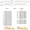
Similarities between the organization of the predicted replication initiation region of plasmid pPagL15_3 and the replication initiation region of plasmid-prophage P1. Arrows in the regions flanking the repA gene indicate the organization and direction of iteron sequences. Sequences of iterons of pPagL15_3 and prophage P1 and their logos are below the comparative scheme.
Figure 5
Figure 5
Curing of P. agglomerans L15 strain from the pPagL15_1 plasmid. Curing was based on screening for colonies that lost the ability to produce yellow stain (zeaxanthin) and to synthesize thiamine encoded by crtX and thiOSGF genes of pPagL15_1, respectively. The phenotypes of pPagL15_1-cured clones of L15 that were obtained after incubation of P. agglomerans L15 at 35°C for about 22 generations, plated on LB medium to obtain single colonies and screened for the presence of white clones. (Upper plate) shows differences in the colony color of pPagL15_1-cured clones and the parental L15 strain. Two plates at the bottom show the inability of pPagL15_1-cured clones to grow on a minimal M9 medium supplemented with glucose but without thiamine (left plate) and their ability to grow on a similar medium supplemented with thiamine (right plate).
Figure 6
Figure 6
Plaques formed by P1 c1-100 Tn9 IS1::kmR bacteriophage released from P. agglomerans L15 and P. agglomerans IPAG312 lysogens on the layer of E. coli N99 indicator strain cells. (A) IPAG312/P1. (B) L15/P1.

Similar articles

References

    1. Abby S. S., Néron B., Ménager H., Touchon M., Rocha E. P. (2014). MacSyFinder: a program to mine genomes for molecular systems with an application to CRISPR-Cas systems. PLoS ONE 9:e110726. 10.1371/journal.pone.0110726 - DOI - PMC - PubMed
    1. Abeles A. L., Friedman S. A., Austin S. J. (1985). Partition of unit-copy miniplasmids to daughter cells. III. The DNA sequence and functional organization of the P1 partition region. J. Mol. Biol. 185, 261–272. 10.1016/0022-2836(85)90402-4 - DOI - PubMed
    1. Adams M. H. (1959). Bacteriophages. New York, NY: Interscience Publishers Inc.
    1. Ahmed A. (1984). A deletion analysis of transposon Tn9. J. Mol. Biol. 173, 523–529. 10.1016/0022-2836(84)90395-4 - DOI - PubMed
    1. Altschul S. F., Madden T. L., Schäffer A. A., Zhang J., Zhang Z., Miller W., et al. . (1997). Gapped BLAST and PSI-BLAST: a new generation of protein database search programs. Nucleic Acids Res. 25, 3389–3402. 10.1093/nar/25.17.3389 - DOI - PMC - PubMed