. 2024 Jan 18;4(4):100467.
doi: 10.1016/j.xops.2024.100467. eCollection 2024 Jul-Aug.

Bispecific VEGF-A and Angiopoietin-2 Antagonist RO-101 Preclinical Efficacy in Model of Neovascular Eye Disease

Affiliations

Bispecific VEGF-A and Angiopoietin-2 Antagonist RO-101 Preclinical Efficacy in Model of Neovascular Eye Disease

Li Xu et al. Ophthalmol Sci. .

Abstract

Objective: To investigate preclinical data regarding the efficacy and biocompatibility of a bispecific protein, RO-101, with effects on VEGF-A and angiopoietin-2 (Ang-2) for use in retinal diseases.

Design: Experimental study.

Subjects: Brown Norway rats and New Zealand White Cross rabbits.

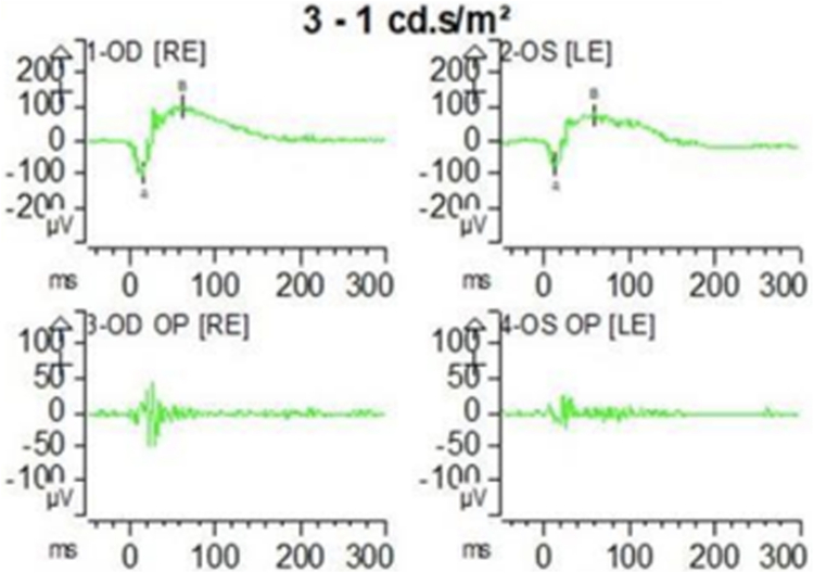
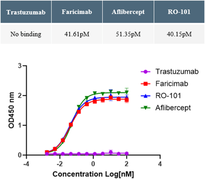
Methods: Preclinical study data of RO-101 in terms of target-specific enzyme-linked immunosorbent assay binding affinity to VEGF-A and Ang-2, vitreous half-life, inhibition of target-receptor interaction, laser choroidal neovascular membrane animal model, human umbilical vein endothelial cell migration, and biocompatibility was obtained. Where applicable, study data were compared with other anti-VEGF agents.

Main outcome measures: Binding affinity, half-life, biocompatibility, and efficacy of RO-101. Neovascularization prevention by RO-101.

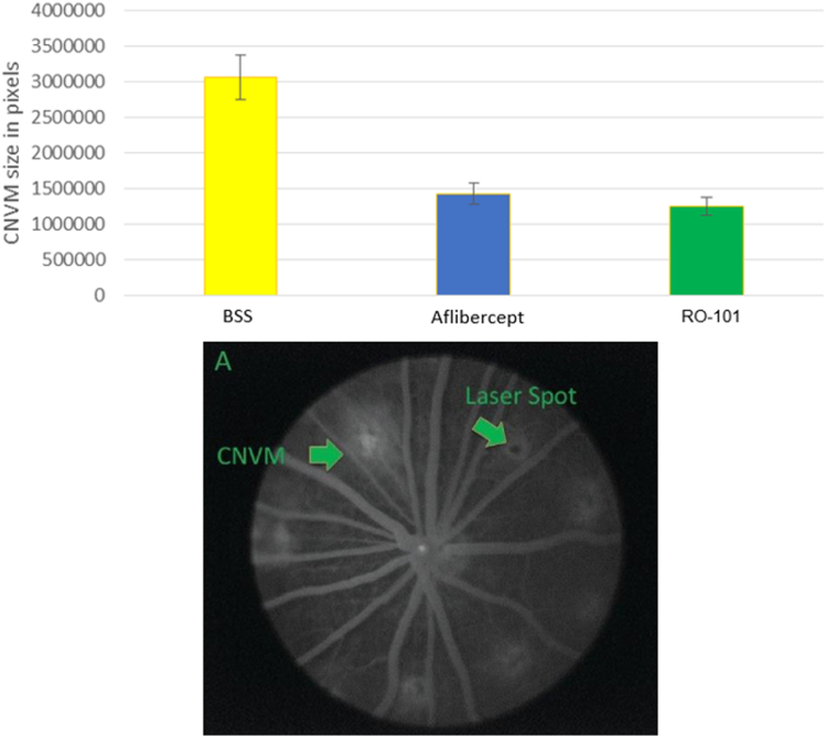
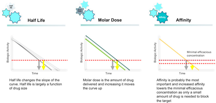
Results: RO-101 demonstrated a strong binding affinity for VEGF-A and Ang-2 and in vitro was able to inhibit binding to the receptor with higher affinity than faricimab. The half-life of RO-101 is comparable to or longer than current VEGF inhibitors used in retinal disease. RO-101 was found to be biocompatible with retinal tissue in Brown Norway rats. RO-101 was as effective or more effective than current anti-VEGF therapeutics in causing regression of neovascular growth in vivo.

Conclusions: RO-101 is a promising candidate for use in retinal diseases. In preclinical models, RO-101 demonstrated similar or higher regression of neovascular growth to current anti-VEGF therapeutics with comparable or longer half-life. It also demonstrates a strong binding affinity for VEGF-A and Ang-2. It also was shown to be biocompatible with retinal tissue in animal studies, indicating potential compatibility for use in humans.

Financial disclosures: Proprietary or commercial disclosure may be found in the Footnotes and Disclosures at the end of this article.

Keywords: AMD; Ang-2; Anti-VEGF; Bispecific; Surrobody.


Figures

Figure 1
Figure 1
The typical monoclonal antibody has a variable light chain (VL, green) that is unique and required for specific target binding. The Surrobody structure uses an invariant surrogate light chain (yellow), providing a universal partner to all heavy chain partners in the Surrobody library to streamline drug discovery and manufacturing.
Figure 2
Figure 2
Choroidal neovascular membrane (CNVM) size in pixels post injection of therapeutic agent was similar among RO-101 and aflibercept whereas both were shown to be superior to control (balanced saline solution, BSS) at preventing choroidal neovascularization (P value = 0.0015) using 2-way analysis of variance. There was no statistically significant difference between aflibercept and RO-101.
Figure 3
Figure 3
Human umbilical vein endothelial cell (HUVEC) wound closure analysis. Quantitative analysis of VEGF-A induced cell migration and effects of aflibercept, bevacizumab, or RO-101 treatment A, Representative images of wound closure of HUVECs after exposure to VEGF-A and either bevacizumab, aflibercept, or RO-101 at 0 and 12 hours B, Replicate analysis of images at 0 hours were deemed to be comparable.
Figure 4
Figure 4
Average cell counts in the ganglion cell layer, inner nuclear cells layer (INL), and outer nuclear cell layer (ONL) of the retina in Brown Norway rats after control or RO-101 treatment.
Figure 5
Figure 5
Electroretinogram readings were taken after exposure to RO-101 in the right eye (OD) and balanced saline solution (BSS) as the control in the left eye (OS). The left image shows the amplitude and response times of the experimental eye. The right images show the amplitude and response times of the control eye with BSS.
Figure 6
Figure 6
Binding affinities to VEGF as demonstrated by half maximal effective concentration of RO-101, aflibercept, faricimab, and negative control trastuzumab are shown.
Figure 7
Figure 7
Binding affinities to angiopoietin-2 as demonstrated by half maximal effective concentration of RO-101, aflibercept, faricimab, and negative control trastuzumab.
Figure 8
Figure 8
The inhibition of VEGF-A and VEGF R2/kinase insert domain receptor (receptor of VEGF-A) binding in enzyme-linked immunosorbent assay; is shown. IC50 = half-maximal inhibitory concentration.
Figure 9
Figure 9
The inhibition of angiopoietin-2 (Ang-2) and Tie-2 (receptor of Ang-2) binding in enzyme-linked immunosorbent assay is shown. IC50 = half-maximal inhibitory concentration.
Figure 10
Figure 10
Three different strategies to extend disease remission are to increase the molar dose, affinity, and half-life.

References

    1. Campochiaro P.A. Ocular neovascularization. J Mol Med (Berl) 2013;91:311–321. doi: 10.1007/s00109-013-0993-5. - DOI - PMC - PubMed
    1. Kvanta A., Algvere P.V., Berglin L., Seregard S. Subfoveal fibrovascular membranes in age-related macular degeneration express vascular endothelial growth factor. Invest Ophthalmol Vis Sci. 1996;37:1929–1934. - PubMed
    1. Lopez P.F., Sippy B.D., Lambert H.M., Thach A.B., Hinton D.R. Transdifferentiated retinal pigment epithelial cells are immunoreactive for vascular endothelial growth factor in surgically excised age-related macular degeneration-related choroidal neovascular membranes. Invest Ophthalmol Vis Sci. 1996;37:855–868. - PubMed
    1. Adamis A.P., Miller J.W., Bernal M.T., et al. Increased vascular endothelial growth factor levels in the vitreous of eyes with proliferative diabetic retinopathy. Am J Ophthalmol. 1994;118:445–450. doi: 10.1016/s0002-9394(14)75794-0. - DOI - PubMed
    1. Aiello L.P., Avery R.L., Arrigg P.G., et al. Vascular endothelial growth factor in ocular fluid of patients with diabetic retinopathy and other retinal disorders. N Engl J Med. 1994;331:1480–1487. doi: 10.1056/NEJM199412013312203. - DOI - PubMed