Molecular basis of hippopotamus ACE2 binding to SARS-CoV-2
- PMID: 38591877
- PMCID: PMC11092335
- DOI: 10.1128/jvi.00451-24
Molecular basis of hippopotamus ACE2 binding to SARS-CoV-2
Abstract
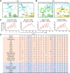
Severe acute respiratory syndrome coronavirus 2 (SARS-CoV-2) has a wide range of hosts, including hippopotami, which are semi-aquatic mammals and phylogenetically closely related to Cetacea. In this study, we characterized the binding properties of hippopotamus angiotensin-converting enzyme 2 (hiACE2) to the spike (S) protein receptor binding domains (RBDs) of the SARS-CoV-2 prototype (PT) and variants of concern (VOCs). Furthermore, the cryo-electron microscopy (cryo-EM) structure of the SARS-CoV-2 PT S protein complexed with hiACE2 was resolved. Structural and mutational analyses revealed that L30 and F83, which are specific to hiACE2, played a crucial role in the hiACE2/SARS-CoV-2 RBD interaction. In addition, comparative and structural analysis of ACE2 orthologs suggested that the cetaceans may have the potential to be infected by SARS-CoV-2. These results provide crucial molecular insights into the susceptibility of hippopotami to SARS-CoV-2 and suggest the potential risk of SARS-CoV-2 VOCs spillover and the necessity for surveillance.
Importance: The hippopotami are the first semi-aquatic artiodactyl mammals wherein SARS-CoV-2 infection has been reported. Exploration of the invasion mechanism of SARS-CoV-2 will provide important information for the surveillance of SARS-CoV-2 in hippopotami, as well as other semi-aquatic mammals and cetaceans. Here, we found that hippopotamus ACE2 (hiACE2) could efficiently bind to the RBDs of the SARS-CoV-2 prototype (PT) and variants of concern (VOCs) and facilitate the transduction of SARS-CoV-2 PT and VOCs pseudoviruses into hiACE2-expressing cells. The cryo-EM structure of the SARS-CoV-2 PT S protein complexed with hiACE2 elucidated a few critical residues in the RBD/hiACE2 interface, especially L30 and F83 of hiACE2 which are unique to hiACE2 and contributed to the decreased binding affinity to PT RBD compared to human ACE2. Our work provides insight into cross-species transmission and highlights the necessity for monitoring host jumps and spillover events on SARS-CoV-2 in semi-aquatic/aquatic mammals.
Keywords: ACE2; SARS-CoV-2; cross-species recognition; cryo-EM structure; hippopotamus; receptor binding domain (RBD); spike (S).
Conflict of interest statement
The authors declare no conflict of interest.
Figures


Similar articles
-
Structural basis and analysis of hamster ACE2 binding to different SARS-CoV-2 spike RBDs.J Virol. 2024 Mar 19;98(3):e0115723. doi: 10.1128/jvi.01157-23. Epub 2024 Feb 2. J Virol. 2024. PMID: 38305152 Free PMC article.
-
The binding and structural basis of fox ACE2 to RBDs from different sarbecoviruses.Virol Sin. 2024 Aug;39(4):609-618. doi: 10.1016/j.virs.2024.06.004. Epub 2024 Jun 10. Virol Sin. 2024. PMID: 38866203 Free PMC article.
-
V367F Mutation in SARS-CoV-2 Spike RBD Emerging during the Early Transmission Phase Enhances Viral Infectivity through Increased Human ACE2 Receptor Binding Affinity.J Virol. 2021 Jul 26;95(16):e0061721. doi: 10.1128/JVI.00617-21. Epub 2021 Jul 26. J Virol. 2021. PMID: 34105996 Free PMC article.
-
Structural basis of severe acute respiratory syndrome coronavirus 2 infection.Curr Opin HIV AIDS. 2021 Jan;16(1):74-81. doi: 10.1097/COH.0000000000000658. Curr Opin HIV AIDS. 2021. PMID: 33186231 Review.
-
Mutations in the SARS-CoV-2 spike receptor binding domain and their delicate balance between ACE2 affinity and antibody evasion.Protein Cell. 2024 May 28;15(6):403-418. doi: 10.1093/procel/pwae007. Protein Cell. 2024. PMID: 38442025 Free PMC article. Review.
Cited by
-
Receptor binding and structural basis of raccoon dog ACE2 binding to SARS-CoV-2 prototype and its variants.PLoS Pathog. 2024 Dec 5;20(12):e1012713. doi: 10.1371/journal.ppat.1012713. eCollection 2024 Dec. PLoS Pathog. 2024. PMID: 39637248 Free PMC article.
References
MeSH terms
Substances
LinkOut - more resources
Full Text Sources
Miscellaneous

