. 2024 May 23;206(5):e0002424.
doi: 10.1128/jb.00024-24. Epub 2024 Apr 9.

Siderophore-mediated iron acquisition by Klebsiella pneumoniae

Affiliations

Siderophore-mediated iron acquisition by Klebsiella pneumoniae

Ashish Kumar et al. J Bacteriol. .

Abstract

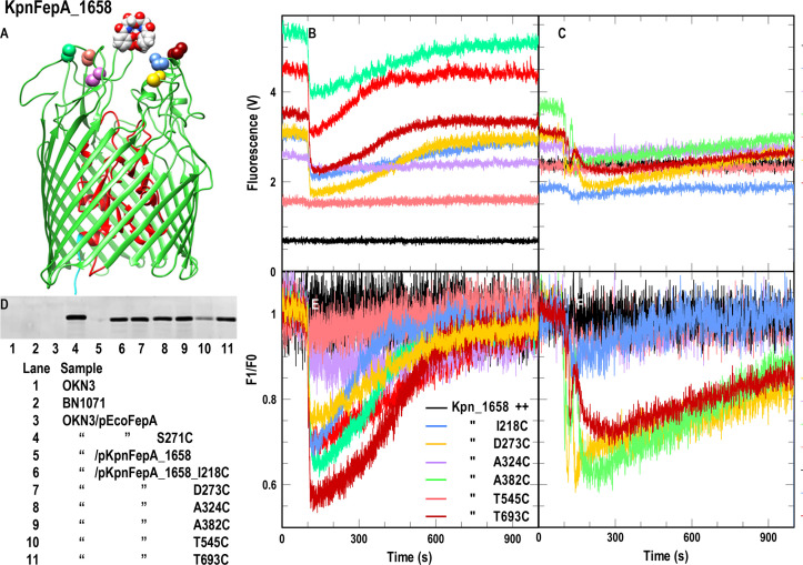
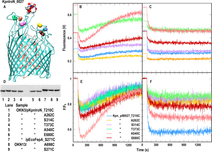
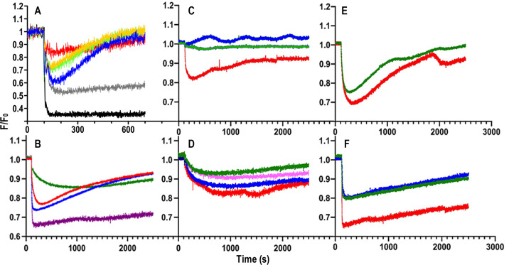
Microbes synthesize and secrete siderophores, that bind and solubilize precipitated or otherwise unavailable iron in their microenvironments. Gram (-) bacterial TonB-dependent outer membrane receptors capture the resulting ferric siderophores to begin the uptake process. From their similarity to fepA, the structural gene for the Escherichia coli ferric enterobactin (FeEnt) receptor, we identified four homologous genes in the human and animal ESKAPE pathogen Klebsiella pneumoniae (strain Kp52.145). One locus encodes IroN (locus 0027 on plasmid pII), and three other loci encode other FepA orthologs/paralogs (chromosomal loci 1658, 2380, and 4984). Based on the crystal structure of E. coli FepA (1FEP), we modeled the tertiary structures of the K. pneumoniae FepA homologs and genetically engineered individual Cys substitutions in their predicted surface loops. We subjected bacteria expressing the Cys mutant proteins to modification with extrinsic fluorescein maleimide (FM) and used the resulting fluorescently labeled cells to spectroscopically monitor the binding and transport of catecholate ferric siderophores by the four different receptors. The FM-modified FepA homologs were nanosensors that defined the ferric catecholate uptake pathways in pathogenic strains of K. pneumoniae. In Kp52.145, loci 1658 and 4984 encoded receptors that primarily recognized and transported FeEnt; locus 0027 produced a receptor that principally bound and transported FeEnt and glucosylated FeEnt (FeGEnt); locus 2380 encoded a protein that bound ferric catecholate compounds but did not detectably transport them. The sensors also characterized the uptake of iron complexes, including FeGEnt, by the hypervirulent, hypermucoviscous K. pneumoniae strain hvKp1.

Importance: Both commensal and pathogenic bacteria produce small organic chelators, called siderophores, that avidly bind iron and increase its bioavailability. Klebsiella pneumoniae variably produces four siderophores that antagonize host iron sequestration: enterobactin, glucosylated enterobactin (also termed salmochelin), aerobactin, and yersiniabactin, which promote colonization of different host tissues. Abundant evidence links bacterial iron acquisition to virulence and infectious diseases. The data we report explain the recognition and transport of ferric catecholates and other siderophores, which are crucial to iron acquisition by K. pneumoniae.

Keywords: ESKAPE pathogen; Klebsiella pneumoniae; TonB-dependent iron acquisition; ferric enterobactin; fluorescent sensors; iron transport; siderophore antibiotic conjugates; siderophores; site-directed mutagenesis.


Conflict of interest statement

The authors declare no conflict of interest.

Figures

Fig 1
Fig 1
FeEnt transport by KpnFepA_1658. (A and D) Site-directed fluorescence labeling of KpnFepA_1658 Cys mutants. (A) Location of engineered Cys residues in KpnFepA_1568 (see the legend in panel E for color-coding). (D) After growth of E. coli OKN3/pKpnFepA_1658 and its Cys mutants in iron-deficient MOPS media, we exposed the cells to FM and evaluated labeling by a fluorescence scan of their SDS-PAGE-resolved cell envelopes. All the mutant KpnFepAs were well labeled, T545C slightly less intensely than the others. (B and E) Spectroscopic observations of FeEnt uptake by E. coli OKN3/pKpnFepA_1658 Cys mutants. To 5 × 10⁶ cells in a stirred 3-mL cuvette containing 2-mL of PBS + 0.4% glucose, we added FeEnt to 10 nM at 100 s and collected the raw data (B) for 1,000 s. FeEnt binding quenched the fluorescence of most mutant proteins, but fluorescence intensity recovered as the bacteria transported FeEnt and depleted it from the ambient solution, The plotted data represent the mean values from triplicate time courses of each of individual mutant. (E) We also normalized the data to the initial cellular fluorescence (F/F0). (C and F) Spectroscopic observations of FeEnt uptake by pKpnFepA_1658 Cys mutants in K. pneumoniae. We repeated the experiment for the same Cys mutants in K. pneumoniae strain KKN5, with similar results. Panels (B andAND C) depict the fluorescence quenching time courses as raw data: each of the fluorescently labeled Cys mutants had a different initial intensity and different extents of quenching in response to the binding/transport of FeEnt. In panels (E andAND F), the initial intensity of each of the fluoresceinated Cys mutants is normalized to an arbitrary value of 1.0, to better compare their responses to the subsequent binding and transport of FeEnt.
Fig 2
Fig 2
FeEnt transport and FeGEnt binding by KpnIroN_0027. (A and D) Site-directed fluorescence labeling of KpnIroN Cys mutants. (AND) Location of engineered Cys residues in KpnIroN_0027 (see the legend in panel E for color-coding). (D) We repeated the protocol described in Fig. 1 with E. coli OKN3/pKpn_0027 and its Cys derivatives; all the mutants were well labeled. (B and E) Spectroscopic observations of FeEnt uptake by E. coli OKN3/pKpnIroN_0027. Following the protocol described in Fig. 1, FM-labeled KpnIroN Cys mutants efficiently transported FeEnt, even when expressed in E. coli: raw (B) and normalized (E) data. (C and F) Spectroscopic observations of FeGEnt binding, but not uptake, by E. coli OKN3/pKpnIroN_0027 and its Cys mutants. We repeated the experiment described in panels (B and E) with FeGEnt: raw (C) and normalized (F) data. FeGEnt binding rapidly quenched fluoresceinated KpnIroN Cys mutants, but the absence of lactone esterases IroD and IroE from the E. coli periplasm prevented its cellular uptake.
Fig 3
Fig 3
Recognition of ferric catecholates by different KpnFepAs. We engineered Cys substitutions in the structural genes of KpnIroN_0027, KpnFepA_1658, KpnFepA_2380 and KpnFepA_4984, carried on pITS23 (see Results and Supplemental Materials). After labeling E. coli OKN13 cells expressing the Cys mutant proteins with FM, we assessed their fluorescence intensity and extent of quenching during ligand binding to find the best candidates for further study: KpnFepA_IroN_T210C-FM (panel A); KpnFepA_1658_A382-FM (B); KpnFepA_2380_T255C-FM (C); KpnFepA_4984_A390C-FM (D). With these constructs, we measured the adsorption of ferric catecholate siderophores: FeEnt, Ent, FeEnt*, FeGEnt, FeDHBA, FeCrn. Each data point represents the mean of triplicate repetitions, with associated standard deviations. For KpnIroN_0027 the KD values (nM) from the fitted curves are FeEnt 0.4 ± 0.1, FeGEnt 4 ± 1, FeEnt* 9.4 ± 3.5, Ent 39 ± 15. For KpnFepA_1658 the KD values (nM) from the fitted curves are FeEnt 0.6 ± 0.05, FeEnt* 27 ± 6, Ent 92 ± 21, FeGEnt 112 ± 15. For KpnFepA_2380, the KD values (μM) from the fitted curves are FeEnt 16 ± 3, FeCrn 13 ± 2.5, and FeDHBA 19 ± 2.8. For KpnFepA_4984, the KD values (nM) from the fitted curves are FeEnt 14 ± 4.5, FeEnt* 178 ± 51, FeGEnt 872 ± 64, Ent 3,171 ± 808.
Fig 4
Fig 4
FD sensor analysis of ferric siderophore uptake by Gram (−) ESKAPE bacteria. (A) Uptake of FeEnt. 107 cells of the decoy sensor strain E. coli OKN13/pKpnfepA_1658-FM were mixed with 107 cells of individual test bacteria in 2 mL of PBS + 0.4% glucose in a quartz cuvette. At t = 0 we began monitoring fluorescence emissions at 520 nm; at 100 s we added FeEnt to 5 nM and observed the ensuing changes in fluorescence intensity: black, sensor only; gray, K. pneumoniae KKN5; blue, E. coli OKN3/pKpnFepA_1658; red, K. pneumoniae 52.145; gold, A. baumannii; green, P. aeruginosa PA01. (B) Ferric siderophore uptake by hypermucoviscous K. pneumoniae. 107 cells of transport-defective E. coli OKN13, harboring plasmids expressing appropriate decoy sensors, were mixed with 107 cells of K. pneumoniae hvKp1 and monitored as described above in response to the addition of the following ferric siderophores at 100 s: blue, FeEnt; green, FeGEnt; red, FeAbn; purple, FeYbt. Panels (C–F) Effects of SCN on uptake of ferric siderophores by hvKp1. In each study 107 cells of E. coli OKN13, harboring plasmids expressing appropriate decoy sensors, were mixed with 107 cells of K. pneumoniae hvKp1, in the absence (red) or presence of human SCN (blue, 0.005 µM; magenta, 0.05; green, 0.5 µM). We monitored fluorescence emissions in response to the addition of 10 nM ferric siderophores at t = 100 s: (C) FeEnt; (D) FeGEnt; (E) FeAbn; (F) FeYbt.

Similar articles

Cited by

References

    1. Holden VI, Bachman MA. 2015. Diverging roles of bacterial siderophores during infection. Metallomics 7:986–995. doi:10.1039/C4MT00333K - DOI - PubMed
    1. Palacios M, Broberg CA, Walker KA, Miller VL. 2017. A serendipitous mutation reveals the severe virulence defect of a Klebsiella pneumoniae fepB mutant. mSphere 2:e00341-17. doi:10.1128/mSphere.00341-17 - DOI - PMC - PubMed
    1. Bailey DC, Drake EJ, Grant TD, Gulick AM. 2016. Structural and functional characterization of aerobactin synthetase Iuca from a hypervirulent pathotype of Klebsiella pneumoniae. Biochemistry 55:3559–35570. doi:10.1021/acs.biochem.6b00409 - DOI - PMC - PubMed
    1. Paczosa MK, Mecsas J. 2016. Klebsiella pneumoniae: going on the offense with a strong defense. Microbiol Mol Biol Rev 80:629–661. doi:10.1128/MMBR.00078-15 - DOI - PMC - PubMed
    1. Navon-Venezia S, Kondratyeva K, Carattoli A. 2017. Klebsiella pneumoniae: a major worldwide source and shuttle for antibiotic resistance. FEMS Microbiol Rev 41:252–275. doi:10.1093/femsre/fux013 - DOI - PubMed

Publication types

MeSH terms

LinkOut - more resources