. 2024 Feb 24;13(5):1286.
doi: 10.3390/jcm13051286.

Navigating Complexity: A Comprehensive Approach to Middle Cerebral Artery Aneurysms

Affiliations

Navigating Complexity: A Comprehensive Approach to Middle Cerebral Artery Aneurysms

Anna Maria Auricchio et al. J Clin Med. .

Abstract

Background: The concept of aneurysm "complexity" has undergone significant changes in recent years, with advancements in endovascular treatments. However, surgical clipping remains a relevant option for middle cerebral artery (MCA) aneurysms. Hence, the classical criteria used to define surgically complex MCA aneurysms require updating. Our objective is to review our institutional series, considering the impacts of various complexity features, and provide a treatment strategy algorithm. Methods: We conducted a retrospective review of our institutional experience with "complex MCA" aneurysms and analyzed single aneurysmal-related factors influencing treatment decisions. Results: We identified 14 complex cases, each exhibiting at least two complexity criteria, including fusiform shape (57%), large size (35%), giant size (21%), vessel branching from the sac (50%), intrasaccular thrombi (35%), and previous clipping/coiling (14%). In 92% of cases, the aneurysm had a wide neck, and 28% exhibited tortuosity or stenosis of proximal vessels. Conclusions: The optimal management of complex MCA aneurysms depends on a decision-making algorithm that considers various complexity criteria. In a modern medical setting, this process helps clarify the choice of treatment strategy, which should be tailored to factors such as aneurysm morphology and patient characteristics, including a combination of endovascular and surgical techniques.

Keywords: complex aneurysms; complexity criteria; endovascular treatment; intracranial aneurysms; middle cerebral artery; surgical clipping; treatment algorithm; unruptured.


Conflict of interest statement

The authors declare no conflicts of interest.

Figures

Figure 1
Figure 1
Flow chart for shape management: fusiform and large/giant complex intracranial aneurysms of the MCA.
Figure 2
Figure 2
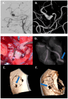
Case 1: Bypass with complete trapping in a large distal fusiform 17 mm aneurysm of M4 in a 24-year-old man. (A,B) Preoperative DSA with 3D reconstruction documenting M4 tract with a distal stenotic vessel. (C,D) Intraoperative view of end-to-side superficial temporal artery–MCA bypass anastomosis with complete trapping of the aneurysm by two clips. (E,F) Postoperative DSA documenting the correct flow replacement with clear view of right superficial temporal artery–MCA bypass through the bone hole (blue arrows).
Figure 3
Figure 3
Flow chart for complex aneurysms of the MCA with vessels originating from the dome.
Figure 4
Figure 4
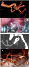
Case 2: complex clipping in a fusiform 7 mm M2 aneurysm with vessel branching from the dome, in a 35-year-old woman. (A) Preoperative CTA with a 3D reconstruction. (B) Intraoperative view of two tandem clips used to reconstruct the parent vessel. (C,D) Complete exclusion of the sac, documented by the postoperative CTA, with a 3D reconstruction.
Figure 5
Figure 5
Flow chart for hemodynamic changes of the sac: thrombosed and/or residual/recurrent in previously treated complex aneurysms of the MCA.
Figure 6
Figure 6
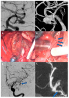
Case 3: Thrombectomy with bypass in a giant, fusiform, and partially thrombosed 21 mm M2 aneurysm carried by a 68-year-old woman with a history of seizures. (A,B) Preoperative DSA with 3D reconstruction reporting the giant, fusiform, and partially thrombosed aneurysm at left M2. (C) Proximal control of M2 performed with a temporary clipping and progressive debulking of intrasaccular thrombus using cavitronic ultrasonic surgical aspirator until visualization of the ostia (blue arrows). (D) An intracranial end-to-side bypass between left temporal M3 branch and distal M2 branch. (E) DSA confirming bypass flow and flow replacement (blue arrows). (F) A 6-month follow-up CTA documenting the complete exclusion of the aneurysm (blue arrows).
Figure 7
Figure 7
Case 4: complex clipping in previously coiled, giant, partially thrombosed 25 mm right MCA bifurcation aneurysm in a 76-year-old woman. (A,B) Preoperative presentation with DSA and 3D model of the previously coiled sac with a recurrence at 12 months. (C) Surgical clipping with progressive removal of thrombus and coils assisted by ultrasonic aspirator. (D) 3D reconstruction DSA confirming the complete exclusion of the sac.

References

    1. Hanel R.A., Spetzler R.F. Surgical Treatment of Complex Intracranial Aneurysms. Neurosurgery. 2008;62:1289–1297; discussion 1297–1299. doi: 10.1227/01.NEU.0000318092.86562.F. - DOI - PubMed
    1. Ravina K., Rennert R.C., Kim P.E., Strickland B.A., Chun A., Russin J.J. Orphaned Middle Cerebral Artery Side-to-Side In Situ Bypass as a Favorable Alternative Approach for Complex Middle Cerebral Artery Aneurysm Treatment: A Case Series. World Neurosurg. 2019;130:e971–e987. doi: 10.1016/j.wneu.2019.07.053. - DOI - PubMed
    1. Etminan N., Brown R.D., Beseoglu K., Juvela S., Raymond J., Morita A., Torner J.C., Derdeyn C.P., Raabe A., Mocco J., et al. The Unruptured Intracranial Aneurysm Treatment Score. Neurology. 2015;85:881–889. doi: 10.1212/WNL.0000000000001891. - DOI - PMC - PubMed
    1. Wessels L., Fekonja L.S., Achberger J., Dengler J., Czabanka M., Hecht N., Schneider U., Tkatschenko D., Schebesch K.-M., Schmidt N.O., et al. Diagnostic Reliability of the Berlin Classification for Complex MCA Aneurysms-Usability in a Series of Only Giant Aneurysms. Acta Neurochir. 2020;162:2753–2758. doi: 10.1007/s00701-020-04565-6. - DOI - PMC - PubMed
    1. Inci S., Karakaya D. Kissing Aneurysms: Radiological and Surgical Difficulties in 30 Operated Cases and a Proposed Classification. World Neurosurg. 2021;155:e83–e94. doi: 10.1016/j.wneu.2021.08.003. - DOI - PubMed

LinkOut - more resources