. 2024 Apr 9;17(831):eadh1922.
doi: 10.1126/scisignal.adh1922. Epub 2024 Apr 9.

Blocking lipid synthesis induces DNA damage in prostate cancer and increases cell death caused by PARP inhibition

Affiliations

Blocking lipid synthesis induces DNA damage in prostate cancer and increases cell death caused by PARP inhibition

Caroline Fidalgo Ribeiro et al. Sci Signal. .

Abstract

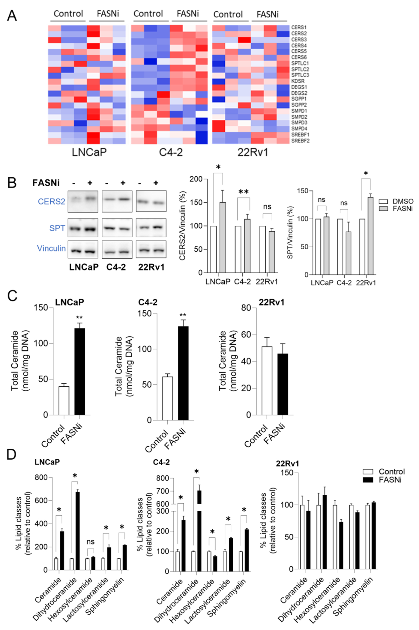
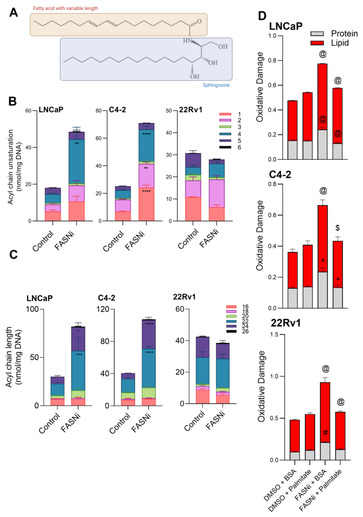
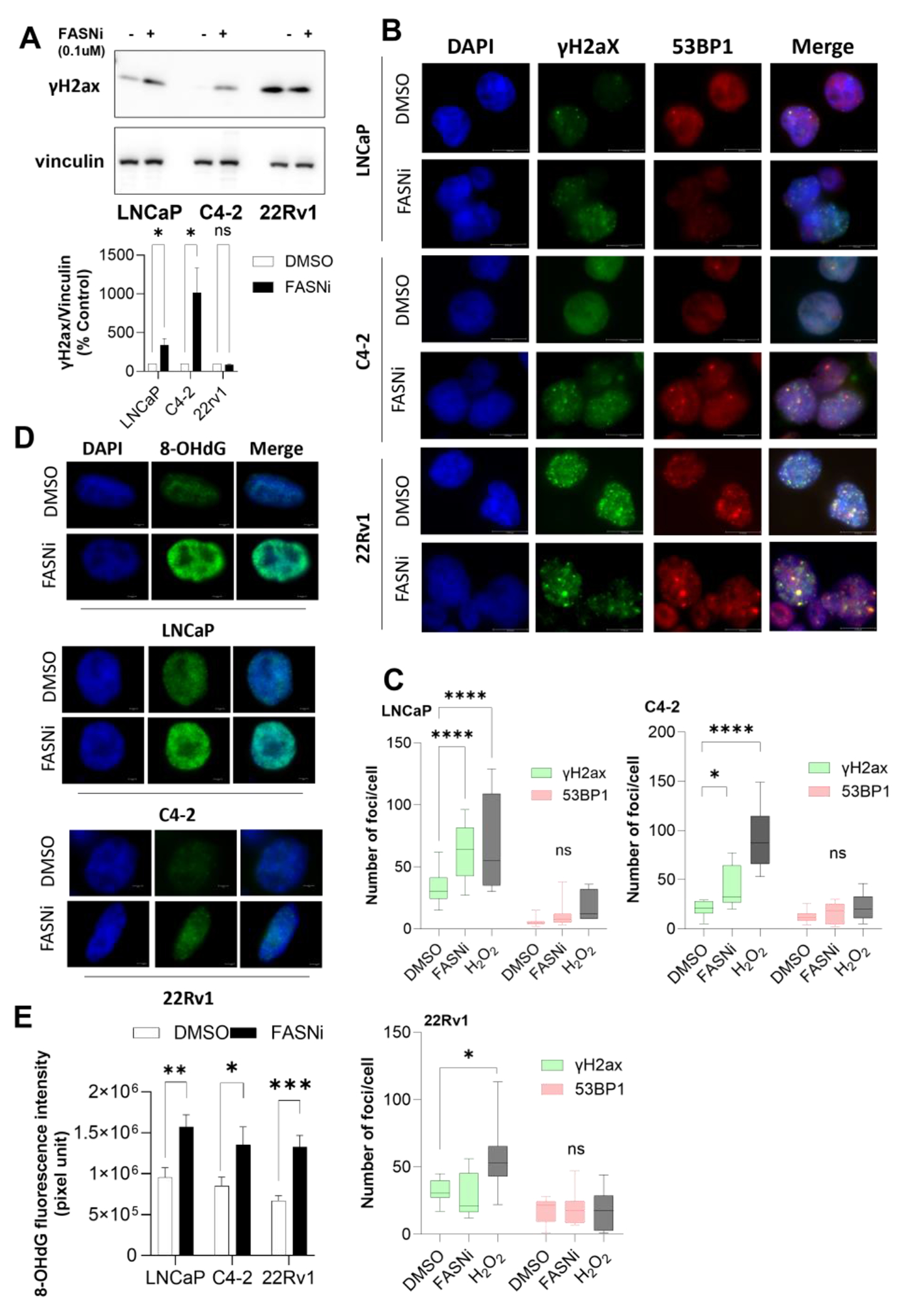
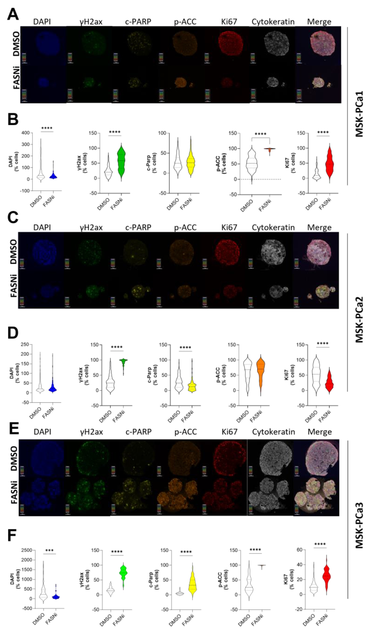
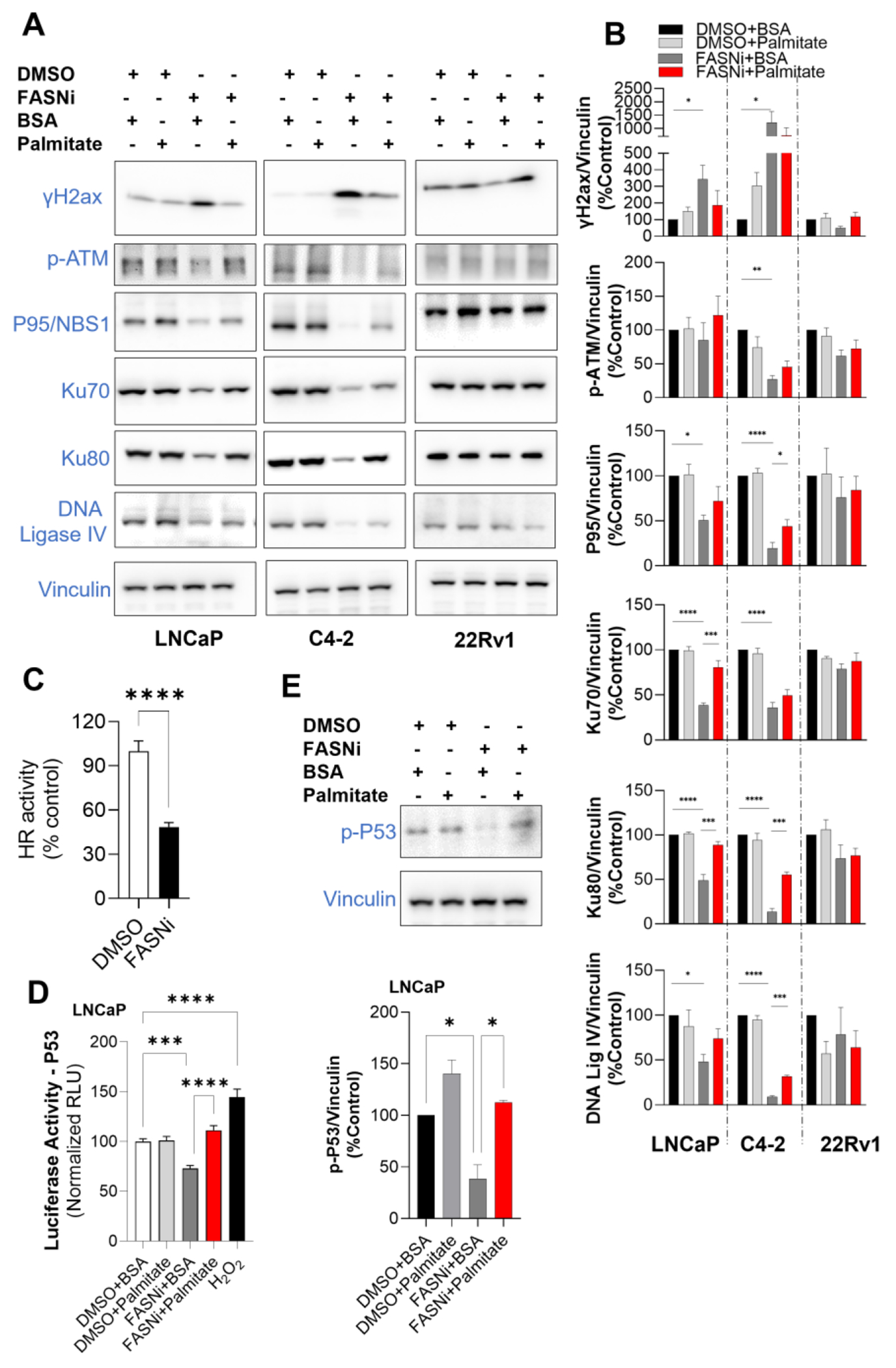
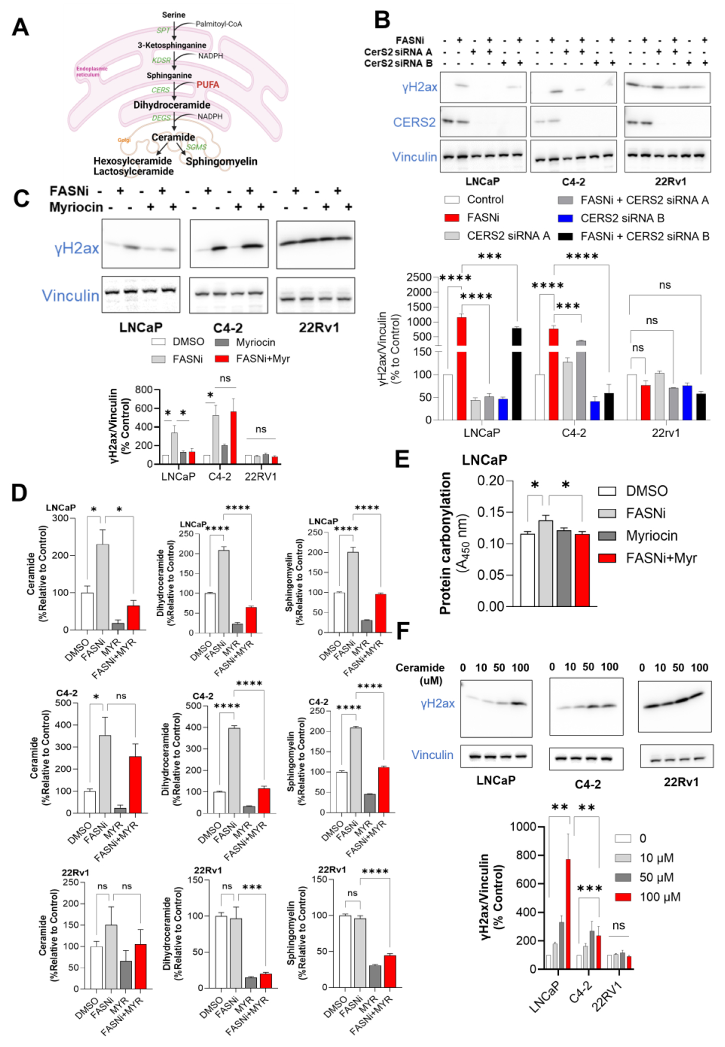
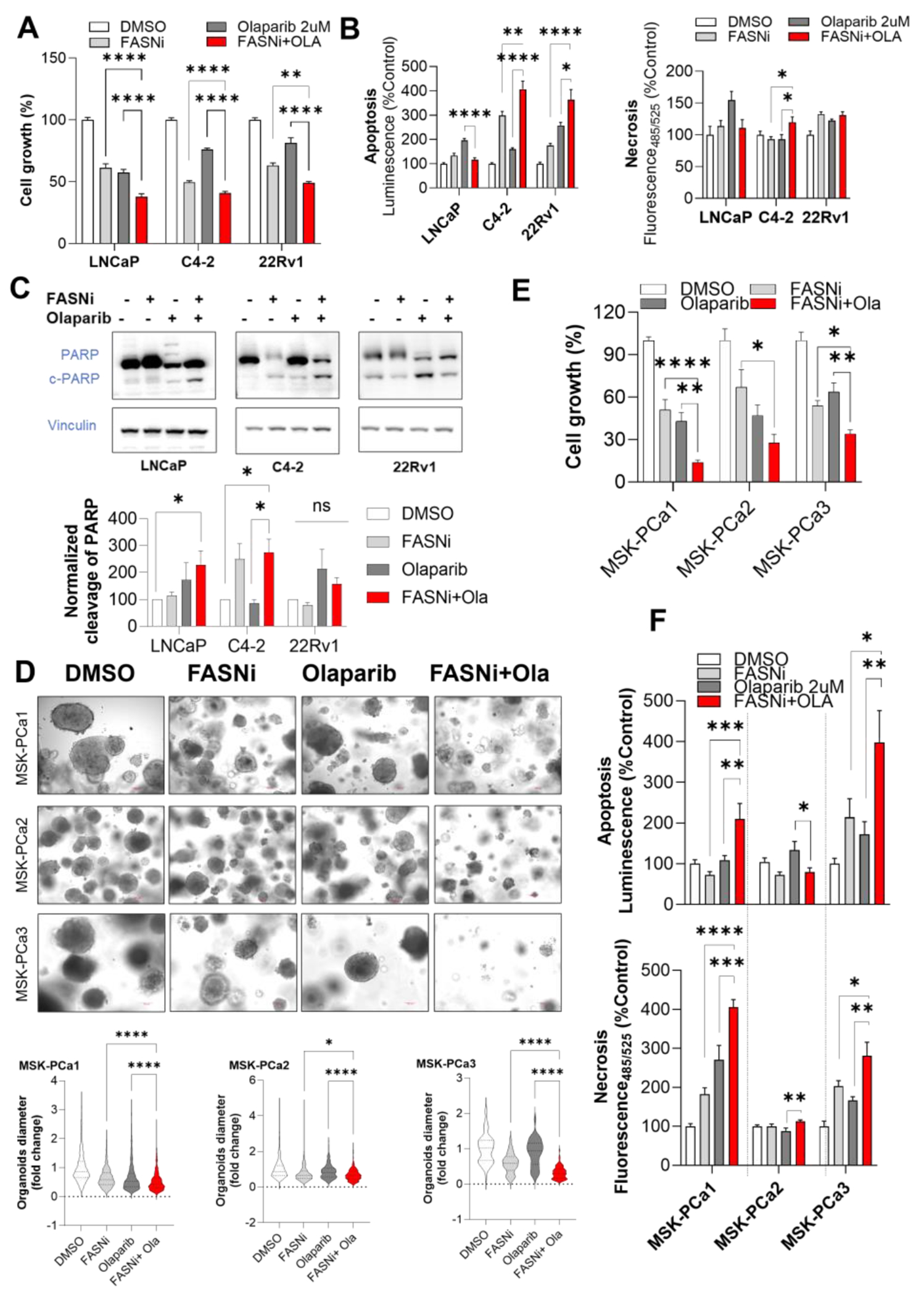
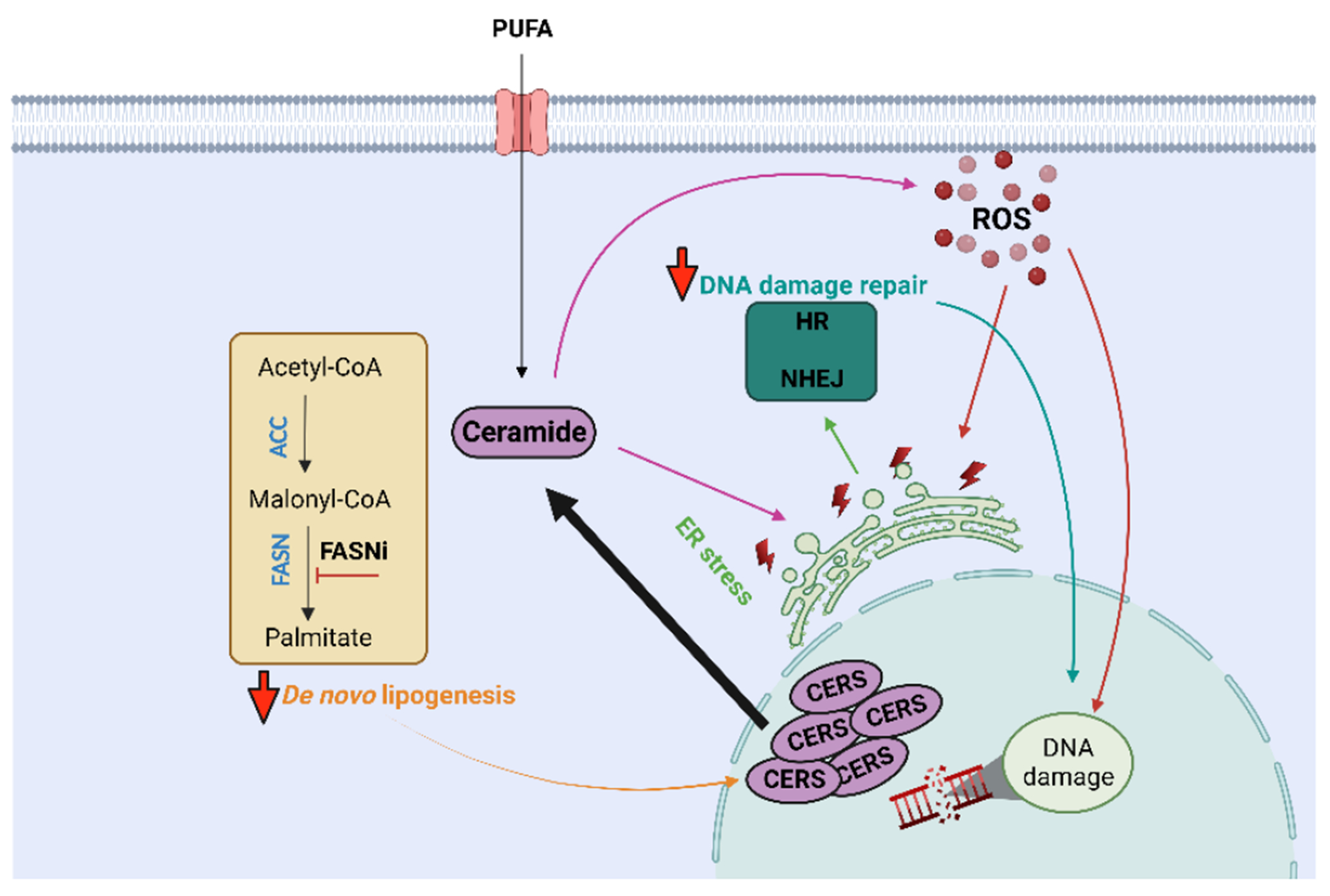
Androgen deprivation therapy (ADT) is the primary treatment for prostate cancer; however, resistance to ADT invariably develops, leading to castration-resistant prostate cancer (CRPC). Prostate cancer progression is marked by increased de novo synthesis of fatty acids due to overexpression of fatty acid synthase (FASN), making this enzyme a therapeutic target for prostate cancer. Inhibition of FASN results in increased intracellular amounts of ceramides and sphingomyelin, leading to DNA damage through the formation of DNA double-strand breaks and cell death. We found that combining a FASNi with the poly-ADP ribose polymerase (PARP) inhibitor olaparib, which induces cell death by blocking DNA damage repair, resulted in a more pronounced reduction in cell growth than that caused by either drug alone. Human CRPC organoids treated with a combination of PARP and FASNi were smaller, had decreased cell proliferation, and showed increased apoptosis and necrosis. Together, these data indicate that targeting FASN increases the therapeutic efficacy of PARP inhibitors by impairing DNA damage repair, suggesting that combination therapies should be explored for CRPC.


Conflict of interest statement

Competing interests

No conflict of interest to be reported.

Figures

Figure 1.
Figure 1.. FASN inhibition and sphingolipid metabolism.
(A) Heatmap of transcriptional regulation of genes involved in de novo synthesis of ceramide, salvage or sphingomyelinase pathways after FASNi treatment. Normalized counts are shown (n=3 biological replicates). Multiple unpaired t-tests, significant differential values reported as * p<0.05. (B) Representative immunoblotting with densitometric analysis (right) of Ceramide Synthase 2 (CERS2) and Serine Palmitoyltransferase (SPT) amounts in cells treated with FASNi. Data plotted as % relative to control of normalized marker expression (marker/vinculin) (n= 3 biological replicates, Mean ± SEM). Multiple unpaired t-tests, *q<0.05, **q<0.01.(C) Total ceramide amounts after FASN inhibition. Lipid amounts are shown as sum notation of ceramide, dihydroceramide, hexosylceramides and lactosylceramides, expressed as nmol/mg of DNA (n=3 biological replicates, mean ± SEM). Unpaired t-test, **p<0.01. (D) Presence of sphingolipid species after FASN blockade. Total combined sum compositions of indicated lipid classes is shown as % relative to control (n=3 biological replicates, mean ± SEM). Multiple unpaired t-tests, *q<0.05.
Figure 2.
Figure 2.. Depletion of palmitate and its effect on ceramide acyl chain unsaturation and length.
(A) Representative chemical structure of ceramide, consisting of a sphingosine and a fatty acid residue that can vary in length and unsaturation level. (B) Number of acyl chain unsaturation in ceramides after FASNi treatment. Ceramides containing fatty acids with the same number of unsaturations (1 to 6) were summed together, data expressed as nmol/mg of DNA (n=3 biological replicates, mean ± SEM). Two-way ANOVA with Šídák’s test, **p<0.01, ****p<0.0001. (C) Carbon length of acyl chain in ceramides following FASNi treatment. Ceramides containing fatty acids with the same chain length (16 to 26) were summed together, data expressed as nmol/mg of DNA (n=3 biological replicates, mean ± SEM). Two-way ANOVA with Šídák’s test, *p<0.05, **p<0.01, ****p<0.0001. (D) Oxidative damage of protein and lipid assessed after FASNi treatment combined with palmitate rescue. Results for protein carbonylation are expressed as normalized absorbance at 450 nm (n=12 replicates over 3 independent experiments, mean ± SEM). Lipid oxidative results are expressed as normalized ratio of fluorescence of the reduced to the oxidized probe (n=12 replicates over 3 independent experiments, mean ± SEM). Two-way ANOVA with Tukey’s test, * p<0.05, # p<0.01, $ p<0.001, @ p<0.0001.
Figure 3.
Figure 3.. FASN inhibition and DNA oxidative damage.
(A) Representative immunoblotting and densitometric analysis (bottom) of DSB marker, phosphorylated-Histone H2A.X (Ser139) (γH2ax ) in cells treated with FASNi. Data plotted as % relative to control of normalized marker expression (n=4-6 biological replicates, Mean ± SEM). Multiple unpaired t-tests, *q<0.05. (B) Representative immunofluorescence of γH2ax (green), 53BP1 (red) and DAPI (blue) in cells treated with FASNi. Scale bar of 15.04 μm. (C) Quantification of γH2ax and 53BP1 foci number per cell (n=20 cells over 3 independent experiments, Min to Max). Positive control (hydrogen peroxide) shown as gray bars. Two-way ANOVA with Dunnett’s test, *p<0.05, ****p<0.0001. (D) Representative immunofluorescence of 8-OHdG (green) and DAPI (blue) in cells treated with FASNi. Images acquired at 40X, scale bar of 3 μm. (E) Quantification of 8-OHdG nuclear fluorescence. Data shown as corrected total nuclear fluorescence (CTNF) normalized to the nuclear area (n=10 cells over 3 independent experiments, mean ± SEM). Multiple unpaired t-test, *q<0.05, **q<0.01, ***q<0.001.
Figure 4.
Figure 4.. FASN inhibition and DNA damage in 3D organoid models of CRPC.
(A) Representative multiplex immunofluorescence of phosphorylated-Histone H2A.X (Ser139) (γH2ax ), cleaved-PARP (c-PARP), phosphorylated Acetyl-CoA Carboxylase (Ser79) (p-ACC), Ki67 and Cytokeratin HMW in MSK-PCa1 organoids treated with FASNi. Scale bar of 50 μm (n=3 independent experiments). (B) Individual marker expression quantification analysis of MSK-PCa1 after FASNi treatment. Whole slide scanned computationally under the same parameters and marker intensity calculated using Halo Image Analysis Platform (Indica Labs). Data shown as % of positively stained cells (organoids analyzed n=94 for DMSO and n=131 for FASNi, over 3 independent experiments, violin plots). Unpaired t-test, ****p<0.0001. (C) Representative multiplex immunofluorescence of γH2ax, c-PARP, p-ACC, Ki67 and Cytokeratin HMW (n=3 independent experiments) and (D) individual marker expression quantification analysis of MSK-PCa2 organoids after FASNi treatment. Data shown as % of positively stained cells (organoids analyzed n=184 for DMSO and n=95 for FASNi, over 3 independent experiments, violin plots). Unpaired t-test, ****p<0.0001. (E) Representative multiplex immunofluorescence of γH2ax, c-PARP, p-ACC, Ki67 and Cytokeratin HMW (n=3 independent experiments) and (F) individual marker expression quantification analysis of MSK-PCa3 organoids after FASNi treatment. Data shown as % of positively stained cells (organoids analyzed n=41 for DMSO and n=48 for FASNi, over 3 independent experiments, violin plots). Unpaired t-test, ***p<0.001, ****p<0.0001.
Figure 5.
Figure 5.. DNA damage repair pathways HR and NHEJ following FASN blockade.
(A) Representative immunoblotting of HR and NHEJ markers with (B) densitometric analysis of cells treated with FASNi and palmitate. Data plotted as % relative to control of normalized marker expression (marker/vinculin) (n=3-9 over 3 independent experiments, Mean ± SEM). One-way ANOVA with Tukey’s test, *p<0.05, **p<0.01, ***p<0.001, ****p<0.0001. (C) Homologous Recombination activity in cells treated with FASNi. Data shown as % relative to control of relative recombination. (n=12 over 3 independent experiments, Mean ± SEM). Unpaired t-test, ****p<0.0001. (D) Luciferase activity of P53 response element in cells treated with FASNi and palmitate. Data shown as normalized Firefly/Renilla luminescence (n=8 over 2 independent experiments, Mean ± SEM). One-way ANOVA with Tukey’s test, ***p<0.001, ****p<0.0001. (E) Representative immunoblotting of phosphorylated P53 (Ser15) (p-P53) in cells treated with FASNi and palmitate. Densitometric analysis (bottom) is also shown, with data plotted as % relative to control of normalized p-P53 to vinculin (n= 3 biological replicates , Mean ± SEM). One-way ANOVA with Tukey’s test, *p<0.05.
Figure 6.
Figure 6.. Ceramide synthesis and FASNi-induced DNA damage.
(A) Schematic representation of de novo synthesis of ceramide. Serine palmitoyltransferase (SPT), 3-Ketodihydrosphingosine Reductase (KDSR), Ceramide Synthase (CERS), Dihydroceramide Desaturase (DEGS), Sphingomyelin Synthase (SGMS). (B) Representative immunoblotting and densitometric analysis (bottom) of γH2ax following FASNi treatment in cells with CE RS2 knock-down. Data plotted as % relative to control of normalized marker expression (n= 3 biological replicates, Mean ± SEM). One-way ANOVA with Bartlett’s test, *p<0.05, ***p<0.001, ****p<0.0001 (C) Representative immunoblotting and densitometric analysis (bottom) of γH2ax following treatment with FASNi and myriocin. Data shown as % relative to control (n = 3 biological replicates, mean ± SEM). One-way ANOVA with Tukey’s test, *p<0.05, ***p<0.001, ****p<0.0001 (D) Modulation of sphingolipid species in cells co-treated with FASNi and myriocin. Total combined sum compositions of ceramide, dihydroceramide and sphingomyelin is shown. Data represented as % relative to control (n = 3, mean ± SEM). One-way ANOVA with Tukey’s test, *p<0.05, ***p<0.001, ****p<0.0001. (E) Protein carbonylation abundance in cells treated with FASNi and myriocin. Results are expressed as absorbance at 450 nm (n=12 over 3 independent experiments, mean ± SEM). One-way ANOVA with Tukey’s test, *p<0.05, **p<0.01, ***p<0.001. (F) Representative immunoblotting and densitometric analysis (bottom) of γH2ax following treatment with exogenous ceramide (FA 24:1). Data shown as % relative to control (n = 3 biological replicates, mean ± SEM). One-way ANOVA with Dunnett’s test, **p<0.01, ***p<0.001
Figure 7.
Figure 7.. FASN inhibitor in combination with PARP inhibition to target prostate cell growth.
(A) Cell growth assessment after co-treatment with FASNi and Olaparib. Data shown as number of viable cells plotted as % relative to control (n=12-30 replicates over 3 independent experiments, mean ± SEM). One-way ANOVA with Tukey’s test, **p<0.01,****p<0.0001. (B) Apoptosis and necrosis abundance in cells following FASNi and Olaparib co-treatment. Data shown as % relative to control (n=6, mean ± SEM). One-way ANOVA with Tukey’s test, *p<0.05, **p<0.01, ****p<0.0001. (C) Representative immunoblotting of total and cleaved-PARP and densitometric analysis (bottom) after co-treatment with FASNi and Olaparib. Results expressed as % relative to control (n = biological replicates, mean ± SEM). One-way ANOVA with Tukey’s test, *p<0.05, ***p<0.001, ****p<0.0001 (D) Representative images and statistical analysis (bottom) of MSK-PCa1, MSK-PCa2 and MSK-PCa3 organoids treated with FASNi and Olaparib. Images acquired in brightfield at 10X, scale bar of 100 μm. Diameters of organoids plotted as fold-change to DMSO (n=33-766 organoids, violin plots). One-way ANOVA with Tukey’s test, *p<0.05, ****p<0.0001. (E) Organoid cell growth assessment after co-treatment with FASNi and Olaparib. Data shown as number of viable cells plotted as % relative to control (n=6-9, mean ± SEM). One-way ANOVA with Tukey’s test, *p<0.05, **p<0.01, ***p<0.001. (F) Apoptosis (RLU) and necrosis (RFU) abundance in organoids treated with FASNi and Olaparib. Data shown as % relative to control (n=14-21, mean ± SEM). One-way ANOVA with Tukey’s test, *p<0.05, **p<0.01, ***p<0.001,****p<0.0001.
Figure 8.
Figure 8.. Schematic mechanism of FASN-inhibition-induced DNA damage through rewiring of sphingolipid metabolism and increased oxidative damage.
De novo lipogenesis inhibition by FASN blockade transcriptionally enhances ceramide synthase (CERS), leading to increased abundance of ceramide. Due to polyunsaturated fatty acids (PUFA) utilization by CERS, highly unsaturated and long acyl chains are observed in ceramides and other sphingolipids, inducing oxidative damage through reactive oxygen species (ROS). Reduced protein synthesis is observed, leading to lower expression abundance of enzymes involved in non-homologous end joining (NHEJ) and homologous recombination (HR) pathways. This reduced DNA damage repair capability, allied to increased oxidative damage, culminates in DNA damage in prostate cancer cells (Created with BioRender.com).

Similar articles

Cited by

References

    1. Swinnen JV, Roskams T, Joniau S, Van Poppel H, Oyen R, Baert L, Heyns W, Verhoeven G, Overexpression of fatty acid synthase is an early and common event in the development of prostate cancer. Int J Cancer 98, 19–22 (2002); published online EpubMar 01 (10.1002/ijc.10127). - DOI - PubMed
    1. Migita T, Ruiz S, Fornari A, Fiorentino M, Priolo C, Zadra G, Inazuka F, Grisanzio C, Palescandolo E, Shin E, Fiore C, Xie W, Kung AL, Febbo PG, Subramanian A, Mucci L, Ma J, Signoretti S, Stampfer M, Hahn WC, Finn S, Loda M, Fatty acid synthase: a metabolic enzyme and candidate oncogene in prostate cancer. J Natl Cancer Inst 101, 519–532 (2009); published online EpubApr (10.1093/jnci/djp030). - DOI - PMC - PubMed
    1. Shurbaji MS, Kalbfleisch JH, Thurmond TS, Immunohistochemical detection of a fatty acid synthase (OA-519) as a predictor of progression of prostate cancer. Hum Pathol 27, 917–921 (1996); published online EpubSep (10.1016/s0046-8177(96)90218-x). - DOI - PubMed
    1. Zadra G, Photopoulos C, Loda M, The fat side of prostate cancer. Biochim Biophys Acta 1831, 1518–1532 (2013); published online EpubOct (10.1016/j.bbalip.2013.03.010). - DOI - PMC - PubMed
    1. Swinnen JV, Heemers H, van de Sande T, de Schrijver E, Brusselmans K, Heyns W, Verhoeven G, Androgens, lipogenesis and prostate cancer. J Steroid Biochem Mol Biol 92, 273–279 (2004); published online EpubNov (10.1016/j.jsbmb.2004.10.013). - DOI - PubMed

MeSH terms

LinkOut - more resources