. 2024 Apr 9;20(4):e1012121.
doi: 10.1371/journal.ppat.1012121. eCollection 2024 Apr.

Pyrrole-based inhibitors of RND-type efflux pumps reverse antibiotic resistance and display anti-virulence potential

Affiliations

Pyrrole-based inhibitors of RND-type efflux pumps reverse antibiotic resistance and display anti-virulence potential

Nisha Mahey et al. PLoS Pathog. .

Abstract

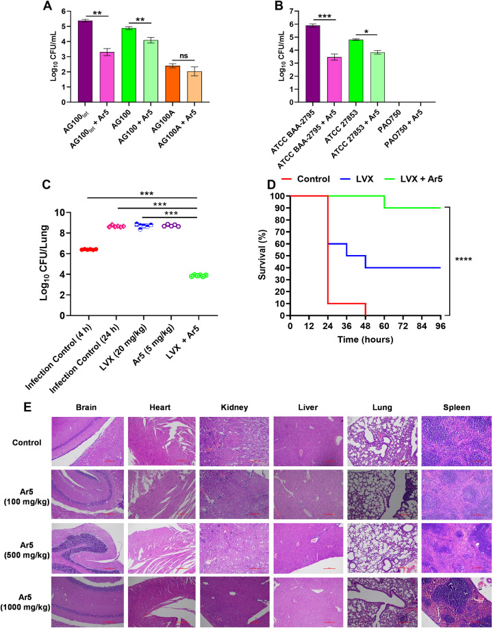
Efflux pumps of the resistance-nodulation-cell division (RND) superfamily, particularly the AcrAB-TolC, and MexAB-OprM, besides mediating intrinsic and acquired resistance, also intervene in bacterial pathogenicity. Inhibitors of such pumps could restore the activities of antibiotics and curb bacterial virulence. Here, we identify pyrrole-based compounds that boost antibiotic activity in Escherichia coli and Pseudomonas aeruginosa by inhibiting their archetype RND transporters. Molecular docking and biophysical studies revealed that the EPIs bind to AcrB. The identified efflux pump inhibitors (EPIs) inhibit the efflux of fluorescent probes, attenuate persister formation, extend post-antibiotic effect, and diminish resistant mutant development. The bacterial membranes remained intact upon exposure to the EPIs. EPIs also possess an anti-pathogenic potential and attenuate P. aeruginosa virulence in vivo. The intracellular invasion of E. coli and P. aeruginosa inside the macrophages was hampered upon treatment with the lead EPI. The excellent efficacy of the EPI-antibiotic combination was evidenced in animal lung infection and sepsis protection models. These findings indicate that EPIs discovered herein with negligible toxicity are potential antibiotic adjuvants to address life-threatening Gram-negative bacterial infections.


Conflict of interest statement

N.M., R.T., R.K., R.I., N.C., S.J., and H.N. are inventors for the patent application of the EPIs disclosed in this study (Indian provisional patent Application no. 202011054425). The remaining authors declare no conflict of interest.

Figures

Fig 1
Fig 1. Representative 2D structures of the 24 pyrrole-based derivatives synthesized for screening.
The structures were drawn using ACD/ChemSketch 2016 2.2 freeware.
Fig 2
Fig 2. The structure of the parent pyrrole and tetracycline boosting activities of compounds Ar1-Ar24 in E.
coli AcrB over-expressing (AG100tet) and AcrAB deletion (AG100A) strains as determined by using the broth microdilution checkerboard synergy method. The assay was performed in three biological replicates having two technical replicates. MIC, Minimum Inhibitory Concentration; TET, Tetracycline; FICI, Fractional Inhibitory Concentration Index; ’-’ represents no fold reduction in tetracycline MIC; PAβN, Phenylalanine-arginine-β-naphthylamide.
Fig 3
Fig 3. Effect of pyrrole derivatives (Ar1-Ar24) on Hoechst 33342 efflux in AcrB-TolC transporter-overproducing E. coli AG100tet strain.
AG100tet cells were pre-loaded with Hoechst 33342 (2.5 μM) and either compounds (Ar1-Ar24 at 16 μg/mL) or PAβN (64 μg/mL, positive control) at ¼ × MIC under the condition that favor maximal accumulation. The data presented correspond to the average of triplicate readings ± SD.
Fig 4
Fig 4. Accumulation of tetracycline (1 × MIC) in the presence of either Ar1-Ar24 at 1/4 × MIC (16 μg/mL) or PAβN at 1/4 × MIC (64 μg/mL) for 25 min in E. coli AG100tet.
The increased accumulation of tetracycline in the bacterial cells was indicated by an augmentation in the tetracycline fluorescence intensity. The experiment was performed in triplicate, and the average data ± SD was plotted.
Fig 5
Fig 5
Structural representation of (A) Ar1 (B) Ar5 (C) Ar11 (D) Ar18 ligands (red spheres) docked in the active site cleft of AcrB (green cartoon). 2D ligand interaction diagram showing several interactions involved in binding of (E) Ar1 (F) Ar5 (G) Ar11 (H) Ar18 to AcrB. BLI study confirms the interaction between purified AcrB and EPIs (I) Ar1 (J) Ar5 (K) Ar11 (L) Ar18; the AcrB protein was immobilized onto an AR2G sensor tip, and interactions using different concentrations (ranging from 5 μM to 1 mM) of compounds were studied.
Fig 6
Fig 6
(A) Time-kill curves of E. coli ATCC BAA-2774 show levofloxacin’s bactericidal effect at 1 × MIC in combination with Ar5 at 16 μg/mL. Killing of (B) E. coli and (C) P. aeruginosa persister cells. The stationary phase cultures of E. coli AG100 and P. aeruginosa ATCC 27853 were treated with carbenicillin at 128 μg/mL and 256 μg/mL, respectively, for 18 h to isolate persister cells. These were re-exposed to carbenicillin in combination with Ar5 (16 μg/mL). Each time point in these experiments represents the mean log10 CFU/mL ± SD of three readings. Post-antibiotic effect (PAE) induced by levofloxacin in combination with (D) Ar1, (E) Ar5, (F) Ar11, and (G) Ar18 in E. coli ATCC BAA-2774 by the turbidimetry-based system. Growth curves of cultures not previously exposed (control) or pre-exposed to levofloxacin (1 × MIC) alone or in combination with EPIs at ¼ × MIC (16 μg/mL) for 2 h. Time 0 h corresponds to the growth monitoring immediately after antibiotic and EPI removal and cell resuspension in a fresh medium. PAE values in the graphs correspond to the growth halt undergone by the treated culture compared to the control in reaching an OD600nm value half of the final OD600nm after 12 h. The data represented is the average value of triplicates. Total ROS accumulation in (H) E. coli AG100tet and (I) P. aeruginosa ATCC BAA-2795 treated with levofloxacin (1 ×MIC) or piperacillin (1 ×MIC) in combination with EPIs (Ar1, Ar5, Ar11, and Ar18) at ¼ × MIC (16 μg/mL) for 2 h. Exogenous addition of NAC (6 mM) reduced the ROS accumulation. The average of triplicates ± SD is shown. (J) The influence of EPIs and levofloxacin on the preformed biofilms. The crystal violet assay assessed the biomass of P. aeruginosa ATCC BAA-2795 after exposure to sub-inhibitory concentrations (1/4 × MIC, 16 μg/mL) of EPIs and levofloxacin (1 × MIC) alone or in combination for 24 h. (K) Quantification of the effect of EPIs on mature biofilm in combination with levofloxacin. The viable bacterial population within the biofilm of P. aeruginosa ATCC BAA-2795 after 24 h exposure to sub-inhibitory concentrations (1/4 × MIC, 16 μg/mL) of EPIs and levofloxacin (1 × MIC) alone or in combination was determined using MTT assay. The results shown here correspond to the average ± SD of three independent readings. Results were considered significant when *p<0.05 and highly significant when **p<0.01 and ***p<0.001.
Fig 7
Fig 7
(A) Effect of EPIs (Ar1-Ar24) on the bacterial outer membrane permeability probed with ANS. E. coli AG100tet was treated with ANS (3 mM) and compounds at (256 μg/mL, 128 μg/mL, 64 μg/mL) for 1 h at 37°C. Colistin (256 μg/mL, 128 μg/mL, 64 μg/mL) and PAβN (256 μg/mL, 128 μg/mL, 64 μg/mL), and polymyxin B (256 μg/mL, 128 μg/mL, 64 μg/mL) were included as positive controls. (B) Effect of EPIs (Ar1-Ar24) on the bacterial membrane depolarization probed with DiSC3(5). E. coli AG100tet was treated with DiSC3(5) (0.4 μM) and compounds at either 1 × MIC (64 μg/mL) or ½ × MIC (32 μg/mL) for 60 min at 37°C. PAβN (64 μg/mL, 32 μg/mL), Polymyxin B (64 μg/mL, 32 μg/mL) and CCCP (64 μg/mL, 32 μg/mL) were included as a positive control. (C) Effect of EPIs (Ar1-Ar24) (16 μg/mL) on E. coli AG100tet ATP levels. The bacterial culture was exposed to compounds or CCCP at ¼ × MIC (16 μg/mL) for 4 h. The ATP levels were quantified using a luciferin-luciferase bioluminescence detection assay. (D) Effect of EPIs (Ar1-Ar24) on the bacterial membrane fluidity probed with laurdan (10 μM). E. coli AG100tet was treated with laurdan (10 μM) in the presence of compounds at 1 × MIC (64 μg/mL). PAβN (1 × MIC), valinomycin (50 μM), and benzyl alcohol (50 mM; a known membrane fluidizer) were included for comparison. The baseline fluorescence for each well was subtracted, and laurdan generalized polarization (GP) was plotted. (E) Mammalian calcium channel inhibition assay with HEK-293 cells. Cytoplasmic Ca2+ measures of carbachol pre- and post-treatment cells in the presence of either Ar1-Ar24 (16 μg/mL), DMSO (vehicle), or calcium channel antagonist verapamil (32 μg/mL). All the results presented here correspond to the average ± SD of triplicate readings. Results were considered significant when **p<0.01 and ***p<0.001.
Fig 8
Fig 8
(A) Growth kinetics of P. aeruginosa PAO1 strain in CA-MHB under sub-inhibitory concentrations (1/4 × MIC (16 μg/mL)) of EPIs (Ar1, Ar5, Ar11, and Ar18). OD600nm values are the means ± SD from three independent readings. (B) Effect of EPIs (Ar1, Ar5, Ar11, and Ar18) on P. aeruginosa PAO1 swimming motility. For treatment conditions, the plates were supplemented with Ar1, Ar5, Ar11, and Ar18 at sub-inhibitory concentrations (16 μg/mL) and incubated for 24 h at 37°C. The control plates were supplemented with an equal volume of DMSO as in treatment plates. Effect of EPIs (Ar1, Ar5, Ar11, and Ar18) on the production of extracellular virulence factors; pyocyanin (C), pyoverdine (D), elastase (E), protease (F), and rhamnolipids (G) levels in the culture supernatants of P. aeruginosa PAO1 in the presence of sub-inhibitory concentrations of EPIs (16 μg/mL). The percentage values were calculated with respect to the bacterial culture without treatment. The results were represented as mean ± SD of three independent readings. (H) Influence of sub-inhibitory concentrations (16 μg/mL) of EPIs (Ar1, Ar5, Ar11, and Ar18) on the invasive capacities of P. aeruginosa PAO1 within RAW264.7 macrophages. The results were presented as mean ± SD log10 CFU/mL of three independent readings. (I) Ar5 inhibits P. aeruginosa PAO1 virulence toward C. elegans. C. elegans were initially applied to E. coli OP50 lawns for feeding, and then the L4 stage worms were transferred to lawns of P. aeruginosa PAO1 grown (for 18 h) in the presence or absence of Ar5 (16 μg/mL). The control (without treatment) plates were supplemented with an equivalent volume of DMSO (vehicle). For survival assay, 10 L4 worms were placed in triplicate plates for each experimental condition. All the results were considered significant when *p<0.05 and highly significant when **p<0.01 and ***p<0.001.
Fig 9
Fig 9
Influence of Ar5 (16 μg/mL) on the invasive capacities of (A) E. coli wild-type AG100, AcrB-TolC overproducing AG100tet, and AcrAB knockout AG100A; (B) P. aeruginosa wild-type ATCC 27853, MexAB-OprM overproducing ATCC BAA-2795, and PAO750 with MexAB deletion within RAW264.7 macrophages. The results were presented as mean ± SD log10 CFU/mL of three independent readings. (C) Ar5-levofloxacin combination is efficacious in mice. Single-dose (subcutaneous; 4 h post-infection, 6 mice/group) treatment with Ar5 (5 mg/kg) and levofloxacin (20 mg/kg) alone or in combination in immunocompetent lung infection model using P. aeruginosa ATCC BAA-2795. For drug-treated animals, lungs CFUs were determined at 24 h post-infection. For controls, CFU in the lungs was determined 4 h and 24 h post-infection. The CFU from each mouse is plotted as individual points, and error bars represent the SD within an experimental group. (D) For the peritonitis survival model, mice (n = 10 per group) were injected with a lethal dose of P. aeruginosa ATCC BAA-2795 and treated with three repetitive dosing of Ar5 (5 mg/kg), levofloxacin (20 mg/kg), and their combination 1, 4, and 7 h post-infection subcutaneously. The percent survival was calculated and represented using the Kaplan-Meier survival plot. Results were considered significant when *p<0.05 and highly significant when **p<0.01, ***p<0.001, and ****p<0.0001. (E) Histopathology analyses of brain, heart, lung, spleen, liver, and kidney of mice when treated with vehicle (control) or Ar5 (100, 500, and 1000 mg/kg). After removal, the organs were fixed in buffered formalin and stained with hematoxylin and eosin.

References

    1. Tacconelli E, Carrara E, Savoldi A, Harbarth S, Mendelson M, Monnet DL, et al.. Discovery, research, and development of new antibiotics: the WHO priority list of antibiotic-resistant bacteria and tuberculosis. The Lancet Infectious Diseases. 2018;18(3):318–27. doi: 10.1016/S1473-3099(17)30753-3 - DOI - PubMed
    1. Murray CJ, Ikuta KS, Sharara F, Swetschinski L, Aguilar GR, Gray A, et al.. Global burden of bacterial antimicrobial resistance in 2019: a systematic analysis. The Lancet. 2022. - PMC - PubMed
    1. Sjuts H, Vargiu AV, Kwasny SM, Nguyen ST, Kim H-S, Ding X, et al.. Molecular basis for inhibition of AcrB multidrug efflux pump by novel and powerful pyranopyridine derivatives. Proceedings of the National Academy of Sciences. 2016;113(13):3509–14. doi: 10.1073/pnas.1602472113 - DOI - PMC - PubMed
    1. Nikaido H, Pagès J-M. Broad-specificity efflux pumps and their role in multidrug resistance of Gram-negative bacteria. FEMS microbiology reviews. 2012;36(2):340–63. doi: 10.1111/j.1574-6976.2011.00290.x - DOI - PMC - PubMed
    1. Nikaido H. Antibiotic resistance caused by gram-negative multidrug efflux pumps. Clinical Infectious Diseases. 1998;27(Supplement_1):S32–S41. doi: 10.1086/514920 - DOI - PubMed

MeSH terms

Substances