. 2024 May 2;111(5):841-862.
doi: 10.1016/j.ajhg.2024.03.007. Epub 2024 Apr 8.

The clinical utility and diagnostic implementation of human subject cell transdifferentiation followed by RNA sequencing

Affiliations

The clinical utility and diagnostic implementation of human subject cell transdifferentiation followed by RNA sequencing

Shenglan Li et al. Am J Hum Genet. .

Abstract

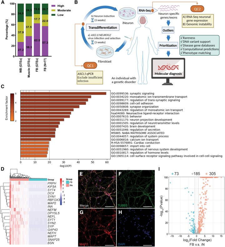
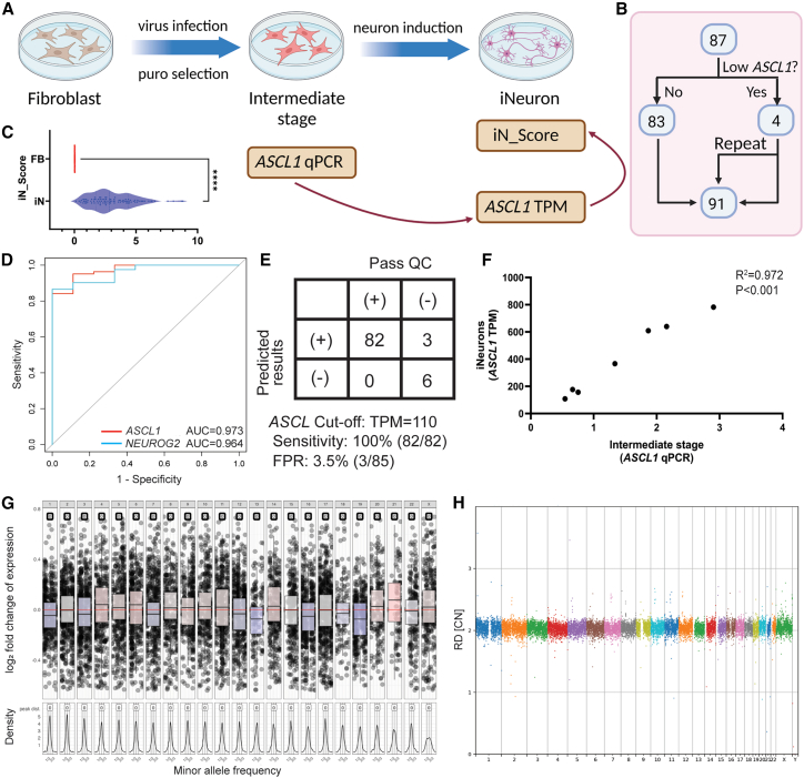
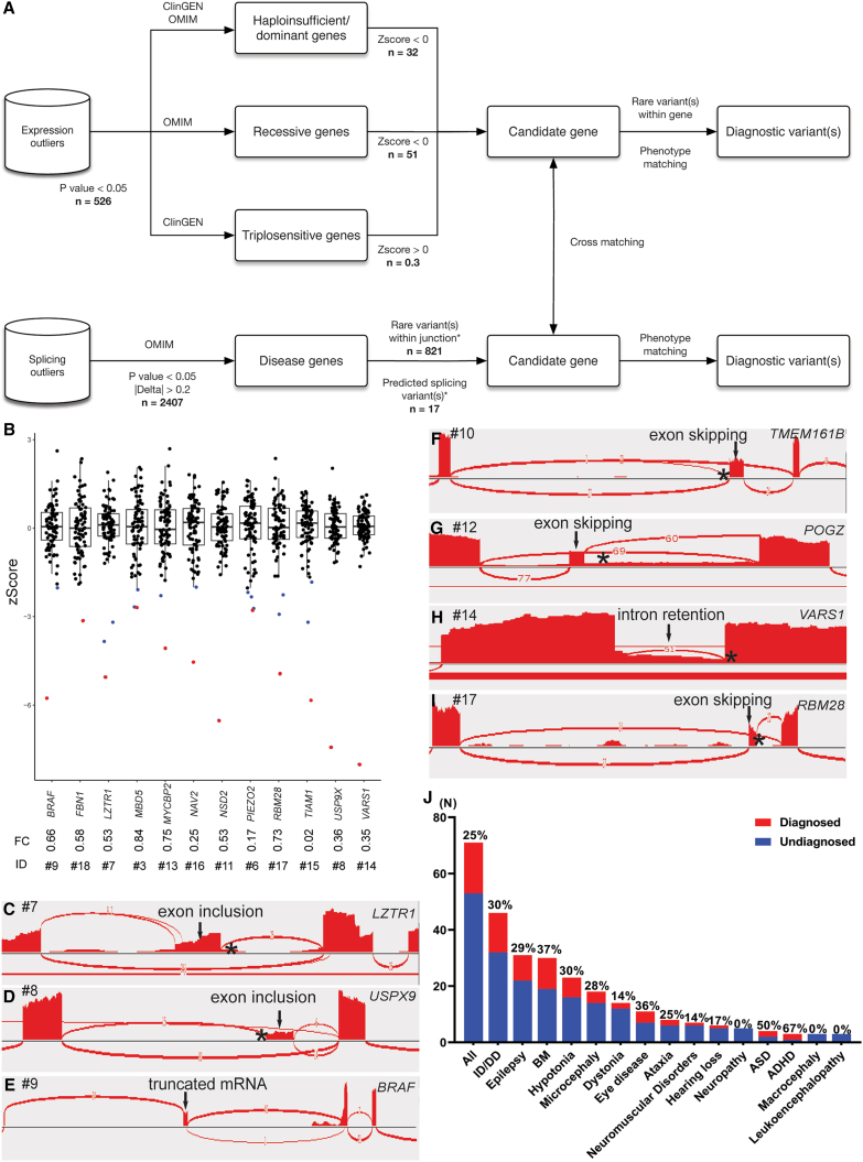
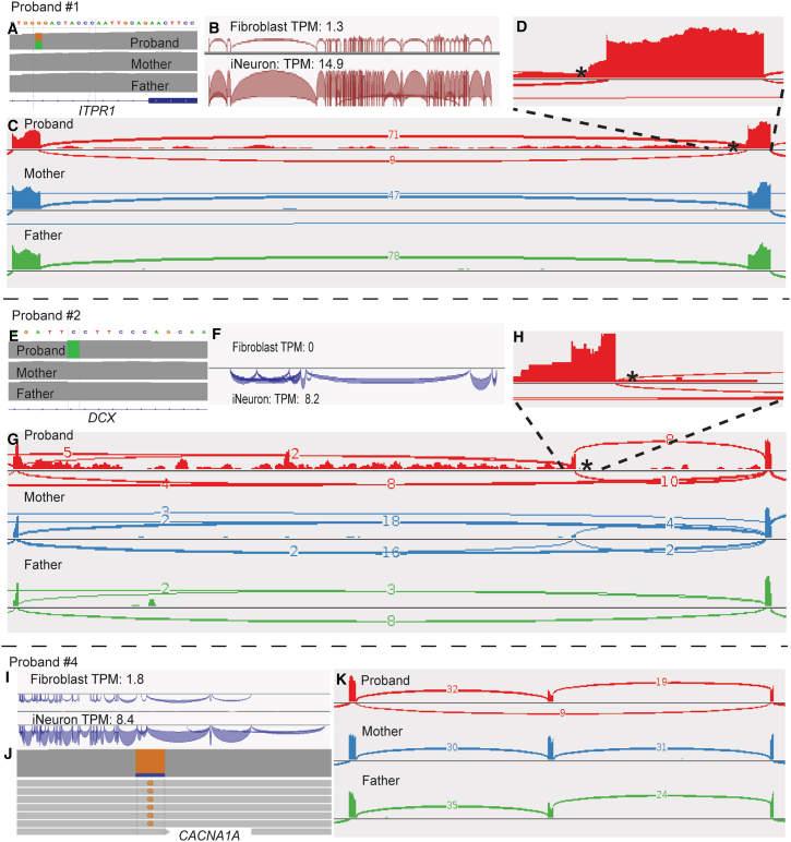
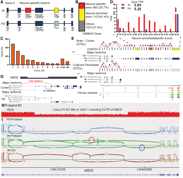
RNA sequencing (RNA-seq) has recently been used in translational research settings to facilitate diagnoses of Mendelian disorders. A significant obstacle for clinical laboratories in adopting RNA-seq is the low or absent expression of a significant number of disease-associated genes/transcripts in clinically accessible samples. As this is especially problematic in neurological diseases, we developed a clinical diagnostic approach that enhanced the detection and evaluation of tissue-specific genes/transcripts through fibroblast-to-neuron cell transdifferentiation. The approach is designed specifically to suit clinical implementation, emphasizing simplicity, cost effectiveness, turnaround time, and reproducibility. For clinical validation, we generated induced neurons (iNeurons) from 71 individuals with primary neurological phenotypes recruited to the Undiagnosed Diseases Network. The overall diagnostic yield was 25.4%. Over a quarter of the diagnostic findings benefited from transdifferentiation and could not be achieved by fibroblast RNA-seq alone. This iNeuron transcriptomic approach can be effectively integrated into diagnostic whole-transcriptome evaluation of individuals with genetic disorders.

Keywords: RNA sequencing; RNA-seq; clinically accessible tissue; fibroblast; genetic diagnosis; induced neuron; isoform; neurological disorder; transcriptome; transdifferentiation.


Conflict of interest statement

Declaration of interests Baylor College of Medicine (BCM) and Miraca Holdings Inc. have formed a joint venture with shared ownership and governance of Baylor Genetics (BG), which performs genetic testing and derives revenue. P.L. and C.M.E. are employees of BCM and derive support through a professional services agreement with BG.

Figures

Figure 1
Figure 1
Activation of low-expression OMIM-N genes in participants' fibroblasts (A) Expression levels of OMIM-N genes in clinically assessable tissues. Gene expression levels were classified as follows: low (TPM < 1), moderate (TPM ≥ 1 and <10), and high (TPM ≥ 10). Fibroblasts (FB) RNA-seq data from GTEx and UDN participants (n = 77) were used for the assessment. (B) Schematic of the workflow of iNeuron transdifferentiation and RNA-seq. Created with BioRender.com. (C) Functional enrichment analysis of DEGs up-regulated in iNeurons. (D) Robust expression of neuron-specific genes in iNeurons. (E–H) Immunofluorescence staining of neuronal makers Tubulin β-III (TUBB3) (F) and MAP2 (G) in iNeurons. Nuclei were stained using DAPI (H). The merged image is shown in (E). Scale bar represents 100 μm. (I) Volcano plot showing activation of low-expression OMIM-N genes.
Figure 2
Figure 2
Multiple quality control measurements during transdifferentiation process (A) Schematic for determining ASCL1 qPCR as a critical step in QC to identify inadequate virus infection. Created with BioRender.com. (B) All iNeurons samples used for QC analysis. (C) iN_Score comparison between iNeurons and fibroblasts, represented by median with violin plot. Statistical significance denoted by ∗∗∗∗p < 0.0001. (D) ROC curve analysis showing expression levels of ASCL1 or NEUROG2 as predictors of iNeuron quality. The area under the ROC curve (AUC) for ASCL1 is 0.973, and for NEUROG2 it is 0.964. There is no statistically significant difference between ASCL1 and NEUROG2. (E) ASCL1 TPM cutoff of 110 in iNeurons with 100% sensitivity and 3.5% false positive rate (FPR). (F) Correlation between ASCL1 qPCR and TPM of iNeurons. ASCL1 qPCR result is normalized to GAPDH. R2 = 0.972, p < 0.001. (G) A representative genome-wide copy number analysis plot assessing post-induction genomic stability. RNA-based CNV calling revealed no aneuploidies or large copy number variations (CNVs) in this iNeuron sample. (H) The post-induction genomic stability assessments are validated by further WGS analysis. WGS also detected no smaller new CNV events.
Figure 3
Figure 3
The interpretation pipeline, molecular diagnostic rates, and selected diagnostic findings from the iNeuron RNA-seq workflow (A) Analytical and interpretation pipelines used in iNeuron RNA-seq analysis. The n values indicate the average number of variants passing filtration at each step. (B) OUTRIDER identified 12 aberrant expression outliers as diagnostic findings (BRAF, FBN1, LZTR1, MBD5, MYCBP2, NAV2, NSD2, PIEZO2, RBM28, TIAM1, USP9X, and VARS1). Each dot represents an individual participant. The blue dots represent expression outliers that fulfill the analytical filtration standards defined in (A). The red dots represent molecular diagnostic findings with integrated considerations from analytical filtrations (blue dots), phenotypic matching, and correlation with DNA variant findings. For each molecular diagnostic expression outlier (red dot), its ID and expression fold change compared to controls are listed underneath. (C–I) FRASER detected 10 aberrant splicing events, with 7 representative events (LZTR1, USP9X, BRAF, TEME161B, POGZ, VARS1, and RBM28) illustrated here. Aberrant junctions are shown in the Sashimi plot. The asterisk represents the position of the causal variant. (J) Molecular diagnostic yields in individuals with different neurological phenotypes.
Figure 4
Figure 4
Molecular diagnoses achieved by iNeuron RNA-seq through elevated expression of OMIM-N genes (A) Trio WGS reveals a de novo heterozygous intronic variant in ITPR1 (GenBank: NM_001378452.1; c.5980−17G>A). (B) Significant increase in expression of ITPR1 after transdifferentiation. (C) iNeuron RNA-seq demonstrates a 15-nucleotide retention from intron 45 in approximately 50% of the reads, as well as complete intron 45 retention in a small fraction of reads. (D) Zoomed in view of the 15-nucleotide retention. (E) Trio WGS identifies a novel hemizygous deep intronic variant (GenBank: NM_001195553.2; c.946+4588G>T) in DCX. (F) DCX is not expressed in fibroblasts but is activated in iNeurons. (G) Sashimi plot displays inclusion of a 13,549 bp cryptic exon from intron, positioned −31 bp from the intronic variant of DCX. (H) Zoomed-in view of the new splice junction at the intron retention. (I) Significant increase in expression of CACNA1A after transdifferentiation. (J) Skewed expression of the variant allele in CACNA1A. (K) Sashimi plot reveals exon skipping located 124 kb away from the missense variant. Variants are indicated by asterisks.
Figure 5
Figure 5
Enhanced molecular diagnosis with iNeuron RNA-seq through detection of neuron-enriched exons (A) Schematic diagram illustrating that neuron-enriched and/or neuron-specific isoforms can be detected in iNeurons. (B) Identification from iNeuron RNA-seq of 9,356 neuron-enriched exons (from 1,690 OMIM-N genes), including 936 neuron-specific exons (from 563 OMIM-N genes). The term “other” refers to the remaining OMIM-N genes that do not include neuron-enriched exons. (C) Distribution of OMIM-N genes based on the number of neuron-enriched exons when comparing iNeurons with fibroblasts. (D–F) The neuron-specific isoform in CAMTA1 is detected by iNeuron RNA-seq. The expected distribution of neuron- and fibroblast-specific isoforms are illustrated in (E) based on data from GTEx. Multiple exons toward the end of the transcript are shown to be highly enriched (yellow) or specific (red) to neurons. Interrogation of these exons in the neuronal isoform is important as illustrated by the distribution of clinically relevant variants from ClinVar in (F). Exon-level RNA-seq data from iNeurons, summarized in (D), demonstrate successful activation of the neuron-enriched and neuron-specific exons. Note that gene-level TPM (2.64 for iNeurons and 5.25 for fibroblasts) can disguise the activation of tissue-specific isoforms. (G) Schematic showing the aberrant junction (red) caused by the 5′ UTR deletion of MBD5, which can be detected only in the neuron-specific long isoform. The alternative junction (blue) corresponds to the normal allele with no deletion. (H) Detection of a heterozygous deletion (0.25 Mb) on 2q23.1, involving noncoding exons of MBD5 through WGS. (I) Sashimi plot depicting the presence of abnormal junctions (red circle) exclusively in proband’s iNeurons, but not in fibroblasts. The number of junction reads in the red and the blue circles represent abundance of alleles with and without the deletion.

Similar articles

Cited by

  • Perspective: Pathological transdifferentiation-a novel therapeutic target for cardiovascular diseases and chronic inflammation.
    Yang WY, Ben Issa M, Saaoud F, Xu K, Shao Y, Lu Y, Dornas W, Cueto R, Jiang X, Wang H, Yang X. Yang WY, et al. Front Cardiovasc Med. 2024 Nov 26;11:1500775. doi: 10.3389/fcvm.2024.1500775. eCollection 2024. Front Cardiovasc Med. 2024. PMID: 39660114 Free PMC article.
  • An outlier approach: advancing diagnosis of neurological diseases through integrating proteomics into multi-omics guided exome reanalysis.
    Chui MM, Kwong AK, Leung HYC, Pang C, Scheller IF, Wong SS, Fung CW, Yépez VA, Gagneur J, Mak CC, Chung BH. Chui MM, et al. NPJ Genom Med. 2025 May 3;10(1):36. doi: 10.1038/s41525-025-00493-5. NPJ Genom Med. 2025. PMID: 40319040 Free PMC article.
  • Translating Muscle RNAseq Into the Clinic for the Diagnosis of Muscle Diseases.
    Segarra-Casas A, Domínguez-González C, Natera-de Benito D, Kapetanovic S, Hernández-Laín A, Estévez-Arias B, Llansó L, Ortez C, Jou C, Martí-Carrera I, López-Márquez A, Rodríguez MJ, González-Mera L, Nedkova V, Fernández-Torrón R, Rodríguez-Santiago B, Jimenez-Mallebrera C, Juntas-Morales R, López-de Munain A, Surrallés J, Nascimento A, Gallardo E, Olivé M, Gallano P, González-Quereda L. Segarra-Casas A, et al. Ann Clin Transl Neurol. 2025 Jul;12(7):1465-1479. doi: 10.1002/acn3.70078. Epub 2025 May 25. Ann Clin Transl Neurol. 2025. PMID: 40413734 Free PMC article.
  • RNA variant assessment using transactivation and transdifferentiation.
    Nicolas-Martinez EC, Robinson O, Pflueger C, Gardner A, Corbett MA, Ritchie T, Kroes T, van Eyk CL, Scheffer IE, Hildebrand MS, Barnier JV, Rousseau V, Genevieve D, Haushalter V, Piton A, Denommé-Pichon AS, Bruel AL, Nambot S, Isidor B, Grigg J, Gonzalez T, Ghedia S, Marchant RG, Bournazos A, Wong WK, Webster RI, Evesson FJ, Jones KJ; PERSYST Investigator Team; Cooper ST, Lister R, Gecz J, Jolly LA. Nicolas-Martinez EC, et al. Am J Hum Genet. 2024 Aug 8;111(8):1673-1699. doi: 10.1016/j.ajhg.2024.06.018. Epub 2024 Jul 30. Am J Hum Genet. 2024. PMID: 39084224 Free PMC article.
  • Combined genomics and proteomics unveils elusive variants and vast aetiologic heterogeneity in dystonia.
    Zech M, Dzinovic I, Skorvanek M, Harrer P, Necpal J, Kopajtich R, Kittke V, Tilch E, Zhao C, Tsoma E, Sorrentino U, Indelicato E, Stehr A, Saparov A, Abela L, Adamovicova M, Afenjar A, Assmann B, Baloghova J, Baumann M, Berutti R, Brezna Z, Brugger M, Brunet T, Cogne B, Colangelo I, Conboy E, Distelmaier F, Eckenweiler M, Garavaglia B, Geerlof A, Graf E, Hackenberg A, Harvanova D, Haslinger B, Havrankova P, Hoffmann GF, Janzarik WG, Keren B, Kolnikova M, Kolokotronis K, Kosutzka Z, Koy A, Krenn M, Krygier M, Kusikova K, Maier O, Meitinger T, Mertes C, Milenkovic I, Monfrini E, Santos Dias Mourao A, Musacchio T, Nizon M, Ostrozovicova M, Pavlov M, Prihodova I, Rektorova I, Romito LM, Rybanska B, Sadr-Nabavi A, Schwenger S, Shoeibi A, Sitzberger A, Smirnov D, Svantnerova J, Tautanova R, Toelle SP, Ulmanova O, Vetrini F, Vill K, Wagner M, Weise D, Zorzi G, Di Fonzo A, Oexle K, Berweck S, Mall V, Boesch S, Schormair B, Prokisch H, Jech R, Winkelmann J. Zech M, et al. Brain. 2025 Aug 1;148(8):2827-2846. doi: 10.1093/brain/awaf059. Brain. 2025. PMID: 39937650 Free PMC article.

References

    1. Liu P., Meng L., Normand E.A., Xia F., Song X., Ghazi A., Rosenfeld J., Magoulas P.L., Braxton A., Ward P., et al. Reanalysis of Clinical Exome Sequencing Data. N. Engl. J. Med. 2019;380:2478–2480. doi: 10.1056/NEJMc1812033. - DOI - PMC - PubMed
    1. Yang Y., Muzny D.M., Reid J.G., Bainbridge M.N., Willis A., Ward P.A., Braxton A., Beuten J., Xia F., Niu Z., et al. Clinical whole-exome sequencing for the diagnosis of mendelian disorders. N. Engl. J. Med. 2013;369:1502–1511. doi: 10.1056/NEJMoa1306555. - DOI - PMC - PubMed
    1. Splinter K., Adams D.R., Bacino C.A., Bellen H.J., Bernstein J.A., Cheatle-Jarvela A.M., Eng C.M., Esteves C., Gahl W.A., Hamid R., et al. Effect of Genetic Diagnosis on Patients with Previously Undiagnosed Disease. N. Engl. J. Med. 2018;379:2131–2139. doi: 10.1056/NEJMoa1714458. - DOI - PMC - PubMed
    1. Wright C.F., Campbell P., Eberhardt R.Y., Aitken S., Perrett D., Brent S., Danecek P., Gardner E.J., Chundru V.K., Lindsay S.J., et al. Genomic Diagnosis of Rare Pediatric Disease in the United Kingdom and Ireland. N. Engl. J. Med. 2023;388:1559–1571. doi: 10.1056/NEJMoa2209046. - DOI - PMC - PubMed
    1. Lionel A.C., Costain G., Monfared N., Walker S., Reuter M.S., Hosseini S.M., Thiruvahindrapuram B., Merico D., Jobling R., Nalpathamkalam T., et al. Improved diagnostic yield compared with targeted gene sequencing panels suggests a role for whole-genome sequencing as a first-tier genetic test. Genet. Med. 2018;20:435–443. doi: 10.1038/gim.2017.119. - DOI - PMC - PubMed

Publication types

LinkOut - more resources