

Plasma and synovial fluid extracellular vesicles display altered microRNA profiles in horses with naturally occurring post-traumatic osteoarthritis: an exploratory study
- PMID: 38593834
- PMCID: PMC11132921
- DOI: 10.2460/javma.24.02.0102
Plasma and synovial fluid extracellular vesicles display altered microRNA profiles in horses with naturally occurring post-traumatic osteoarthritis: an exploratory study
Abstract
Objective: The objective of this study was to characterize extracellular vesicles (EVs) in plasma and synovial fluid obtained from horses with and without naturally occurring post-traumatic osteoarthritis (PTOA).
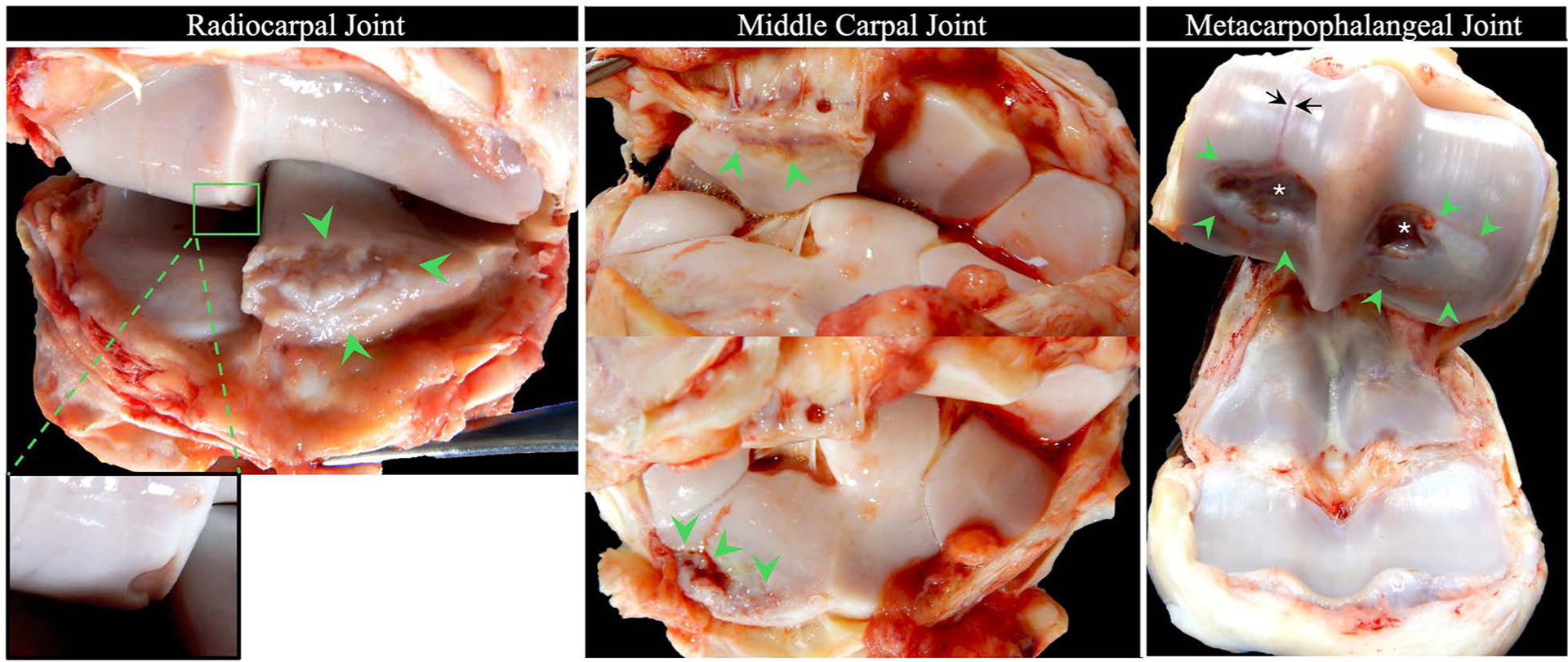
Animals: EVs were isolated from plasma and synovial fluid from horses with (n = 6) and without (n = 6) PTOA.
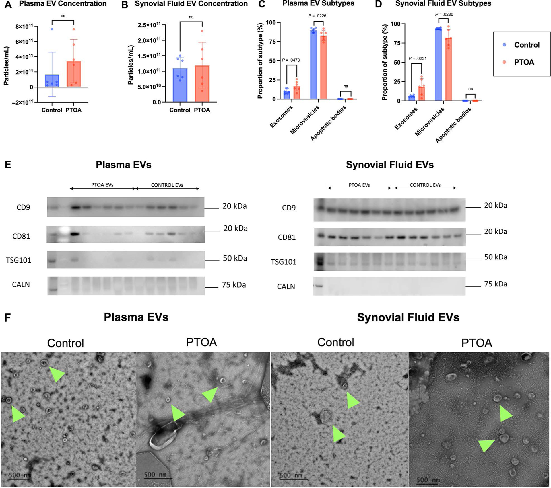
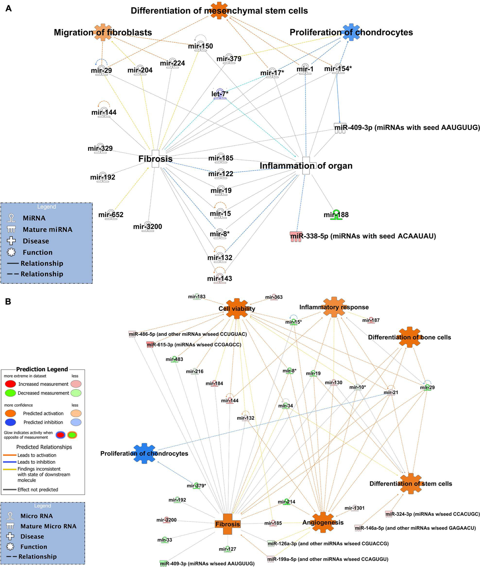
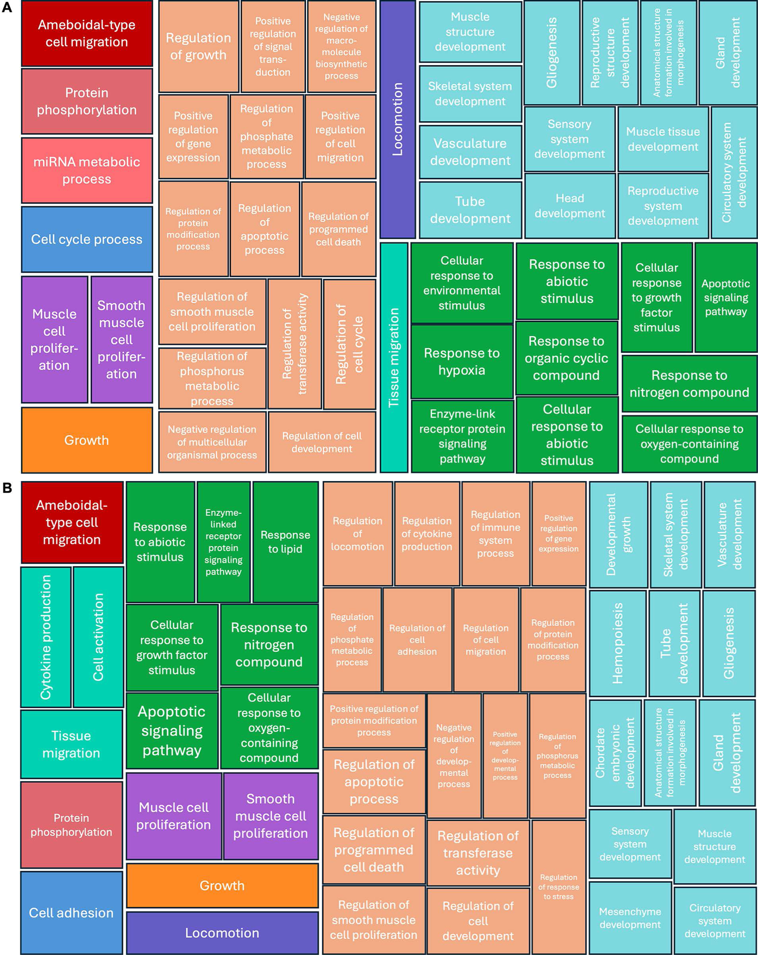
Methods: Plasma and synovial fluid EVs were characterized with respect to quantity, size, and surface markers. Small RNA sequencing was performed, and differentially expressed microRNAs (miRNAs) underwent bioinformatic analysis to identify putative targets and to explore potential associations with specific biological processes.
Results: Plasma and synovial fluid samples from horses with PTOA had a significantly higher proportion of exosomes and a lower proportion of microvesicles compared to horses without PTOA. Small RNA sequencing revealed several differentially expressed miRNAs, including miR-144, miR-219-3p, and miR-199a-3l in plasma and miR-199a-3p, miR-214, and miR-9094 in synovial fluid EVs. Bioinformatics analysis of the differentially expressed miRNAs highlighted their potential role in fibrosis, differentiation of chondrocytes, apoptosis, and inflammation pathways in PTOA.
Clinical relevance: We have identified dynamic molecular changes in the small noncoding signatures of plasma and synovial fluid EVs in horses with naturally occurring PTOA. These findings could serve to identify promising biomarkers in the pathogenesis of PTOA, to facilitate the development of targeted therapies, and to aid in establishing appropriate translational models of PTOA.
Keywords: equine; exosomes; joint disease; miRNA; small non-coding RNA.
Conflict of interest statement
Disclosures
Dr. Ortved served as guest editor for this JAVMA supplemental issue. She declares that she had no role in the editorial direction of this manuscript. All authors declare that the research was conducted in the absence of any commercial or financial relationships that could be construed as a potential conflict of interest.
All claims expressed in this article are solely those of the authors and do not necessarily represent those of their affiliated organizations or those of the publisher, the editors, and the reviewers. Any product that may be evaluated in this article, or claim that may be made by its manufacturer, is not guaranteed or endorsed by the publisher.
No AI assisted technologies were used in the generation of this manuscript.
Figures
Similar articles
-
Identification and characterisation of temporal abundance of microRNAs in synovial fluid from an experimental equine model of osteoarthritis.Equine Vet J. 2025 Jul;57(4):1138-1150. doi: 10.1111/evj.14456. Epub 2025 Jan 8. Equine Vet J. 2025. PMID: 39775906 Free PMC article.
-
Equine synovial fluid small non-coding RNA signatures in early osteoarthritis.BMC Vet Res. 2021 Jan 9;17(1):26. doi: 10.1186/s12917-020-02707-7. BMC Vet Res. 2021. PMID: 33422071 Free PMC article.
-
Metabolism and global protein glycosylation are differentially expressed in healthy and osteoarthritic equine carpal synovial fluid.Equine Vet J. 2022 Mar;54(2):323-333. doi: 10.1111/evj.13440. Epub 2021 Mar 18. Equine Vet J. 2022. PMID: 33587757 Free PMC article.
-
An Evidence-Based Systematic Review of Human Knee Post-Traumatic Osteoarthritis (PTOA): Timeline of Clinical Presentation and Disease Markers, Comparison of Knee Joint PTOA Models and Early Disease Implications.Int J Mol Sci. 2021 Feb 17;22(4):1996. doi: 10.3390/ijms22041996. Int J Mol Sci. 2021. PMID: 33671471 Free PMC article.
-
Extracellular vesicles in osteoarthritis of peripheral joint and temporomandibular joint.Front Endocrinol (Lausanne). 2023 Mar 6;14:1158744. doi: 10.3389/fendo.2023.1158744. eCollection 2023. Front Endocrinol (Lausanne). 2023. PMID: 36950682 Free PMC article. Review.
Cited by
-
Synovial fluid as a complex molecular pool contributing to knee osteoarthritis.Nat Rev Rheumatol. 2025 Aug;21(8):447-464. doi: 10.1038/s41584-025-01271-4. Epub 2025 Jul 7. Nat Rev Rheumatol. 2025. PMID: 40624394 Review.
-
The Impact of the Competition on miRNA, Proteins, and Metabolites in the Blood Exosomes of the Yili Horse.Genes (Basel). 2025 Feb 15;16(2):224. doi: 10.3390/genes16020224. Genes (Basel). 2025. PMID: 40004554 Free PMC article.
-
Extracellular Vesicles in Sport Horses: Potential Biomarkers and Modulators of Exercise Adaptation and Therapeutics.Int J Mol Sci. 2025 May 3;26(9):4359. doi: 10.3390/ijms26094359. Int J Mol Sci. 2025. PMID: 40362597 Free PMC article. Review.
References
-
- Charalambous CP. The response of articular cartilage to mechanical injury. In: Banaszkiewicz PA, Kader DF, eds. Classic Papers in Orthopaedics. London: Springer London, 2014;381–383.
MeSH terms
Substances
Grants and funding
LinkOut - more resources
Full Text Sources
Medical
Molecular Biology Databases
