

Enzymatic phosphatization of fish scales-a pathway for fish fossilization
- PMID: 38594297
- PMCID: PMC11003971
- DOI: 10.1038/s41598-024-59025-3
Enzymatic phosphatization of fish scales-a pathway for fish fossilization
Abstract
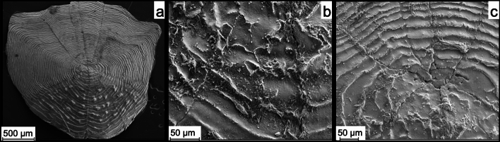
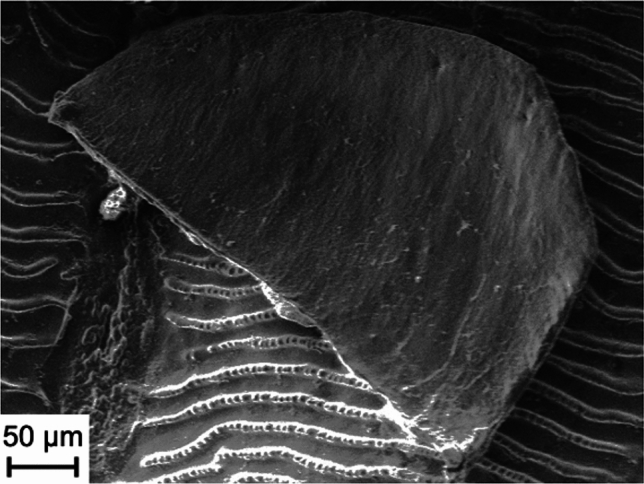
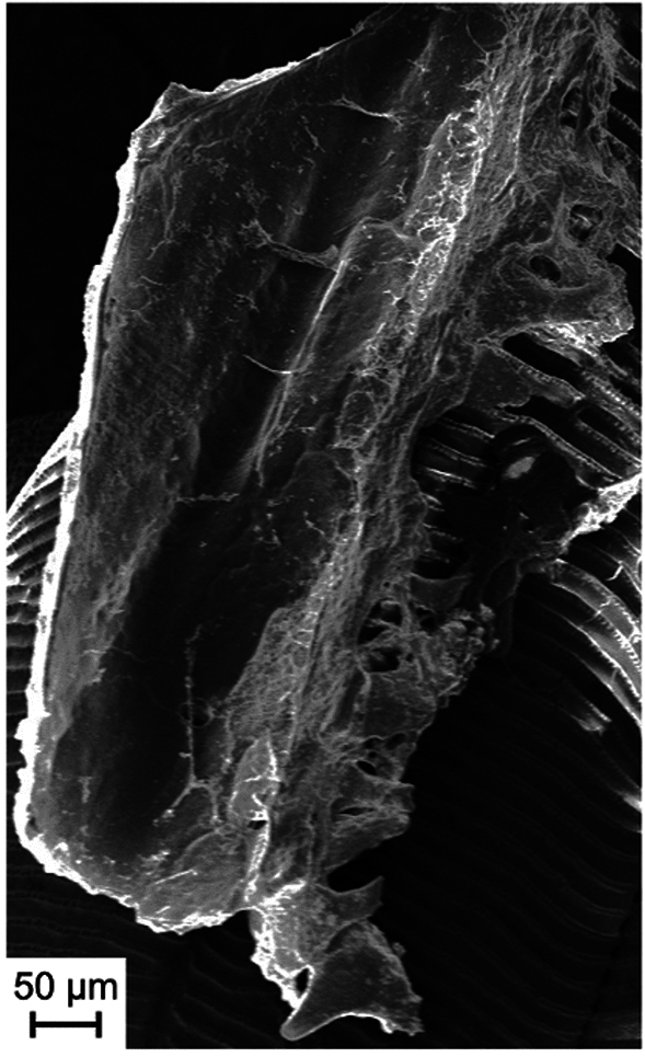
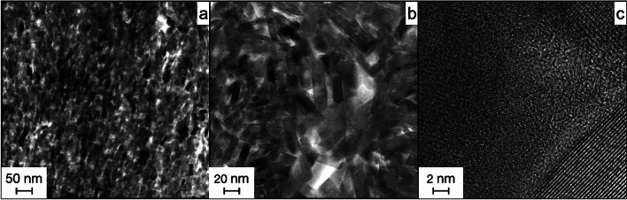
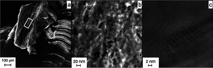
Phosphatized fish fossils occur in various locations worldwide. Although these fossils have been intensively studied over the past decades they remain a matter of ongoing research. The mechanism of the permineralization reaction itself remains still debated in the community. The mineralization in apatite of a whole fish requires a substantial amount of phosphate which is scarce in seawater, so the origin of the excess is unknown. Previous research has shown that alkaline phosphatase, a ubiquitous enzyme, can increase the phosphate content in vitro in a medium to the degree of saturation concerning apatite. We applied this principle to an experimental setup where fish scales were exposed to commercial bovine alkaline phosphatase. We analyzed the samples with SEM and TEM and found that apatite crystals had formed on the remaining soft tissue. A comparison of these newly formed apatite crystals with fish fossils from the Solnhofen and Santana fossil deposits showed striking similarities. Both are made up of almost identically sized and shaped nano-apatites. This suggests a common formation process: the spontaneous precipitation from an oversaturated solution. The excess activity of alkaline phosphatase could explain that effect. Therefore, our findings could provide insight into the formation of well-preserved fossils.
© 2024. The Author(s).
Conflict of interest statement
The authors declare no competing interests.
Figures


Similar articles
-
Control of vertebrate skeletal mineralization by polyphosphates.PLoS One. 2009 May 20;4(5):e5634. doi: 10.1371/journal.pone.0005634. PLoS One. 2009. PMID: 19492083 Free PMC article.
-
Fossil preservation in the Neoproterozoic Doushantuo phosphorite Lagerstatte, South China.Lethaia. 1999 Sep;32(3):219-40. doi: 10.1111/j.1502-3931.1999.tb00541.x. Lethaia. 1999. PMID: 11543524
-
Effects of non-collagenous proteins on the formation of apatite in calcium beta-glycerophosphate solutions.Arch Oral Biol. 1992 Jan;37(1):15-21. doi: 10.1016/0003-9969(92)90147-z. Arch Oral Biol. 1992. PMID: 1596204
-
A review of phosphate mineral nucleation in biology and geobiology.Calcif Tissue Int. 2013 Oct;93(4):382-96. doi: 10.1007/s00223-013-9784-9. Calcif Tissue Int. 2013. PMID: 24077874 Free PMC article. Review.
-
The role of brushite and octacalcium phosphate in apatite formation.Crit Rev Oral Biol Med. 1992;3(1-2):61-82. doi: 10.1177/10454411920030010601. Crit Rev Oral Biol Med. 1992. PMID: 1730071 Review.
References
-
- Pan Y, Fürsich FT, Chellouche P, Hu L. Taphonomy of fish concentrations from the upper Jurassic Solnhofen Plattenkalk of Southern Germany. njgpa. 2019;292:73–92. doi: 10.1127/njgpa/2019/0809. - DOI
-
- Long JA, Trinajstic K. The Late Devonian Gogo formation Lägerstatte of Western Australia: Exceptional early vertebrate preservation and diversity. Annu. Rev. Earth Planet. Sci. 2010;38:255–279. doi: 10.1146/annurev-earth-040809-152416. - DOI
-
- Martill DM. Preservation of fish in the Cretaceous Santana formation of Brazil. Palaeontology. 1988;31:1–18.
-
- Grande L. An updated review of the fish faunas from the Green River formation, the World’s most productive freshwater Lagerstätten. In: Gunnell GF, editor. Eocene Biodiversity: Unusual Occurrences and Rarely Sampled Habitats. Boston: Springer; 2001. pp. 1–38.
-
- Grandi F, et al. Exceptional preservation of large fossil vertebrates in a volcanic setting (Camp dels Ninots, Spain) Hist. Biol. 2023;35:1234–1249. doi: 10.1080/08912963.2022.2085570. - DOI
MeSH terms
Substances
Grants and funding
LinkOut - more resources
Full Text Sources
Miscellaneous
