. 2024 Jul;19(7):1940-1983.
doi: 10.1038/s41596-024-00977-1. Epub 2024 Apr 9.

Control of DNA replication in vitro using a reversible replication barrier

Affiliations

Control of DNA replication in vitro using a reversible replication barrier

Emma J Vontalge et al. Nat Protoc. 2024 Jul.

Abstract

A major obstacle to studying DNA replication is that it involves asynchronous and highly delocalized events. A reversible replication barrier overcomes this limitation and allows replication fork movement to be synchronized and localized, facilitating the study of replication fork function and replication coupled repair. Here we provide details on establishing a reversible replication barrier in vitro and using it to monitor different aspects of DNA replication. DNA template containing an array of lac operator (lacO) sequences is first bound to purified lac repressor (LacR). This substrate is then replicated in vitro using a biochemical replication system, which results in replication forks stalled on either side of the LacR array regardless of when or where they arise. Once replication forks are synchronized at the barrier, isopropyl-β-D-thiogalactopyranoside can be added to disrupt LacR binding so that replication forks synchronously resume synthesis. We describe how this approach can be employed to control replication fork elongation, termination, stalling and uncoupling, as well as assays that can be used to monitor these processes. We also explain how this approach can be adapted to control whether replication forks encounter a DNA lesion on the leading or lagging strand template and whether a converging fork is present. The required reagents can be prepared in 1-2 weeks and experiments using this approach are typically performed over 1-3 d. The main requirements for utilizing the LacR replication barrier are basic biochemical expertise and access to an in vitro system to study DNA replication. Investigators should also be trained in working with radioactive materials.


Conflict of interest statement

Competing interests

No, I declare the authors have no competing interests as defined by Nature Research

Figures

Fig 1.
Fig 1.. Control of replication fork movement by a lac repressor (LacR) array
a, Plasmid DNA harboring a lac operator array (p[lacO]) is incubated with LacR (in blue) to assemble a site-specific replication barrier (‘LacR barrier’) Regardless of when and where replication is initiated on the plasmid, replication forks ultimately end up arrested either side of the LacR barrier. Addition of IPTG disrupts the LacR barrier, which allows replication forks to restart and ultimately complete DNA synthesis. Nascent DNA strands can be radiolabeled by inclusion of radioactive dATP ([32P]dATP) to allow replication intermediates and products to be detected. b, Schematic of a replication fork stalled at a LacR barrier. c, Schematic of two terminating forks converging after disruption of the LacR barrier. d, Schematic of an elongating fork after disruption of the LacR barrier. Analysis of fork progression without termination can be readily achieved by increasing the size of the LacR barrier to delay the encounter with a converging fork. e, Schematic of an ‘uncoupled’ replication fork after disruption of the LacR barrier and inhibition of DNA polymerases (e.g., using aphidicolin). ‘Uncoupling’ occurs when DNA polymerase activity is inhibited while activity of the replicative helicase is allowed to proceed. This results in unwinding of the parental DNA strands (black) without synthesis of daughter DNA strands (red). f, Schematic of a replication fork encountering a DNA lesion on the leading strand template. The LacR barrier blocks the replication fork moving in the opposite direction to ensure only a single fork encounters the lesion on a specific strand. g, Schematic of a replication fork encountering a DNA lesion on the leading strand template. Strand specific encounter by a single fork is enforced by the LacR barrier (as in (f)). h, Schematic of a single replication fork encountering a DNA lesion that involves both DNA strands. Encounter by a single fork is enforced by the LacR barrier (as in (f)). i, Schematic of two replication forks converging upon a DNA lesion that involves both DNA strands. This can be enforced by omitting the LacR barrier or adding IPTG to disrupt it.
Fig 2.
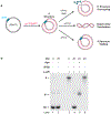
Fig 2.. Use of a LacR barrier to control replication fork termination, uncoupling, and stalling.
a p[lacO] was replicated in Xenopus egg extracts in the presence of dATP[α-32P] to label the nascent DNA strands. Once forks were stalled at the barrier, the reaction was split and treated with IPTG to induce termination, IPTG plus aphidicolin to induce uncoupling, or vehicle to maintain replication fork stalling. b, Samples treated as described in a were separated on an agarose gel and visualized by autoradiography. Note that the dramatic change in mobility for the θ* species arises from compensatory supercoiling when the DNA is deproteinized and parental DNA strands reanneal (also see Fig 6A).
Fig 3.
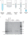
Fig 3.. Purification of biotinylated LacR
a, Schematic of the key steps in the LacR purification procedure. First, a bacterial cell pellet containing biotinylated LacR is solubilized (Procedure 1: Steps 21–34). Next, the biotinylated LacR is precipitated with ammonium sulfate to remove most soluble proteins (Procedure 1: Steps 40–43). Biotinylated LacR is then resuspended and bound to a monomeric avidin column (Procedure 1: Steps 44–48). Non-specifically bound proteins are washed away (Procedure 1: Step 49) and, finally, biotinylated LacR is eluted by addition of biotin (Procedure 1: Steps 50–53). b, Samples from a were separated on an SDS-PAGE gel and visualized by Coomassie staining. Note that elution 4 (lane 11) was performed overnight.
Fig 4.
Fig 4.. Quality control of subcloned lacO array plasmids
a, Cartoon depicting pJD161 and the expected fragment sizes after SacI/KpnI digestion. SacI and KpnI restriction sites on the plasmid are depicted in green, lacO array is depicted in blue. b, Eight subclones of pJD161 were restriction digested with SacI and KpnI, and separated on an agarose gel and detected by SYBR gold-staining (lanes 5–12). Undigested DNA (lane 2) and single digested DNA (lanes 3 and 4) were also included as a positive control for restriction enzyme activity (lanes 2–4). Note that clones in lanes 5, 7, 8 did not pass plasmid validation. Red arrows indicate altered lacO array sizes within a subset of molecules in subclones shown in lanes 5 and 8.
Fig 5.
Fig 5.. Preparation of plasmids containing modified nucleotides
a, Schematic of the procedure for preparing plasmid DNA containing modified nucleotides. First, a host plasmid is nicked with Nb.BsmI either side of the sequence 5’ – CATTCACCGGTATCCTTACGAGCG – 3’ (orange) and the intervening DNA is melted off using heat (Procedure 1: Steps 121–126). Next, a modified oligonucleotide (green) is annealed into the gapped host plasmid and ligated (Procedure 1: Steps 127–131). The products of ligation include plasmids that contain the modified oligonucleotide (green) as well as those containing the parental DNA sequence (orange). To distinguish these, the modified oligonucleotide is engineered so that the DNA damage is within the AgeI recognition site (ACCGGT). This allows the ligated products to be treated with AgeI to linearize plasmids containing the parental DNA sequence, while leaving those containing the modified oligonucleotide untouched (Procedure 1: Steps 132–133). Treatment with T5 Exonuclease is then used to degrade the linearized molecules that contain the parental DNA (Procedure 1: Steps 134–135). Note that T5 Exonuclease also degrades any nicked DNA molecules arising from incomplete ligation. b, Samples from (a) were separated on an agarose gel and visualized by ethidium bromide staining. The modified oligonucleotide in this case (SDO2) contained a single nucleotide mismatch (5’ – CATTCACTGGTATCCTTACGAGCG – 3’) within the AgeI recognition site (ACCGGT). Note that the parental plasmid (lane 2) is negatively supercoiled while the ligated products (lanes 4–7) are not. Thus, the parental plasmid (lane 2) undergoes less positive supercoiling in response to ethidium bromide compared to the ligated products (lanes 4–7).
Fig 6.
Fig 6.. Replication fork uncoupling using a LacR barrier.
a, p[lacO] containing XmnI and AlwnI restriction digest sites (in green) was replicated in Xenopus egg extracts in the presence of dATP[α-32P] to label the nascent DNA strands. In parallel a control plasmid without a lacO array was replicated (p[Ctrl]). Once forks are stalled at the barrier, IPTG plus aphidicolin was added to induce uncoupling on p[lacO]. p[Ctrl] was unaffected by IPTG and aphidicolin addition. Uncoupling resulted in θ* structures where the parental DNA strands were unwound. After samples were withdrawn from the reaction they were deproteinized by addition of SDS and Proteinase K. For θ* structures this resulted in compensatory catenane formation due to reannealing of the parental DNA strands. b, Samples from a were separated on an agarose gel and visualized by autoradiography to visualize the different DNA intermediates of DNA replication. c, Quantification of θ structures, which correspond to replication fork structures, from b. Loss of θ structures is due to replication fork uncoupling, which generates θ* structures.
Fig 7.
Fig 7.. Native gel analysis of replication fork structures
a, The DNA intermediates of DNA replication from 6a were purified and digested with XmnI to identify replication intermediates (RI) and linear products (lin) of replication. b, Samples from a were separated on an agarose gel and visualized by autoradiography. RI species increase in mobility over time and few linear products of replication are produced, suggesting that degradation takes place. c, Quantification of RI and lin species from b. RI species decrease without a commensurate increase in the linear products of replication, which shows that the RI species are degraded.
Fig 8.
Fig 8.. 2D gel analysis of replication fork structures
a, The DNA intermediates of DNA replication from 6a were purified and digested with XmnI to yield double Ys (DYs) and reversed forks (RFs). b, Samples from a were separated by 2D electrophoresis to and visualized by autoradiography. Prior to addition of IPTG and aphidicolin (0 min) most of the signal is present in DYs because replication fork structure is unaltered. After addition of IPTG and aphidicolin (60 min), uncoupling was induced, resulting in most of the signal shifting to RFs because fork reversal took place. c, Quantification of DYs and RFs from b. Between 0 and 60 min DYs decrease in abundance while RFs increase in abundance due to fork reversal. d, Cartoon indicating XhoI and DraIII restriction digest sites on pJD161 used to excise individual replication forks for 2D gel analysis. e, The DNA intermediates of DNA replication from 6a were purified and digested with XhoI and DraIII to yield a single replication fork (Ys) and reversed forks (RFs). f, Samples from e were separated by 2D electrophoresis and visualized by autoradiography. Prior to addition of IPTG and aphidicolin (0 min) most of the signal is present in Ys because replication fork structure is unaltered. After addition of IPTG and aphidicolin (60 min), uncoupling was induced, resulting in most of the signal shifting to RFs because fork reversal took place. Note that multiple RF species are present due to degradation by the DNA2 nuclease (as in). g, Quantification of Ys and RFs from f. Between 0 and 60 min Ys decrease in abundance while RFs increase in abundance due to fork reversal.
Fig 9.
Fig 9.. Denaturing gel analysis of nascent DNA strands
a, The DNA intermediates of DNA replication from 6a were purified and digested with AlwNI to yield leftward strands (LWS) and rightward strands (RWS) of different sizes. b, Samples from a were separated on an alkaline denaturing gel and visualized by autoradiography. LWS strands are visible while the much smaller RWS are not. LWS decrease in abundance over time due to degradation and smears of degradation products (‘deg’) become visible. LWS are rapidly lost upon the onset of degradation because they decrease in size, which allows LWS abundance to serve as a read-out for the onset of degradation. c, Quantification of LWS from b. LWS decrease in abundance due to degradation. Note that degradation is more rapid than when total signal is monitored (compare to Fig 7C) because loss of LWS is a measure of the onset of degradation.

Similar articles

  • Adapting Safety Plans for Autistic Adults with Involvement from the Autism Community.
    Goodwin J, Gordon I, O'Keeffe S, Carling S, Berresford A, Bhattarai N, Heslop P, Nielsen E, O'Connor RC, Ogundimu E, Pelton M, Ramsay SE, Rodgers J, Townsend E, Vale L, Wilson C, Cassidy S. Goodwin J, et al. Autism Adulthood. 2025 May 28;7(3):293-302. doi: 10.1089/aut.2023.0124. eCollection 2025 Jun. Autism Adulthood. 2025. PMID: 40539213
  • Short-Term Memory Impairment.
    Cascella M, Al Khalili Y. Cascella M, et al. 2024 Jun 8. In: StatPearls [Internet]. Treasure Island (FL): StatPearls Publishing; 2025 Jan–. 2024 Jun 8. In: StatPearls [Internet]. Treasure Island (FL): StatPearls Publishing; 2025 Jan–. PMID: 31424720 Free Books & Documents.
  • The Black Book of Psychotropic Dosing and Monitoring.
    DeBattista C, Schatzberg AF. DeBattista C, et al. Psychopharmacol Bull. 2024 Jul 8;54(3):8-59. Psychopharmacol Bull. 2024. PMID: 38993656 Free PMC article. Review.
  • Sexual Harassment and Prevention Training.
    Cedeno R, Bohlen J. Cedeno R, et al. 2024 Mar 29. In: StatPearls [Internet]. Treasure Island (FL): StatPearls Publishing; 2025 Jan–. 2024 Mar 29. In: StatPearls [Internet]. Treasure Island (FL): StatPearls Publishing; 2025 Jan–. PMID: 36508513 Free Books & Documents.
  • The Lived Experience of Autistic Adults in Employment: A Systematic Search and Synthesis.
    Thorpe D, McKinlay M, Richards J, Sang K, Stewart ME. Thorpe D, et al. Autism Adulthood. 2024 Dec 2;6(4):495-509. doi: 10.1089/aut.2022.0114. eCollection 2024 Dec. Autism Adulthood. 2024. PMID: 40018061 Review.

Cited by

References

    1. Berti M, Cortez D & Lopes M The plasticity of DNA replication forks in response to clinically relevant genotoxic stress. Nat Rev Mol Cell Biol 21, 633–651, doi:10.1038/s41580-020-0257-5 (2020). - DOI - PubMed
    1. Chen YH et al. Transcription shapes DNA replication initiation and termination in human cells. Nat Struct Mol Biol 26, 67–77, doi:10.1038/s41594-018-0171-0 (2019). - DOI - PMC - PubMed
    1. Czajkowsky DM, Liu J, Hamlin JL & Shao Z DNA combing reveals intrinsic temporal disorder in the replication of yeast chromosome VI. J Mol Biol 375, 12–19, doi:10.1016/j.jmb.2007.10.046 (2008). - DOI - PMC - PubMed
    1. McGuffee SR, Smith DJ & Whitehouse I Quantitative, genome-wide analysis of eukaryotic replication initiation and termination. Mol Cell 50, 123–135, doi:10.1016/j.molcel.2013.03.004 (2013). - DOI - PMC - PubMed
    1. Wang W et al. Genome-wide mapping of human DNA replication by optical replication mapping supports a stochastic model of eukaryotic replication. Mol Cell 81, 2975–2988.e2976, doi:10.1016/j.molcel.2021.05.024 (2021). - DOI - PMC - PubMed

Publication types

LinkOut - more resources