

Focal ischemic myocardial fibrosis assessed by late gadolinium enhancement cardiovascular magnetic resonance in patients with hypertrophic cardiomyopathy
- PMID: 38594610
- PMCID: PMC11003119
- DOI: 10.1186/s12872-024-03859-2
Focal ischemic myocardial fibrosis assessed by late gadolinium enhancement cardiovascular magnetic resonance in patients with hypertrophic cardiomyopathy
Abstract
Background: In patients with hypertrophic cardiomyopathy (HCM), ischemic myocardial fibrosis assessed by late gadolinium enhancement (I-LGE) using cardiovascular magnetic resonance (CMR) have been reported. However, the clinical significance of I-LGE has not been completely understood. We aim to evaluate the I-LGE differ phenotypically from HCM without LGE or nonischemic myocardial fibrosis assessed by late gadolinium enhancement (NI-LGE) in the left ventricle (LV).
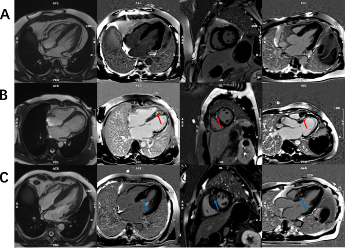
Methods: The patients with HCM whom was underwent CMR were enrolled, using cine cardiac magnetic resonance to evaluate LV function and LGE to detect the myocardial fibrosis. Three groups were assorted: 1) HCM without LGE; 2) HCM with LGE involved the subendocardial layer was defined as I-LGE; 3) HCM with LGE not involved the subendocardial layer was defined as NI-LGE.
Results: We enrolled 122 patients with HCM in the present study. LGE was detected in 58 of 122 (48%) patients with HCM, and 22 (18%) of patients reported I-LGE. HCM with I-LGE had increased higher left ventricular mass index (LVMI) (P < 0.0001) than HCM with NI-LGE or without LGE. In addition, HCM with I-LGE had a larger LV end- systolic volume (P = 0.045), lower LV ejection fraction (LVEF) (P = 0.026), higher LV myocardial mass (P < 0.001) and thicker LV wall (P < 0.001) more than HCM without LGE alone. The I-LGE were significantly associated with LVEF (OR: 0.961; P = 0.016), LV mass (OR: 1.028; P < 0.001), and maximal end-diastolic LVWT (OR: 1.567; P < 0.001). On multivariate analysis, LVEF (OR: 0.948; P = 0.013) and maximal end-diastolic LVWT (OR: 1.548; P = 0.001) were associated with higher risk for I-LGE compared to HCM without LGE. Noticeably, the maximal end-diastolic LVWT (OR: 1.316; P = 0.011) was the only associated with NI-LGE compared to HCM without LGE.
Conclusions: I-LGE is not uncommon in patients with HCM. HCM with I-LGE was associated with significant LV hypertrophy, extensive LGE and poor LV ejection fraction. We should consider focal ischemic myocardial fibrosis when applying LGE to risk stratification for HCM.
Keywords: Cardiac magnetic resonance imaging; Fibrosis; Hypertrophic cardiomyopathy; Late gadolinium enhancement.
© 2024. The Author(s).
Conflict of interest statement
The authors declare no competing interests.
Figures
Similar articles
-
Association between cardiac dysfunction and late gadolinium enhancement confined to the LV intramural region in patients with hypertrophic cardiomyopathy.Ann Med. 2025 Dec;57(1):2533425. doi: 10.1080/07853890.2025.2533425. Epub 2025 Jul 23. Ann Med. 2025. PMID: 40698385 Free PMC article.
-
The relationship between the quantitative extent of late gadolinium enhancement and burden of nonsustained ventricular tachycardia in hypertrophic cardiomyopathy: A delayed contrast-enhanced magnetic resonance study.J Cardiovasc Electrophysiol. 2019 May;30(5):651-657. doi: 10.1111/jce.13855. Epub 2019 Feb 2. J Cardiovasc Electrophysiol. 2019. PMID: 30680853
-
Progression of Myocardial Fibrosis in Hypertrophic Cardiomyopathy: A Cardiac Magnetic Resonance Study.JACC Cardiovasc Imaging. 2021 May;14(5):947-958. doi: 10.1016/j.jcmg.2020.09.037. Epub 2020 Nov 25. JACC Cardiovasc Imaging. 2021. PMID: 33248971
-
The current and emerging role of cardiovascular magnetic resonance imaging in hypertrophic cardiomyopathy.J Cardiovasc Transl Res. 2009 Dec;2(4):415-25. doi: 10.1007/s12265-009-9136-3. Epub 2009 Nov 7. J Cardiovasc Transl Res. 2009. PMID: 20560000 Review.
-
Clinical utility of cardiovascular magnetic resonance in hypertrophic cardiomyopathy.J Cardiovasc Magn Reson. 2012 Feb 1;14(1):13. doi: 10.1186/1532-429X-14-13. J Cardiovasc Magn Reson. 2012. PMID: 22296938 Free PMC article. Review.
Cited by
-
Mexican guidelines 2024 for the diagnosis and treatment of hypertrophic cardiomyopathy.Arch Cardiol Mex. 2024;94(Supl 4):1-75. doi: 10.24875/ACM.M25000098. Arch Cardiol Mex. 2024. PMID: 39928711 Free PMC article. English. No abstract available.
-
Reduction in myofilament Ca2+ sensitivity partially ameliorates the cardiac phenotype in hypertrophic cardiomyopathy linked to a TnT-R92Q mutation.Front Physiol. 2025 May 23;16:1600117. doi: 10.3389/fphys.2025.1600117. eCollection 2025. Front Physiol. 2025. PMID: 40488145 Free PMC article.
-
Scanner-generated native T1 mapping: a novel approach for assessing myocardial fibrosis in coronary heart disease.Front Cardiovasc Med. 2025 May 1;12:1553919. doi: 10.3389/fcvm.2025.1553919. eCollection 2025. Front Cardiovasc Med. 2025. PMID: 40376144 Free PMC article.
-
[Clincial Research Progress in Using Magnetic Resonance Imaging to Assess Myocardial Fibrosis in Hypertrophic Cardiomyopathy].Sichuan Da Xue Xue Bao Yi Xue Ban. 2024 Nov 20;55(6):1357-1363. doi: 10.12182/20241160601. Sichuan Da Xue Xue Bao Yi Xue Ban. 2024. PMID: 39990836 Free PMC article. Review. Chinese.
References
-
- Shintani Y, Nakayama T, Masaki A, Yokoi M, Wakami K, Ito T, et al. Clinical impact of the pathological quantification of myocardial fibrosis and infiltrating t lymphocytes using an endomyocardial biopsy in patients with hypertrophic cardiomyopathy. Int J Cardiol. 2022;362:110–117. doi: 10.1016/j.ijcard.2022.05.068. - DOI - PubMed
Publication types
MeSH terms
Substances
Grants and funding
LinkOut - more resources
Full Text Sources
