. 2024 Apr 9;22(1):221.
doi: 10.1186/s12964-024-01567-0.

Proteomic studies of VEGFR2 in human placentas reveal protein associations with preeclampsia, diabetes, gravidity, and labor

Affiliations

Proteomic studies of VEGFR2 in human placentas reveal protein associations with preeclampsia, diabetes, gravidity, and labor

Shannon J Ho et al. Cell Commun Signal. .

Abstract

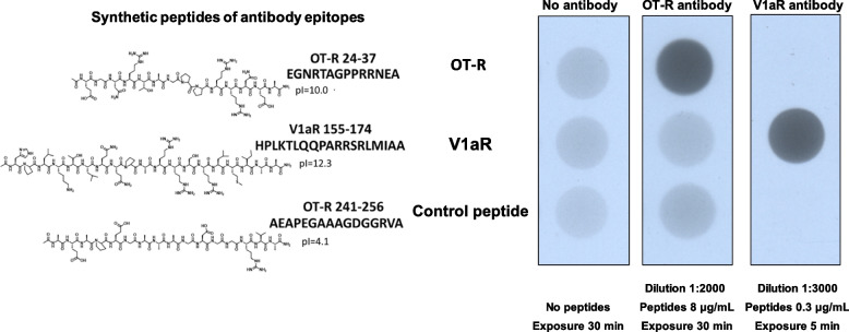
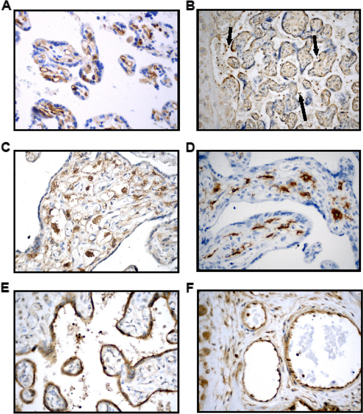
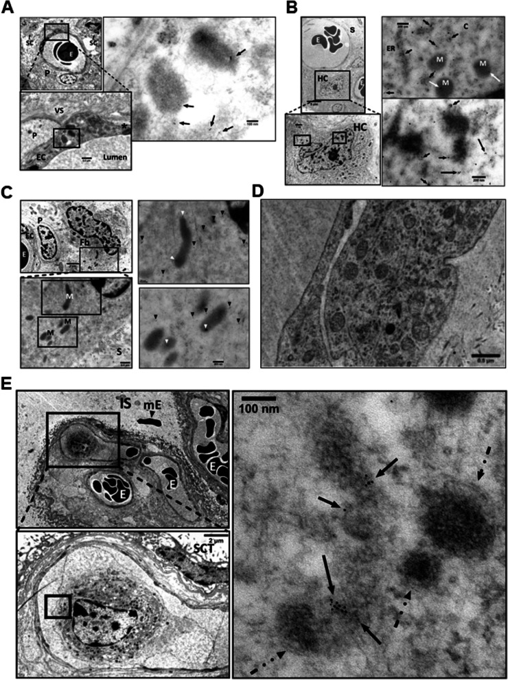
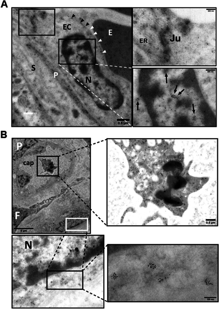
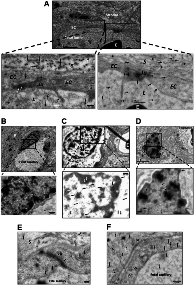
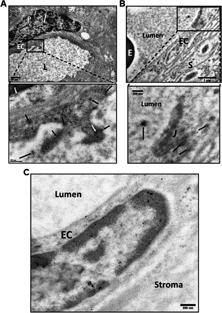
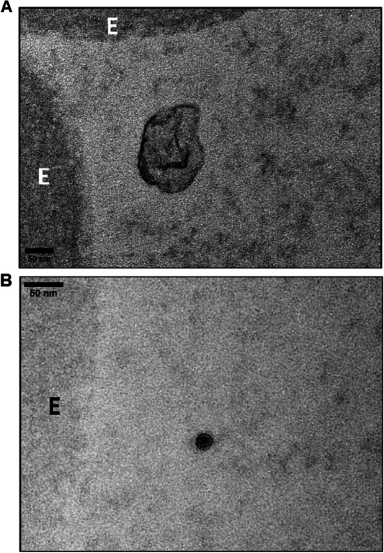
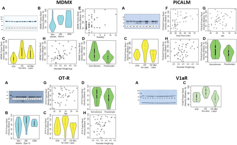
VEGFR2 (Vascular endothelial growth factor receptor 2) is a central regulator of placental angiogenesis. The study of the VEGFR2 proteome of chorionic villi at term revealed its partners MDMX (Double minute 4 protein) and PICALM (Phosphatidylinositol-binding clathrin assembly protein). Subsequently, the oxytocin receptor (OT-R) and vasopressin V1aR receptor were detected in MDMX and PICALM immunoprecipitations. Immunogold electron microscopy showed VEGFR2 on endothelial cell (EC) nuclei, mitochondria, and Hofbauer cells (HC), tissue-resident macrophages of the placenta. MDMX, PICALM, and V1aR were located on EC plasma membranes, nuclei, and HC nuclei. Unexpectedly, PICALM and OT-R were detected on EC projections into the fetal lumen and OT-R on 20-150 nm clusters therein, prompting the hypothesis that placental exosomes transport OT-R to the fetus and across the blood-brain barrier. Insights on gestational complications were gained by univariable and multivariable regression analyses associating preeclampsia with lower MDMX protein levels in membrane extracts of chorionic villi, and lower MDMX, PICALM, OT-R, and V1aR with spontaneous vaginal deliveries compared to cesarean deliveries before the onset of labor. We found select associations between higher MDMX, PICALM, OT-R protein levels and either gravidity, diabetes, BMI, maternal age, or neonatal weight, and correlations only between PICALM-OT-R (p < 2.7 × 10⁻⁸), PICALM-V1aR (p < 0.006), and OT-R-V1aR (p < 0.001). These results offer for exploration new partnerships in metabolic networks, tissue-resident immunity, and labor, notably for HC that predominantly express MDMX.

Keywords: Hofbauer cells; MDMX; Oxytocin receptor; PICALM; Placenta; Proteomics; TRIM21; TRPM7; VEGFR2; Vasopressin receptor V1aR.


Conflict of interest statement

DATC declares a grant from Merck for research unrelated to this manuscript. The remaining authors declare no competing interests.

Figures

Fig. 1
Fig. 1
Validation of OT-R and V1aR antibodies. The synthesized peptides and their pI are shown next to the scans of the coated Nunc C-bottom strips probed with the antibodies being validated
Fig. 2
Fig. 2
Protein elution profiles in VEGFR2, MDMX and PICALM IP. Detergent-extracted villous sample X was incubated with magnetic bead suspension charged with the target Ab (see Methods). E denotes eluted protein fractions. Western blots detected the target proteins eluted in the uniformly performed IP experiments. A VEGFR2 in VEGFR2-IP. B MDMX in VEGFR2-IP. C V1aR in VEGFR2-IP. D OT-R in VEGFR2-IP. E MDMX in MDMX-IP. F PICALM in PICALM-IP. G Left-side, OT-R in samples from PICALM-IP, and right-side OT-R in samples from MDMX-IP. H Left-side, V1aR in samples from MDMX-IP, and right-side samples from PICALM-IP. Panel (C) shows the membrane in (B) reprobed for V1aR without stripping
Fig. 3
Fig. 3
Immunohistochemical staining of placental sections. A VEGFR2 immunostaining is strongly positive in villous endothelial cells (patient Q3). B TRIM21 is uniformly positive in villous trophoblast (patient N4). Very strong staining is seen of maternal leukocytes (arrows). C MDMX is strongly positive in the cytoplasm of HC and moderately positive in endothelial cells (patient W2). D CD163 is strongly positive in the cytoplasm of the HC (patient U1). E PICALM is strongly positive in the trophoblast (patient R4). F PICALM positivity seen in the villous endothelial cells and fetal blood leukocytes (patient R4). Magnifications in A-F were 10x, 20x  and 40x
Fig. 4
Fig. 4
Detection of VEGFR2 by IGEM in chorionic villi of placenta Ho-73 (preeclamptic). A Top left micrograph shows a villous capillary. P: pericyte, E: RBC, SC: stromal cells. Bottom image details area outlined on the top micrograph and shows endothelial cell (EC) cytoplasm, pericyte (P), tight junction (*), collagen fibers in villous stroma (VS), and capillary lumen (L). Image at right: Enlargement of area outlined on the bottom left shows VEGFR2 in mitochondria (arrows). B Micrograph on top left shows villous capillary, stroma (S), luminal RBC (E), and Hofbauer cell (HC). Enlargement of rectangle at bottom shows prominent nucleolus, mitochondria, and endoplasmic reticulum of HC. Micrographs on the right show enlargement of areas outlined on the bottom left micrograph that detail clusters of VEGFR2 (arrows) in the cytosol (C), along segments of endoplasmic reticulum (ER), and mitochondria (M). Enlargement of image at bottom right shows diffuse localization of VEGFR2 clusters throughout the nucleoplasm (arrows). C Top left micrograph: Villus stroma depicting a fibroblast (Fb), and a pericyte (P) associated with fetal capillary in partial profile, RBC (E), and EC cytoplasm. Bottom left is enlargement of area outlined on the top graph and shows fibroblast cytosol, mitochondria (M), and villous stroma (S). To the right, enlargement of two areas show VEGFR2 in cytoplasm and in partial profiles of mitochondrial matrix (white arrow heads). D A TEM micrograph after osmication provided a better outline of mitochondria in an EC, compared to (A, B and C) panels. E Fetal capillaries with luminal RBC (E) are shown in the top left image, IS: intervillous space, mE: maternal RBC. Bottom: enlargement of outlined area shows a fetal macrophage in greater detail, a syncytiotrophoblast (SCT) is also present. Right micrograph: enlargement of area in bottom left micrograph, points to VEGFR2 labelling in an incomplete profile of a mitochondrion. Dashed arrows outline the outer double membrane of the labeled and unlabeled mitochondria. The gold particle diameter is 6 nm
Fig. 5
Fig. 5
Detection of MDMX by IGEM in chorionic villi of placentas Ho-71 (normotensive) and Ho-73 (preeclamptic). A Left micrograph: Nucleus (N) of endothelial cell (EC) from Ho-73. Arrow heads point to small MDMX clusters, parallel to the inner aspect of the EC plasma membrane. Area outlined, top left: EC junction, P: pericyte process, E: RBC in capillary lumen, S: stroma. This area is enlarged on the top right graph and shows MDMX clusters along the junction (Ju), and endoplasmic reticulum (ER). Bottom right: enlargement of outlined area shows diffuse pattern of MDMX clusters throughout the nucleoplasm. B Top left micrograph shows an outline of apoptotic macrophage in the lumen of fetal capillary (cap) from Ho-71. P: pericyte, F: partial profile of fibroblast (F) and HC partially marked by a white outline. Enlargement on the top right image shows diffuse MDMX localization in the vacuolated cytoplasm of luminal macrophage. Image at bottom left is enlargement of area outlined in white and shows MDMX clustering in the nucleus (N) and cytosol of the HC. Clusters, averaging 50–100 nm in diameter, run along the nuclear membrane and appear to be associated with mitochondrial and endoplasmic reticulum membranes. Bottom right: enlarged area shows MDMX clusters of approximately 100 nm. The gold particle diameter is 6 nm
Fig. 6
Fig. 6
Detection of PICALM in chorionic villi of placentas Ho-71 (normotensive) and Ho-73 (preeclamptic). A Top IGEM micrograph: partial profile of villus capillary from Ho-71. EC: endothelial cells. E: RBC, stroma. Bottom images: enlargement of two selected areas of endothelium show PICALM localization (arrows) at EC junctions (Ju), along EC plasma membrane, in cytoplasmic projections into lumen (L) and adjacent stroma (S). B Top micrograph: the nuclei of endothelium of a villus capillary from Ho-71, and the adjacent pericyte (P) are patent. Bottom image: enlargement of nucleus, shows PICALM in nucleoplasm near chromatin, on EC cytoplasmic membrane (white arrows) and in luminal space (black arrow). C Top micrograph: HC in stroma of Ho-73. E: RBC in capillary. At bottom: enlargement shows PICALM in HC nucleus (arrows). D Top micrograph: HC cell in the stroma of placenta Ho-71. Bottom image: enlargement shows PICALM in nucleoplasm in association with chromatin. E PICALM is shown (arrows) at the junction (Ju), and plasma membrane of EC and in adjacent stroma (S) of fetal capillary from Ho-73. F PICALM is shown (arrows) in cytoplasm and plasma membrane of EC of capillary from Ho-71, and in basal lamina (BL), stroma (S) and capillary lumen. The gold particle diameter is 10 nm
Fig. 7
Fig. 7
Detection of Oxytocin Receptor (OT-R) in chorionic villi of placentas of normotensive Ho-71, and preeclamptic Ho-73 patients. A Top IGEM micrograph: Villus capillary from Ho-71. P: pericyte adjacent to the endothelial cell (EC), L: lumen. White arrow shows cluster of OT-R on P. Bottom: enlargement shows OT-R in endoplasmic reticulum (ER) and cytoplasm projections of EC into the lumen. (arrows). B Top micrograph: Partial view of villus capillary from Ho73, E: RBC, S: stroma. Bottom graph shows EC junction (J) and OT-R clusters on J, EC cytoplasm, and lumen (arrow). C OT-R is seen in the nucleus of an EC from Ho-73.The gold particle diameter is 10 nm
Fig. 8
Fig. 8
Detection of Vasopressin Receptor V1aR in chorionic villus of placenta Ho-72 (diabetic). A Composite IGEM image shows nucleus of HC in stroma on the left and enlargements of three demarcated regions of its nucleus on the right. V1aR clusters, 20–100 nm, in the nucleus are indicated by arrows. B Top micrograph shows EC aspect of a villus capillary. Bottom image is enlargement that shows V1aR on EC membrane, nucleus, and stroma (arrows). C Top micrograph shows aspect of EC of villus capillary, an RBC is seen in the lumen. Bottom image is enlargement that shows several 20–100 nm clusters of V1aR on surface of the RBC (arrows). The gold particle diameter is 10 nm
Fig. 9
Fig. 9
Transmission electron microscopy of osmicated chorionic villus from placenta Ho-73 (preeclamptic). A An exosome [9] is shown in the lumen with the characteristic lipid bilayer adjacent to RBC (E). B A dense particle with a lipid bilayer, perhaps an exomere [12], is shown in the lumen next to an RBC (E). Scale in A and B is 50 μm
Fig. 10
Fig. 10
Representative western blots (A) of MDMX, PICALM, OT-R, and V1aR and their protein levels in relation to diabetes (B), mode of delivery (C), preeclampsia (D), gravidity (E), BMI (F), maternal age (G), and neonatal weight (H). Western blots show the molecular mass (kDa) and the placental extracts from patients identified by a letter-number code shown in Table 1. Internal control for MDMX and PICALM was sample Q1, and sample V1 for OT-R and V1aR

Similar articles

Cited by

References

    1. Burton GJ, Fowden AL. The placenta: a multifaceted, transient organ. Philos Trans R Soc Lond B Biol Sci. 2015;370(1663):20140066. doi: 10.1098/Rstb.2014.0066. - DOI - PMC - PubMed
    1. Burton GJ, Redman CW, Roberts JM, Moffett A. Pre-Eclampsia: pathophysiology and clinical implications. BMJ. 2019;366:L2381. doi: 10.1136/Bmj.L2381. - DOI - PubMed
    1. Aplin JD, Myers JE, Timms K, Westwood M. Tracking placental development in health and disease. Nat Rev Endocrinol. 2020;16(9):479–494. doi: 10.1038/S41574-020-0372-6. - DOI - PubMed
    1. Shibuya M. Vegfr and type-V Rtk activation and signaling. Cold Spring Harb Perspect Biol. 2013;5(10):A009092. doi: 10.1101/Cshperspect.A009092. - DOI - PMC - PubMed
    1. Peach, C. J., Mignone, V. W., Arruda, M. A., Alcobia, D. C., Hill, S. J., Kilpatrick, L. E., Et Al. (2018). Molecular Pharmacology Of Vegf-A Isoforms: Binding And Signalling At Vegfr2. Int J Mol Sci, 19(4) 10.3390/Ijms19041264. - PMC - PubMed

Publication types

Substances