. 2024 Aug 2;23(8):3012-3024.
doi: 10.1021/acs.jproteome.3c00649. Epub 2024 Apr 9.

Analysis of Vascular Smooth Muscle Cells from Thoracic Aortic Aneurysms Reveals DNA Damage and Cell Cycle Arrest as Hallmarks in Bicuspid Aortic Valve Patients

Affiliations

Analysis of Vascular Smooth Muscle Cells from Thoracic Aortic Aneurysms Reveals DNA Damage and Cell Cycle Arrest as Hallmarks in Bicuspid Aortic Valve Patients

Ariadna Martin-Blazquez et al. J Proteome Res. .

Abstract

Thoracic aortic aneurysm (TAA) is mainly sporadic and with higher incidence in the presence of a bicuspid aortic valve (BAV) for unknown reasons. The lack of drug therapy to delay TAA progression lies in the limited knowledge of pathophysiology. We aimed to identify the molecular hallmarks that differentiate the aortic dilatation associated with BAV and tricuspid aortic valve (TAV). Aortic vascular smooth muscle cells (VSMCs) isolated from sporadic TAA patients with BAV or TAV were analyzed by mass spectrometry. DNA oxidative damage assay and cell cycle profiling were performed in three independent cohorts supporting proteomics data. The alteration of secreted proteins was confirmed in plasma. Stress phenotype, oxidative stress, and enhanced DNA damage response (increased S-phase arrest and apoptosis) were found in BAV-TAA patients. The increased levels of plasma C1QTNF5, LAMA2, THSB3, and FAP confirm the enhanced stress in BAV-TAA. Plasma FAP and BGN point to an increased inflammatory condition in TAV. The arterial wall of BAV patients shows a limited capacity to counteract drivers of sporadic TAA. The molecular pathways identified support the need of differential molecular diagnosis and therapeutic approaches for BAV and TAV patients, showing specific markers in plasma which may serve to monitor therapy efficacy.

Keywords: DNA damage; aortic aneurysm; bicuspid aortic valve; cardiovascular disease; oxidative stress; proteomics; vascular smooth muscle cells.


Conflict of interest statement

The authors declare no competing financial interest.

Figures

Figure 1
Figure 1
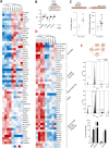
Stress phenotype and altered DNA replication and repair machinery in thoracic aortic aneurysm associated with bicuspid aortic valve. Aortic VSMCs from TAA patients were analyzed by quantitative high-throughput proteomics, and protein-abundant changes are compiled here. (A) VSMC characterization was performed by staining with αSMA and calponin and negative staining for endothelial cells (CD31) and fibroblasts (S100A4). (B) Contractile, proliferative, and stress phenotypes were found in BAV-TAA and TAV-TAA VSMCs, but a differential stress phenotype was evident in BAV-TAA cells. (C) The differential protein profile in BAV-TAA versus TAV-TAA VSMCs includes 52 proteins with nP ≥ 3, −2 ≥ ΔZq ≥ 2 and p value ≤ 0.01. Individual examination of these proteins indicates a deregulation in DNA machinery in BAV-TAA patients. Color scale: ΔZq = −3 (green), through 0 (white), to +3 (orange). * p value ≤ 0.05, ** p value < 0.01. Heatmap proteins: p value ≤ 0.05.
Figure 2
Figure 2
DNA replication machinery and signal transduction are altered biological processes in BAV-associated thoracic aortic aneurysm. Coordinated protein analysis was conducted with the SBT algorithm to identify biological processes altered beyond individual protein changes. Processes showing an alteration of −3.0 > ΔZc > 3.0, homogeneous intragroup behavior, and p value < 0.001 were considered the most relevant altered pathways in TAA associated with BAV. The top eight altered processes are represented together with the quantitative difference between BAV-TAA and TAV-TAA patients (bar chart ΔZc). These eight processes indicate impairment of the DNA replication machinery in BAV-TAA VSMCs accompanied by an increase in G-protein-mediated signal transduction. The proteins driving these process changes were further investigated and grouped into families. The figure shows the outstanding protein abundance changes from the most representative protein families for BAV-TAA vs TAV-TAA. BAV-TAA VSMCs showed relatively lowered levels of DNA-related proteins, including MCM (minichromosome maintenance), CHD (chromodomain helicase DNA binding), DPO (DNA polymerase), ORC (origin recognition complex), and RFC (replication factor C). In contrast, several G proteins, including Ras-superfamily proteins and heterodimeric G proteins, showed significantly increased abundance. * p value ≤ 0.05, ** p value < 0.01; *** p value < 0.001; **** p value < 0.0001.
Figure 3
Figure 3
Enhanced oxidative stress environment, DNA oxidative damage, and cell cycle arrest in BAV-TAA patients. The individual protein and coordinated protein alterations in BAV-TAA patients suggest elevated oxidative stress driven by the significantly altered VSMC proteins listed. (A) Heatmap of proteins related to oxidative stress identified by coordinated response analysis. (B) Altered proteins related to antioxidant defense between BAV-TAA and TAV-TAA VSMCs. (C) 8-Hydroxy-2′-deoxyguanosine (8-OHdG) quantification from DNA extracted from VSMCs (left chart) and urine samples (right chart) in BAV-TAA patients. (D) Heatmap of proteins related to cell cycle progression: histones, histone-related proteins, proliferation markers, mismatch repair (MMR) proteins, and replication factor proteins. (E) Cell cycle profiling in TAV-TAA patients (gray histogram) and BAV-TAA patients (black histogram); cell percentage in apoptosis, G0-G1, S, and G2-M phases is shown in the bar chart. Color scale: ΔZq = −3 (blue), through 0 (white), to +3 (red). * p value ≤ 0.05, ** p value < 0.01.
Figure 4
Figure 4
Transferable secreted proteins in VSMCs to plasma. VSMC proteins showing altered abundance in the proteomics analysis and identified as extracellularly secreted were quantified and further investigated in their soluble form in human plasma from TAV-TAA and BAV-TAA patients by ELISA. (A) Changes in protein abundance in VSMCs for C1QTNF5, LAMA2, FAP, BGN, CCDC80, SERPINE2, and THBS3 showing an increase in BAV-TAA. (B) Respective plasma alteration showing significantly diminished levels in plasma from TAA patients. Protein differences between clinical groups were analyzed by t test or Mann–Whitney test according to the results of the normality test. * p value ≤ 0.05, ** p value < 0.01, *** p value < 0.001, **** p value < 0.0001.
Figure 5
Figure 5
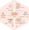
Bicuspid valve-associated thoracic aortic aneurysm is driven by specific mechanisms and should be treated as a distinct clinical entity. Individual and coordinated protein changes in TAA patients with BAV provide evidence for alterations to six biological pathways that could be potential therapeutic targets. The aortic wall is subject to greater shear stress in BAV-TAA than in TAV-TAA. This is accompanied by exposure to more severe oxidative stress, resulting in enhanced DNA oxidative damage. DNA damage response is one of the most evident mechanisms operating in BAV-TAA according to our analysis of human VSMCs. Treatments used in other cardiovascular conditions limit DNA damage and thus may help in the management of BAV-TAA patients. Plasma analysis of the soluble form of altered VSMC proteins confirmed an increased stress phenotype (LAMA2, FAP, and THBS3) and increased oxidative stress (C1QTNF5) in BAV-TAA patients. Vascular remodeling is a hallmark of aneurysm generally. THSB3, FAP, BGN, CCDC80, and SERPINE 2 evidence the differential vascular remodeling in the aorta during TAA, pointing to inflammation in TAV-TAA and to protein homeostasis and diminished repair in BAV-TAA subjects. Our analysis reveals vascular remodeling, protein homeostasis, cell cycle arrest, and enhanced cell death, partly mediated by G proteins. Because G proteins are involved in so many processes, their use as therapeutic targets is challenging and requires further research.

References

    1. Pinard A.; Jones G. T.; Milewicz D. M. Genetics of Thoracic and Abdominal Aortic Diseases. Circ. Res. 2019, 124 (4), 588–606. 10.1161/CIRCRESAHA.118.312436. - DOI - PMC - PubMed
    1. Saeyeldin A. A.; Velasquez C. A.; Mahmood S. U. B.; Brownstein A. J.; Zafar M. A.; Ziganshin B. A.; Elefteriades J. A. Thoracic aortic aneurysm: unlocking the ″silent killer″ secrets. Gen Thorac Cardiovasc Surg 2019, 67 (1), 1–11. 10.1007/s11748-017-0874-x. - DOI - PubMed
    1. Martin-Blazquez A.; Heredero A.; Aldamiz-Echevarria G.; Martin-Lorenzo M.; Alvarez-Llamas G. Non-syndromic thoracic aortic aneurysm: cellular and molecular insights. J. Pathol 2021, 254 (3), 229–238. 10.1002/path.5683. - DOI - PMC - PubMed
    1. Verma S.; Siu S. C. Aortic dilatation in patients with bicuspid aortic valve. N Engl J. Med. 2014, 370 (20), 1920–1929. 10.1056/NEJMra1207059. - DOI - PubMed
    1. Michelena H. I.; Khanna A. D.; Mahoney D.; Margaryan E.; Topilsky Y.; Suri R. M.; Eidem B.; Edwards W. D.; Sundt T. M. 3rd; Enriquez-Sarano M. Incidence of aortic complications in patients with bicuspid aortic valves. Jama 2011, 306 (10), 1104–1112. 10.1001/jama.2011.1286. - DOI - PubMed

Publication types

MeSH terms