. 2025 Jan;57(1):217-231.
doi: 10.1111/evj.14093. Epub 2024 Apr 9.

Closure of the neuro-central synchondrosis and other physes in foal cervical spines

Affiliations

Closure of the neuro-central synchondrosis and other physes in foal cervical spines

Kristin Olstad et al. Equine Vet J. 2025 Jan.

Abstract

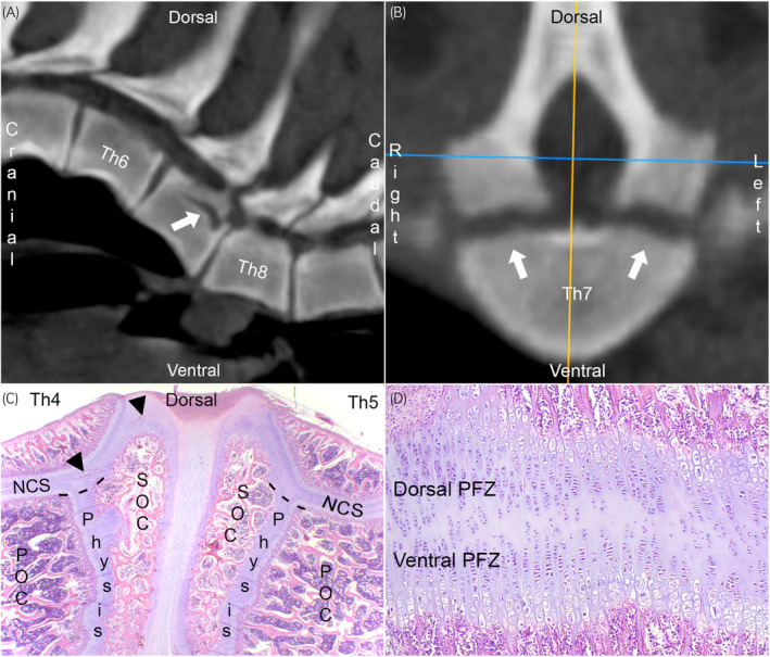
Background: The neuro-central synchondrosis (NCS) is a physis responsible for the growth of the dorsal third of the vertebral body and neural arches. When the NCS of pigs is tethered to model scoliosis, stenosis also ensues. It is necessary to describe the NCS for future evaluation of its potential role in equine spinal cord compression and ataxia (wobbler syndrome).

Objectives: To describe the NCS, including when it and other physes closed in computed tomographic (CT) scans of the cervical spine of foals, due to its potential role in vertebral stenosis.

Study design: Post-mortem cohort study.

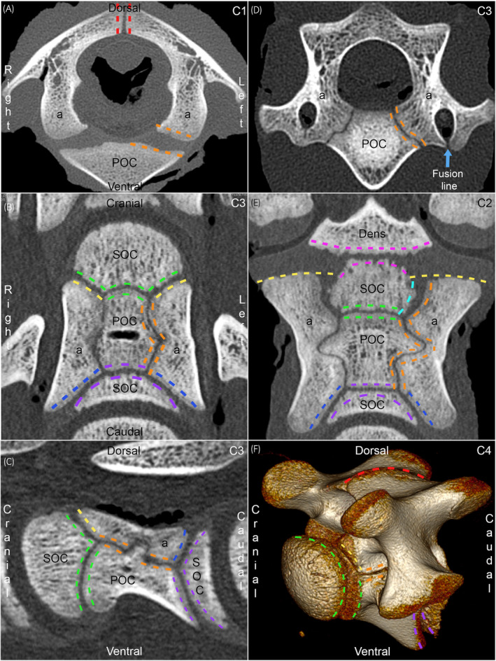
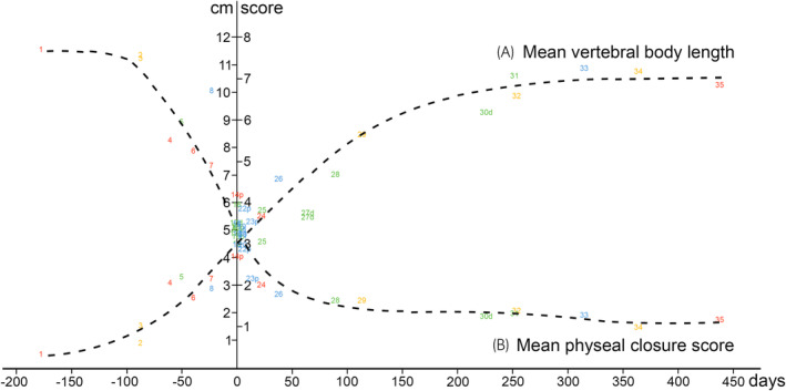
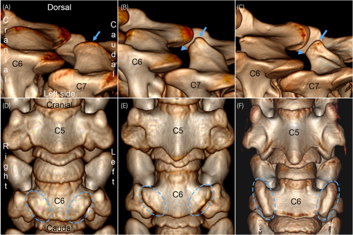
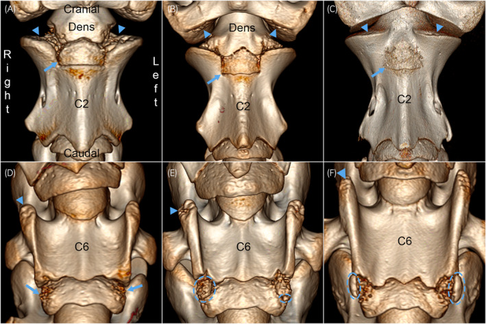
Methods: The cervical spine of 35 cases, comprising both sexes and miscellaneous breeds from 153 gestational days to 438 days old, was examined with CT and physes scored from 6: fully open to 0: fully closed. The dorsal physis, physis of the dens and mid-NCS were scored separately, whereas the cranial and caudal NCS portions were scored together with the respective cranial and caudal vertebral body physes.

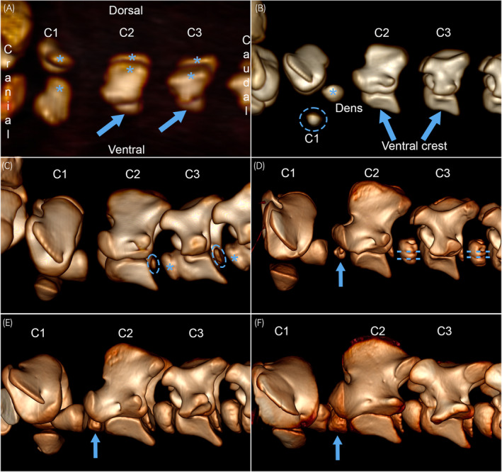
Results: The NCS was a pair of thin physes located in a predominantly dorsal plane between the vertebral body and neural arches. The mid-NCS was closed in C1 from 115 days of age, and in C2-C7 from 38 days of age. The dorsal physis closed later than the NCS in C1, and earlier than the NCS in C2-C7. The dens physis was closed from 227 days of age. The cranial and caudal physes were closing, but not closed from different ages in the different vertebrae of the oldest cases.

Main limitations: Hospital population.

Conclusions: The NCS was a thin physis that contributed mainly to height-wise growth, but also width- and length-wise growth of the vertebral body and neural arches. The mid-NCS was closed in all cervical vertebrae from 115 days of age. The NCS warrants further investigation in the pathogenesis of vertebral stenosis.

Historial: La sincondrosis neuro‐central (NCS) es la fisis responsable del crecimiento del tercio dorsal del cuerpo vertebral y de los arcos neurales. Cuando la NCS en cerdos se asocia a un modelo de escoliosis, también se produce estenosis. Es necesario describir la NCS para la futura evaluación de su rol potencial en la comprensión de la medula espinal equina y ataxia (síndrome de Wobbler).

Objetivos: Describir la NCS incluyendo cuando ella y otras fisis se cierran, por tomografía computarizada (CT) de la columna cervical de potrillos, debido a su rol potencial en la estenosis vertebral. DISEÑO DEL ESTUDIO: Estudio de cohorte post‐mortem. MÉTODOS: La columna cervical de 35 casos, incluyendo ambos sexos y diferentes razas, desde 153 días gestacionales hasta 438 días de edad, fueron examinadas por CT y las fisis fueron dadas un puntaje de, 6: completamente abiertas a, 0: completamente cerradas. La fisis dorsal, la fisis del hueso odontoides y NCS media fueron evaluadas en forma separada, mientras las porciones de NCS craneal y caudal fueron evaluadas juntas con las respectiva fisis del cuerpo vertebral craneal y caudal.

Resultados: La NCS es un par de fisis delgadas localizadas predominantemente en el plano dorsal entre el cuerpo vertebral y los arcos vertebrales. La NCS media estaba cerrada en C1 desde los días 115 de edad, y en C2‐C7 a partir de los 38 días de edad. La fisis dorsal se cerró más tarde que la NCS en C1, y antes que la NCS en C2‐C7. La fisis del hueso odontoides estaba cerradas a partir de los 227 días de edad. Las fisis craneal y caudal estaban cerrándose, pero no estaban cerradas a distintas edades en las diferentes vertebras en los casos mayores de edad.

Limitaciones principales: Población de hospital CONCLUSIONES: La NCS es una fisis delgada que contribuye principalmente al crecimiento en altura, pero también en ancho y largo del cuerpo vertebral y arcos vertebrales. La NCS media estaba cerrada en todas las vértebras cervicales a partir de los 115 días de edad. La NCS merece ser investigada más en la patogénesis de la estenosis vertebral. Palabras Clave: ataxia, tomografía computarizada, caballo, osteocondrosis, estenosis, crecimiento vertebral.

Keywords: ataxia; computed tomography; horse; osteochondrosis; stenosis; vertebral growth.


Conflict of interest statement

The authors have declared no conflicting interests.

Figures

FIGURE 1
FIGURE 1
Neuro‐central synchondrosis in pigs. (A) Computed tomographic (CT) scan of a 100 kg Landrace boar, parasagittal slice. The hypoattenuating line in Th7 (arrow) was suspected of representing fracture secondary to adjacent vertebral wedging. (B) Transverse slice through Th7 from (A). The hypoattenuating line is symmetric (arrows) and literature search revealed that it runs through a pair of physes between the vertebral body and each neural arch known as the neuro‐central synchondroses (NCSs). (C) Parasagittal histological section from Th4‐5 of a 12 kg mixed‐breed piglet, haematoxylin and eosin staining. The vertebral bodies have a primary (POC), a cranial and a caudal secondary ossification centre (SOC). The physes between these centres are responsible for growth of the ventral two‐thirds of the vertebral body (below dashed lines), whereas the NCS is responsible for growth of the dorsal third of the vertebral body and the neural arches (above dashed lines). The growth cartilage contains blood vessels (arrowheads). (D) Magnified view from (C). The middle portion of the NCS has a central resting zone and one set of proliferative (PFZ), hypertrophic and mineralised zones in each of the dorsal and ventral directions. Modified and reprinted with permission from Olstad et al.
FIGURE 2
FIGURE 2
Grouping of physes for scoring. All 2D images are from case 16, 0 days/19 h old, figure (F) is from case 27d, 65 days old. (A) Transverse slice from C1. Three physes were scored in C1: the (mid‐)dorsal physis (red dashed lines), left (orange lines) and right neuro‐central synchondroses. POC, primary ossification centre; a, neural arch. (B) Dorsal slice from C3. In C3 (shown) to C7, five physes were scored: the dorsal physis (not shown), left and right NCSs, cranial and caudal physes of the vertebral body. The mid‐portion of the NCS (orange lines) was scored on its own, whereas the cranial portion of the NCS was grouped with the cranial physis (green and yellow lines), and the caudal portion of the NCS was grouped with the caudal physis (blue and purple lines) for scoring. The mid‐portion of the NCS is craniocaudally oriented, whereas the cranial and caudal portions are axio‐laterally obliquely oriented. SOC, secondary ossification centre. Remainder of legend, see 2A. (C) Sagittal slice from C3, same colour codes as (B). The mid‐portion of the NCS is craniocaudally oriented, whereas the cranial and caudal portions are (axio‐)ventro‐dorsally obliquely oriented. Note that the NCS makes a dorsally‐pointing V‐shaped deviation midway between cranial and caudal. (D) Transverse slice from C3, same orientation as (A) and same colour codes as (B). The mid‐portion of the NCS is axio‐dorsally to ventro‐laterally obliquely oriented. There is a hypoattenuating line (arrow) where the lateral portion fuses with the medial portion of the primary ossification centre ventral to the transverse foramen. (E) Dorsal slice from C2, same orientation as (B). Six physes were scored in C2: the dorsal physis (not shown), left and right NCSs, physis of the dens, cranial and caudal physes. The dens scoring category included both the portion between the dens and the SOC cranially in C2 (pink lines), and the portions between the dens and the neural arches (pink and yellow lines), also known as the neuro‐dental synchondroses. The NCS scoring category included the growth cartilage on the left and right sides of the cranial SOC (turquoise and orange lines), whereas the cranial physis of the vertebral body (green lines) was scored on its own, without any portions of NCS. The caudal physis category included the caudal portions of the NCS (blue and purple lines) as for C3–C7, above. Note that the NCS makes a laterally pointing V‐shaped deviation midway between cranial and caudal. (F) 3D volume‐rendered model from C4, same colour codes as (A, B). The figure shows parts of some of the physes in a volume‐rendered model, and a rotating movie of the model is available in Video S1.
FIGURE 3
FIGURE 3
Mean vertebral body length (A) increased on a shallow sigmoid curve, whereas mean physeal closure score (B) decreased on a slightly steeper sigmoid curve, with age.
FIGURE 4
FIGURE 4
Radiological maturity, early group. 3D volume‐rendered CT images of the C1–C2–C3, left lateral views. (A) Case 1, 153 days of gestation. There are large primary ossification centres (asterisks) in the neural arches of C1 (slightly rotated) to C3, and smaller primary centres (arrows) in the bodies of C2–C3, but not C1. (B) Case 3, 244 days of gestation. In addition to the centres in (A), there is a large ossification centre in the dens (asterisk) and a small ossification centre in the ventral arch of C1 (dashed circle). The ventral crest (arrows) is present on the primary ossification centre of the vertebral bodies of C2–C3. (C) Case 6, 289 days of gestation. In addition to the centres in (A, B), there are large secondary centres (asterisks) cranially and smaller ossification nuclei (dashed circles) caudally in C2–C3. Unlike Cases 4–8 and 14p from 271 to 320 days of gestation, there is no secondary centre cranially in C2 and the cranial secondary centres are fused to the primary centres in the bodies of C3–C4. (D) Case 5, 280 days of gestation. There is a small secondary ossification centre (arrow) cranially in C2. There are regularly spaced, craniocaudally oriented grooves (dashed lines; likely vascular grooves) in the contours of the secondary centres cranially in C3–C4. (E) Case 4, 271 days of gestation. There is a properly formed secondary ossification centre cranially in C2 (arrow), and the secondary ossification centres cranially in C3–C4 are smoother than in Case 5 (D). (F) Case 14p, 0 days/16 h old, but 12 days premature, equivalent to 320 days of gestation. The secondary ossification centre cranially in C2 is almost as large and smooth as the ossification centres cranially in C3–C4.
FIGURE 5
FIGURE 5
Radiological maturity, middle group. 3D volume‐rendered CT images of C6–C7 (left dorsolateral view) and C5–C6 (ventral view). (A) Case 16, 0 days/19 h. The dorsal spinous process (arrow) is only slightly taller on C7 than C6. (B) Case 22p, 6 days. There is a protruding focus (arrow) on the dorsal spinous process of C7, giving it a triangular appearance. The cranial border (arrowhead) of the neural arch is relatively low and round. (C) Case 25, 21 days. There is extra bone lipping (thickening) towards the dorsal margin (arrow), and the cranial contour (arrowhead) is taller and squarer, giving the dorsal spinous process a rectangular, mature shape. (D) Case 21, 6 days. The lamina region (dashed circles) protrudes only slightly further ventrally (between arrows) on C6 than C5. (E) Case 13, age not recorded. There are taller ridges (dashed circles) ventrally on C6 and they have bone lipping along part of their margins. (F) Case 24, 20 days old. There are large, mature‐shaped tubercles (dashed circles) ventrally on C6 with bone lipping along all of their margins.
FIGURE 6
FIGURE 6
Radiological maturity, late group. 3D volume‐rendered CT images of C2 and C6 (ventral views). (A) Case 28, 93 days. The cranial ossification fronts of the neural arches in C2 (arrowheads), caudal ossification fronts of the neural arches in C1, and lateral ossification fronts of the dens have generalised dimples and tubes. The secondary centre cranially in C2 (arrow) looks like a regular ossification centre. (B) Case 29, 115 days. The caudal ossification fronts of the neural arches in C1 (arrowheads) and cranial ossification fronts of the neural arches in C2 have central dimples and tubes, whereas the dens only has dimples in small, peripheral regions. The cranial secondary ossification centre (arrow) is a square shape, outlined by linear grooves, in the ventral contour of C2. (C) Case 32, 260 days. The cranial ossification fronts of the neural arches of C2 have dimples in a small, peripheral region (arrowheads), whereas the other ossification fronts are smooth. The entire margin of the ossification centre cranially in C2 (arrow) is granular in appearance. (D) Case 29, 115 days. The cranial tips of the ventral laminae (arrowhead) have shallow dimples and short tubes. The caudal physis of C6 has dimples and tubes, more prominent in the ossification front of the secondary (arrows) than the primary ossification centre. (E) Case 30d, 227 days. The cranial tips of the ventral laminae have deep dimples and tall tubes (arrowhead). The caudal physis of C6 has mineralised bodies, some of which appear to have coalesced to form large, irregular‐shaped mineralised bodies (dashed circles) within the physis. (F) Case 34, 366 days. The cranial tips of the ventral laminae have deep dimples and a tall tube (arrowhead). The caudal physis has large, smooth and regular ellipsoid‐shaped mineralised bodies bilaterally (dashed circles). The ellipsoid bodies are located just lateral to the physis, at the junction with the growth cartilage of the caudal part of the ventral laminae, thus probably represent the secondary ossification centres for this part of the laminae.

Similar articles

Cited by

References

    1. Olstad K, Aasmundstad T, Kongsro J, Grindflek E. Osteochondrosis and other lesions in all intervertebral, articular process and rib joints from occiput to sacrum in pigs with poor back conformation, and relationship to juvenile kyphosis. BMC Vet Res. 2022;18(1):44. 10.1186/s12917-021-03091-6 - DOI - PMC - PubMed
    1. Vital JM, Beguiristain JL, Algara C, Villas C, Lavignolle B, Grenier N, et al. The neurocentral vertebral cartilage: anatomy, physiology and physiopathology. Surg Radiol Anat. 1989;11(4):323–328. 10.1007/BF02098705 - DOI - PubMed
    1. Bozkus H, Crawford NR, Chamberlain RH, Valenzuela TD, Espinoza A, Yuksel Z, et al. Comparative anatomy of the porcine and human thoracic spines with reference to thoracoscopic surgical techniques. Surg Endosc. 2005;19(12):1652–1665. 10.1007/s00464-005-0159-9 - DOI - PubMed
    1. Maierl J, Zechmeister R, Schill W, Gerhards H, Liebich HG. Radiologic description of the growth plates of the atlas and axis in foals. Tierarztl Prax Ausg G Grosstiere Nutztiere. 1998;26(6):341–345. - PubMed
    1. Wissdorf H, Gerhards H, Huskamp B, Deegen E. Praxisorientierte Anatomie und Propädeutik des Pferdes (Practice‐oriented anatomy and propedeutics of the horse). 3rd ed. Hannover, Germany: M. & H. Schaper Verlag GmbH; 2010.