

Glymphatic system: a gateway for neuroinflammation
- PMID: 38595285
- PMCID: PMC11168510
- DOI: 10.4103/1673-5374.391312
Glymphatic system: a gateway for neuroinflammation
Abstract
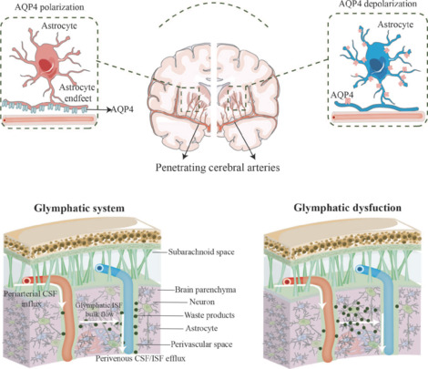
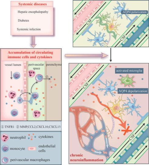
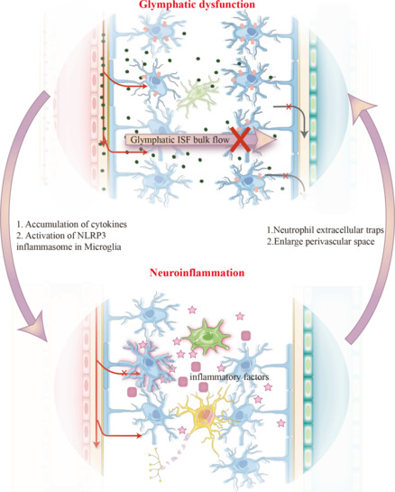
The glymphatic system is a relatively recently identified fluid exchange and transport system in the brain. Accumulating evidence indicates that glymphatic function is impaired not only in central nervous system disorders but also in systemic diseases. Systemic diseases can trigger the inflammatory responses in the central nervous system, occasionally leading to sustained inflammation and functional disturbance of the central nervous system. This review summarizes the current knowledge on the association between glymphatic dysfunction and central nervous system inflammation. In addition, we discuss the hypothesis that disease conditions initially associated with peripheral inflammation overwhelm the performance of the glymphatic system, thereby triggering central nervous system dysfunction, chronic neuroinflammation, and neurodegeneration. Future research investigating the role of the glymphatic system in neuroinflammation may offer innovative therapeutic approaches for central nervous system disorders.
Copyright © 2024 Copyright: © 2024 Neural Regeneration Research.
Conflict of interest statement
Figures
Similar articles
-
The Glymphatic System (En)during Inflammation.Int J Mol Sci. 2021 Jul 13;22(14):7491. doi: 10.3390/ijms22147491. Int J Mol Sci. 2021. PMID: 34299111 Free PMC article. Review.
-
Glymphatic System Pathology and Neuroinflammation as Two Risk Factors of Neurodegeneration.Cells. 2024 Feb 5;13(3):286. doi: 10.3390/cells13030286. Cells. 2024. PMID: 38334678 Free PMC article. Review.
-
Glymphatic System Dysfunction in Central Nervous System Diseases and Mood Disorders.Front Aging Neurosci. 2022 Apr 25;14:873697. doi: 10.3389/fnagi.2022.873697. eCollection 2022. Front Aging Neurosci. 2022. PMID: 35547631 Free PMC article. Review.
-
Glymphatic Dysfunction in Migraine Mice Model.Neuroscience. 2023 Sep 15;528:64-74. doi: 10.1016/j.neuroscience.2023.07.027. Epub 2023 Jul 28. Neuroscience. 2023. PMID: 37516436
-
Glymphatic and lymphatic communication with systemic responses during physiological and pathological conditions in the central nervous system.Commun Biol. 2024 Feb 24;7(1):229. doi: 10.1038/s42003-024-05911-5. Commun Biol. 2024. PMID: 38402351 Free PMC article. Review.
Cited by
-
Functional abnormalities of the glymphatic system in cognitive disorders.Neural Regen Res. 2025 Dec 1;20(12):3430-3447. doi: 10.4103/NRR.NRR-D-24-01049. Epub 2025 Jan 13. Neural Regen Res. 2025. PMID: 39820293 Free PMC article.
-
Perivascular space fluid diffusivity predicts clinical deterioration in prodromal and early-stage Parkinson's disease.NPJ Parkinsons Dis. 2025 Jun 14;11(1):169. doi: 10.1038/s41531-025-01036-6. NPJ Parkinsons Dis. 2025. PMID: 40517151 Free PMC article.
-
Alterations in the Glymphatic System and Association with Brain Structure and Cognitive Function in Moyamoya Disease.Transl Stroke Res. 2025 Aug;16(4):1173-1184. doi: 10.1007/s12975-024-01296-z. Epub 2024 Sep 9. Transl Stroke Res. 2025. PMID: 39245689
-
Glymphatic system clearance and Alzheimer's disease risk: a CSF proteome-wide study.Alzheimers Res Ther. 2025 Jan 31;17(1):31. doi: 10.1186/s13195-024-01612-7. Alzheimers Res Ther. 2025. PMID: 39891246 Free PMC article.
-
OAB-14 Attenuated Glymphatic System Disorder, Neuroinflammation and Dyskinesia in Parkinson's Disease Model Mice Induced by Rotenone.Neurochem Res. 2025 Apr 12;50(2):142. doi: 10.1007/s11064-025-04388-w. Neurochem Res. 2025. PMID: 40220255
References
-
- Abe Y, Ikegawa N, Yoshida K, Muramatsu K, Hattori S, Kawai K, Murakami M, Tanaka T, Goda W, Goto M, Yamamoto T, Hashimoto T, Yamada K, Shibata T, Misawa H, Mimura M, Tanaka KF, Miyakawa T, Iwatsubo T, Hata JI, et al. Behavioral and electrophysiological evidence for a neuroprotective role of aquaporin-4 in the 5xFAD transgenic mice model. Acta Neuropathol Commun. 2020;8:67. - PMC - PubMed
-
- Akhtar A, Sah SP. Insulin signaling pathway and related molecules: role in neurodegeneration and Alzheimer’s disease. Neurochem Int. 2020;135:104707. - PubMed
-
- Althoff GE, Wolfer DP, Timmesfeld N, Kanzler B, Schrewe H, Pagenstecher A. Long-term expression of tissue-inhibitor of matrix metalloproteinase-1 in the murine central nervous system does not alter the morphological and behavioral phenotype but alleviates the course of experimental allergic encephalomyelitis. Am J Pathol. 2010;177:840–853. - PMC - PubMed
LinkOut - more resources
Full Text Sources
