

Use of Adult Dry Human Mandibles to Study of Mandibular Foramen from Various Bony Landmarks
- PMID: 38595526
- PMCID: PMC11000888
- DOI: 10.4103/jpbs.jpbs_927_23
Use of Adult Dry Human Mandibles to Study of Mandibular Foramen from Various Bony Landmarks
Abstract
Background: Precise knowledge of the mandibular foramen's location is essential for clinical and surgical procedures, especially the inferior alveolar nerve block. Variability in its position concerning different bony landmarks can significantly impact clinical outcomes.
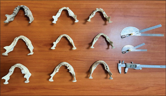
Materials and methods: This study examined 30 Adult dry human mandibles to determine the mandibular foramen's positions in relation to specific bony landmarks: the occlusal plane, posterior border of the ramus, and lingula. Measurements were obtained using a calibrated digital caliper, and statistical analysis was performed.
Results: The study revealed significant variations in the position of the mandibular foramen. In relation to the occlusal plane, the mandibular foramen was found at an average height of approximately 15.2 mm (±2.1 mm). Regarding the posterior border of the ramus, it was situated at an average distance of about 18.5 mm (±3.4 mm). In relation to the lingula, the average distance was approximately 21.8 mm (±4.0 mm). These results underscore the considerable individual differences and anatomical variations in the mandibular foramen's location among the studied specimens.
Conclusion: The observed variations in the position of the mandibular foramen emphasize the need for clinicians and surgeons to be cognizant of these differences when performing procedures involving the inferior alveolar nerve block. Understanding these anatomical variations is crucial for enhancing clinical precision, reducing complications, and ensuring optimal outcomes.
Keywords: Anatomical variations; dental procedures; dry mandibles; inferior alveolar nerve block; lingula; mandibular foramen; occlusal plane; posterior border of the ramus; surgical techniques.
Copyright: © 2023 Journal of Pharmacy and Bioallied Sciences.
Conflict of interest statement
There are no conflicts of interest.
Figures
Similar articles
-
A Morphometric Study of the Mandibular Foramen, Lingula, and the Incidence of Accessory Mandibular Foramina in Dry Mandibles.Cureus. 2025 Mar 24;17(3):e81087. doi: 10.7759/cureus.81087. eCollection 2025 Mar. Cureus. 2025. PMID: 40271342 Free PMC article.
-
Determining the position of the lingula and the mandibular foramen using the antilingula in orthognathic surgery.BMC Oral Health. 2024 Apr 27;24(1):499. doi: 10.1186/s12903-024-04286-7. BMC Oral Health. 2024. PMID: 38678231 Free PMC article.
-
Anatomic study of the mandibular foramen, lingula and antilingula in dry mandibles, and its statistical relationship between the true lingula and the antilingula.Int J Oral Maxillofac Surg. 2012 Jan;41(1):74-8. doi: 10.1016/j.ijom.2011.08.009. Epub 2011 Sep 28. Int J Oral Maxillofac Surg. 2012. PMID: 21955366
-
Locating the Mandibular Lingula Using Cone-Beam Computed Tomography: A Literature Review.J Clin Med. 2023 Jan 22;12(3):881. doi: 10.3390/jcm12030881. J Clin Med. 2023. PMID: 36769529 Free PMC article. Review.
-
Anatomical review of the mandibular lingula for inferior alveolar nerve block.Folia Morphol (Warsz). 2021;80(4):786-791. doi: 10.5603/FM.a2020.0135. Epub 2020 Nov 10. Folia Morphol (Warsz). 2021. PMID: 33169354 Review.
Cited by
-
Morphological Variations of the Mandibular Lingula: Clinical Implications for Surgical and Anesthetic Strategies in Adult Patients.Cureus. 2025 Feb 25;17(2):e79632. doi: 10.7759/cureus.79632. eCollection 2025 Feb. Cureus. 2025. PMID: 40151715 Free PMC article.
References
-
- Smith AC, Barry SE, Chiong AY, Hadzakis D, Kha SL, Mok SC, et al. Inferior alveolar nerve damage following removal of mandibular third molar teeth. A prospective study using panoramic radiography. Aust Dent J. 1997;42:149–52. doi: 10.1111/j.1834-7819.1997.tb00111.x. - PubMed
-
- Von Arx T, Lozanoff S, Sendi P. The mandibular incisive canal: A morphologic study. J Endod. 2014;40:1238–41.
-
- Al-Juboori MJ, Mohammed QZ, Al-Kuraishy HM. Anatomical variations of mandibular canal detected by cone beam computed tomography. Dent Med Probl. 2018;55:241–6. - PubMed
-
- Lopes SL, Neto JM, Verner FS, Dias PC. Topographic anatomy of the mandibular foramen in Brazilians: Clinical relevance for inferior alveolar nerve block. J Appl Oral Sci. 2011;19:308–12.
-
- Jacobs R, Mraiwa N, Van Cleynenbreugel J, Sanderink G, Schutyser F, Suetens P, et al. Neurovascularization of the anterior jaw bones revisited using high-resolution magnetic resonance imaging. Oral Surg Oral Med Oral Pathol Oral Radiol Endod. 2007;103:683–93. - PubMed
LinkOut - more resources
Full Text Sources