. 2024 Feb 28;32(2):200783.
doi: 10.1016/j.omton.2024.200783. eCollection 2024 Jun 20.

Conditionally replicative adenovirus as a therapy for malignant peripheral nerve sheath tumors

Affiliations

Conditionally replicative adenovirus as a therapy for malignant peripheral nerve sheath tumors

Julia A Nikrad et al. Mol Ther Oncol. .

Abstract

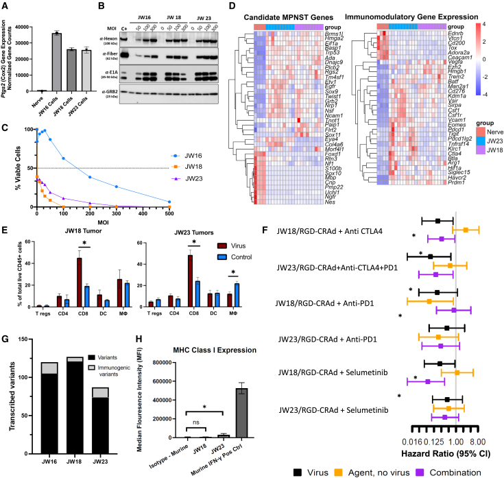
Oncolytic adenoviruses (Ads) stand out as a promising strategy for the targeted infection and lysis of tumor cells, with well-established clinical utility across various malignancies. This study delves into the therapeutic potential of oncolytic Ads in the context of neurofibromatosis type 1 (NF1)-associated malignant peripheral nerve sheath tumors (MPNSTs). Specifically, we evaluate conditionally replicative adenoviruses (CRAds) driven by the cyclooxygenase 2 (COX2) promoter, as selective agents against MPNSTs, demonstrating their preferential targeting of MPNST cells compared with non-malignant Schwann cell control. COX2-driven CRAds, particularly those with modified fiber-knobs exhibit superior binding affinity toward MPNST cells and demonstrate efficient and preferential replication and lysis of MPNST cells, with minimal impact on non-malignant control cells. In vivo experiments involving intratumoral CRAd injections in immunocompromised mice with human MPNST xenografts significantly extend survival and reduce tumor growth rate compared with controls. Moreover, in immunocompetent mouse models with MPNST-like allografts, CRAd injections induce a robust infiltration of CD8+ T cells into the tumor microenvironment (TME), indicating the potential to promote a pro-inflammatory response. These findings underscore oncolytic Ads as promising, selective, and minimally toxic agents for MPNST therapy, warranting further exploration.

Keywords: MT: Regular Issue; conditionally replicative adenovirus (CRAd); cyclooxygenase 2 (COX2); malignant peripheral nerve sheath tumor (MPNST); neurofibromatosis type 1 (NF1); oncolytic adenovirus.


Conflict of interest statement

D.A.L. is the co-founder and co-owner of NeoClone Biotechnologies, Inc., Discovery Genomics, Inc. (acquired by Immusoft, Inc.), B-MoGen Biotechnologies, Inc. (acquired by Bio-Techne corporation), and Luminary Therapeutics, Inc. D.A.L. holds equity in, is a Board of Directors member of, and serves as the Senior Scientific Advisor to Recombinetics, a genome-editing company, and Makana, a xenotransplantation company. D.A.L. consults for Styx Biotechnologies, Inc. and Genentech, Inc., which is funding some of his research. The business of all the companies above is unrelated to the contents of this manuscript.

Figures

None
Graphical abstract
Figure 1
Figure 1
Adenovirus binding affinity to MPNST cell lines and viral receptor expression (A) Schematic of viral genome for Ad vectors used in binding assay. (B) Each cell line was infected with Ad vectors equipped with either WT (Ad5), RGD fiber-modified (RGD), or chimeric Ad5/Ad3 fiber (Ad5/3) at 100 VP/cell. Binding was allowed to proceed for 2 h at 4°C, then was assessed by qPCR. (C) Flow cytometry analysis of CAR and integrin expression. MPNST cell lines were incubated with fluorescent antibodies against αVβ3 and αVβ5 integrins and coxsackie adenovirus receptor (CAR). The data are shown as a relative percentage of positive cells scored among at least 10,000 cells assessed. (D) Flow cytometry plots by cell line with respective isotype control for viral entry receptors in MPNST cell lines and iHSC1λ controls. Error bars represents ± standard deviation. ∗p < 0.05; ∗∗p < 0.01; ∗∗∗p < 0.001; ∗∗∗∗p < 0.0001, Student's t test.
Figure 2
Figure 2
COX2 expression and viral protein production in MPNST cell lines and Schwann cells (A) Analysis of COX2 mRNA in MPNST cell lines (S462-TY and STS26T) and Schwann cells (ipn02.3 2λ, iHSC1λ and pHSC) via RT-PCR. A549 cell line represents a positive control. A total of 500 ng of RNA from each sample was used as template input. (B) Western blot analysis of viral protein production in infected cells. A total of 250,000 cells were infected with RGD-COX2-CRAd at MOI 0 VP/cell (negative control) 10 VP/cell, and 50 VP/cell), 48 h post transduction whole cell lysates were probed by immunoblotting for early (E1A) and late viral proteins (hexon and fiber). GRB2 was used as a loading control.
Figure 3
Figure 3
Selectivity of RGD-COX2-CRAd toward MPNST cells (A) Cell viability curves for MPNST cell lines (S462-TY and STS26T) compared with immortalized human Schwann cells (iHSC1λ and ipn02.3λ), and primary human Schwann cells (pHSC). Cell lines were infected with RGD-COX2-CRAd at various MOIs (0–200 PFU/cell, n = 6), and percent viable cells was determined 5 days post-infection utilizing MTS assay protocol. Error bars represent ± standard deviation. (B) The IC50 values for MPNSTs and Schwann cells were calculated based on cell viability curves. (C) Percent viable cells remaining within monolayer after 5 days of infection with RGD-CRAd-COX2 at 50 MOI, relative to corresponding uninfected controls (0 MOI) were quantified using Cytation5 software. p < 0.0001, Student's t test. (D) Representative images of cell monolayers stained with crystal violet 5 days post-infection with RGD-COX2-CRAds at 50 MOI and corresponding uninfected controls (0 MOI). The COX2-overexpressing lung cancer cell line, A549, was used as a positive control.
Figure 4
Figure 4
In vitro analysis of COX2-CRAd replication and viral progeny spread (A) S462-TY, iHSC1λ (non-cancer control), and A549 (positive COX2-overexpression control) were used. The cells were infected at low MOI of 0.01, 0.1, and 1 VP/cell and cultured without medium change (to allow viral progeny production and spread) for up to 35 days or until cell monolayer was cleared. Cytotoxicity was assessed by affixing viable cell monolayers to the well, staining them with crystal violet dye, and quantifying images using FIJI software. Error bars represent ± standard deviation. The WT promoter-driven viruses (Ad5 and RGD-WT) were utilized as non-selective positive controls and exhibited robust viral spread and cell clearance rate. iHSC1λ cells served as non-cancer controls. Both COX2-CRAd and RGD-COX2-CRAd selectively killed COX-2-positive control cell line, A549 and MPNST cell line, S462-TY while leaving iHSC1λ mainly unharmed. RGD-COX2-CRAd showed a slightly better cytocidal effect compared with its WT fiber counterpart (COX2-CRAd). (B) Schematic representation of the four replication-competent Ad vectors. Vectors were engineered to have either WT or COX2 promoter and either WT or RGD fiber. Four replication-competent vectors were used. Vectors with WT promoter were used as non-selective controls. Vectors with WT fiber-knob domains (Ad5WT and COX2-CRAd) were employed for comparison with RGD fiber-modified vectors (RGD-WT and RGD-COX2-CRAd) to assess the degree of infectivity enhancement compared with WT fiber.
Figure 5
Figure 5
In vivo analysis of tumor growth kinetics and viral protein production (A) NOD-Rag1nullIL2rgnull (NRG) mice were inoculated subcutaneously with MPNST cell lines (S462-TY or STS26T) at 2.0 × 106 tumor cells/mouse. Virus (3.3 × 1010 VP/dose) or PBS was intratumorally injected when nodules reached approximately 200 mm3. The injections were repeated two more times every other day for a total of three injections and cumulative viral dose of 1.0 × 1011 VP/animal. Tumor size is shown in mm3 starting on day 0 (first viral injection). S462-TY/PBS and S462-TY/RGD-COX2-CRAd n = 6; S462-TY/COX2-CRAd and S462-TY/RGD-WT n = 4; all STS26T treatment and control groups n = 5. (B) Tumors injected with any of the viral vectors demonstrated reduced tumor growth rates compared with those injected with PBS. The treatment effects on tumor volume were calculated using a linear mixed effects model where each group was compared with PBS. All treatments had a significant negative effect on tumor volume. An average reduction of tumor volumes, within 95% confidence intervals is between 437 and 938 mm3 (RGD-CRAd-COX2), between 405 and 934 mm3 (RGD-WT), and between 293 and 822 mm3 (CRAd-COX2). For more information on statistical analysis see Figure S5. (C) Mice bearing S462-TY xenografts injected with virus vectors survived significantly longer compared with those injected with PBS (p < 0.0045, Log rank test). There were no significant differences in survival between S462-TY-bearing virus injected groups. Median survival in days was as follows: PBS = 19; COX2-CRAd = 41; RGD-COX2-CRAd = 47; RGD-WT = 45. (D) STS26T xenograft-bearing mice treated with either RGD-COX2-CRAd or RGD-WT demonstrated a statistically significant increase in survival probability compared with PBS (p = 0.002, Log rank test). Median survival of STS26T xenograft-bearing mice was PBS = 16 days; RGD-COX2-CRAd = 21 days COX2-CRAd = 16 days; RGD-WT = 22 days. RGD-WT vector was used as a non-selective positive control. (E) Immunohistochemistry analysis of tumor xenografts resected at endpoint from mice injected with RGD-COX2-CRAd and PBS. All tumors injected with RGD-COX2-CRAd were positive for viral proteins (E1A, fiber, and hexon) and markers of apoptosis (cleaved caspase 3). Some areas within tissues appeared torn due to necrotic tumor tissue. All tumors injected with PBS were uniformly negative for all viral proteins. STS26T tumors injected with PBS demonstrated a positive signal for cleaved caspase 3 in some cases. Representative example (Figure S5A).
Figure 6
Figure 6
Investigation of CRAd in syngeneic models of MPNST (A–C) Application of RGD-COX2-CRAd was investigated for murine MPNST-like syngeneic models. (A) RNA sequencing of JW cell lines shows upregulated Cox2 compared with normal murine sciatic nerve. (B) Western blot of RGD-COX2-CRAd-infected JW cells producing early (E1A) and late (hexon, fiber) viral proteins when infected at high MOIs. The human cell line was used as a positive control (C+) and was infected with RGD-COX2-CRAd at 3 MOI. (C) Both JW18 and JW23 are susceptible to cell killing by RGD-COX2-CRAd (IC50) < 1 as assessed by MTS assay 5 days post-infection, while JW16 is more resistant (six replicates performed per cell line per MOI). (D) Bulk RNA sequencing was performed on JW tumors implanted in C57BL/6 mice, and differential gene expression analysis performed against sciatic nerve of C57BL/6 mice, with all genes represented being significantly differentially expressed (p adjusted <0.05). The gene expression profile for JW cell lines is analogous to the gene expression profile in human MPNST, relative to normal nerve. Additionally, JW cell lines differentially express immunomodulatory genes, with JW23 showing the highest expression of both Pdcd1 (PD1) and Ctla4 (CTLA-4). (E) JW18 and JW23 flank allografts were established and subjected to virus or PBS injection to assess immune infiltration 10 days after implantation (six replicates per treatment group). In both JW18- and JW23-treated tumors, an increase in CD8+ T cell infiltration into the tumor microenvironment is observed (∗p < 0.05). (F) Six in vivo therapeutic trials were conducted (n = 6 mice per treatment group). Compared with intratumoral PBS injection (control), RGD-COX2-CRAd injections delayed tumor growth, although this was significant in only 50% of the trials (∗ represents hazard ratio of p < 0.05). CRAd was combined with immune checkpoint blockade or MEK inhibition with selumetinib, but no synergistic combination was identified possibly due to failure of MHCI presentation. (G) Whole exome and RNA sequencing identify nonsynonymous protein coding variants in these models, including variants predicted to bind to MHC class I H2-kb. (H) The baseline expression of MHC class 1 on the JW cell lines is low relative to IFN-γ stimulated cells.

Similar articles

References

    1. Le Guellec S., Decouvelaere A.V., Filleron T., Valo I., Charon-Barra C., Robin Y.M., Terrier P., Chevreau C., Coindre J.M. Malignant Peripheral Nerve Sheath Tumor Is a Challenging Diagnosis: A Systematic Pathology Review, Immunohistochemistry, and Molecular Analysis in 160 Patients from the French Sarcoma Group Database. Am. J. Surg. Pathol. 2016;40:896–908. doi: 10.1097/PAS.0000000000000655. - DOI - PubMed
    1. Staedtke V., Bai R.Y., Blakeley J.O. Cancer of the Peripheral Nerve in Neurofibromatosis Type 1. Neurotherapeutics. 2017;14:298–306. doi: 10.1007/s13311-017-0518-y. - DOI - PMC - PubMed
    1. Korfhage J., Lombard D.B. Malignant Peripheral Nerve Sheath Tumors: From Epigenome to Bedside. Mol. Cancer Res. 2019;17:1417–1428. doi: 10.1158/1541-7786. - DOI - PMC - PubMed
    1. Upadhyaya M., Kluwe L., Spurlock G., Monem B., Majounie E., Mantripragada K., Ruggieri M., Chuzhanova N., Evans D.G., Ferner R., et al. Germline and Somatic NF1 Gene Mutation Spectrum in NF1-Associated Malignant Peripheral Nerve Sheath Tumors (MPNSTs) Hum. Mutat. 2008;29:74–82. doi: 10.1002/humu.20601. - DOI - PubMed
    1. Pemov A., Li H., Presley W., Wallace M.R., Miller D.T. Genetics of Human Malignant Peripheral Nerve Sheath Tumors. Neurooncol. Adv. 2020;2:i50–i61. doi: 10.1093/noajnl/vdz049. - DOI - PMC - PubMed