

Management of Ptosis in Kearns-Sayre Syndrome: A Case Report and Literature Review
- PMID: 38596148
- PMCID: PMC11001462
- DOI: 10.1055/a-2207-7587
Management of Ptosis in Kearns-Sayre Syndrome: A Case Report and Literature Review
Abstract
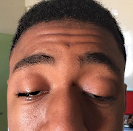
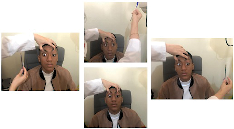
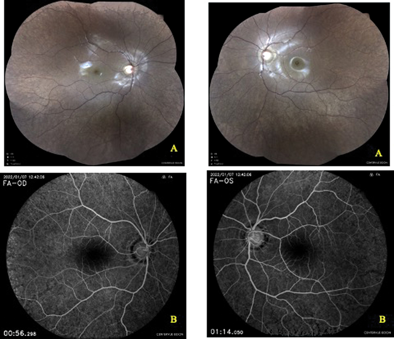
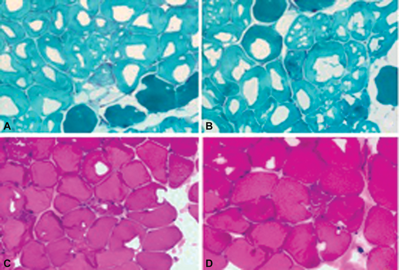
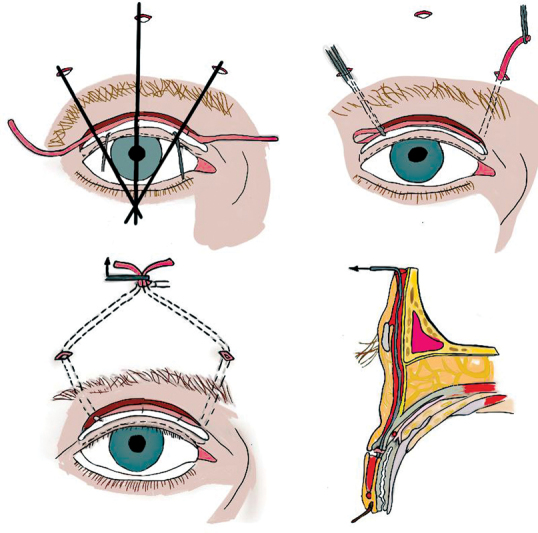
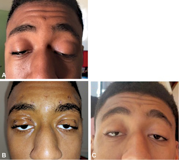
Kearns-Sayre syndrome (KSS) is a rare mitochondrial disease that affects young adults, due to a deletion of mitochondrial DNA and characterized by the triad: age of onset lower than 20 years, chronic progressive external ophthalmoplegia, and an atypical pigmentary retinopathy. It is also characterized by other endocrine, neurological, and especially cardiac impairment with a very high risk of cardiac complications during surgical procedures under all types of anesthesia. We report a case of KSS revealed by severe bilateral ptosis and confirmed by a muscle biopsy with "ragged red fibers." The ptosis was surgically managed by cautious Frontal suspension under local anesthesia "Frontal nerve block." Through this case, we discuss challenges in the management of KSS patients.
Keywords: Frontal suspension; Kearns–Sayre syndrome; chronic progressive external ophthalmoplegia; ptosis.
The Author(s). This is an open access article published by Thieme under the terms of the Creative Commons Attribution License, permitting unrestricted use, distribution, and reproduction so long as the original work is properly cited. ( https://creativecommons.org/licenses/by/4.0/ ).
Conflict of interest statement
Conflict of Interest None declared.
Figures
Similar articles
-
Kearns-Sayre syndrome: An unusual ophthalmic presentation.Oman J Ophthalmol. 2012 May;5(2):115-7. doi: 10.4103/0974-620X.99377. Oman J Ophthalmol. 2012. PMID: 22993469 Free PMC article.
-
[Kearns-Sayre syndrome: ophthalmic manifestations].An Pediatr (Barc). 2015 Jan;82(1):e151-3. doi: 10.1016/j.anpedi.2014.05.012. Epub 2014 Nov 20. An Pediatr (Barc). 2015. PMID: 25441208 Spanish.
-
Ophthalmologic school-based screening revealing Kearns-Sayre syndrome: a case report.Pan Afr Med J. 2022 Mar 18;41:226. doi: 10.11604/pamj.2022.41.226.33085. eCollection 2022. Pan Afr Med J. 2022. PMID: 35721635 Free PMC article.
-
Kearns-Sayre syndrome: a case report and review.Eur J Ophthalmol. 1992 Jan-Mar;2(1):15-20. doi: 10.1177/112067219200200104. Eur J Ophthalmol. 1992. PMID: 1638160 Review.
-
Kearns Sayre Syndrome--case report with review of literature.Indian J Pediatr. 2012 May;79(5):650-4. doi: 10.1007/s12098-011-0618-3. Epub 2012 Jan 10. Indian J Pediatr. 2012. Retraction in: Indian J Pediatr. 2013 Nov;80(11):982. doi: 10.1007/s12098-013-1123-7. PMID: 22231766 Retracted. Review.
References
-
- Zeviani M, Moraes C T, DiMauro S et al.Deletions of mitochondrial DNA in Kearns-Sayre syndrome. Neurology. 1988;38(09):1339–1346. - PubMed
-
- Shemesh A, Margolin E. StatPearls Publishing; Treasure Island (FL): 2022. Kearns-Sayre Syndrome. - PubMed
-
- Kearns T P, Sayre G P. Retinitis pigmentosa, external ophthalmophegia, and complete heart block: unusual syndrome with histologic study in one of two cases. AMA Arch Opthalmol. 1958;60(02):280–289. - PubMed
-
- Tsang S H, Aycinena A RP, Sharma T. Mitochondrial disorder: Kearns-Sayre syndrome. Adv Exp Med Biol. 2018;1085:161–162. - PubMed
LinkOut - more resources
Full Text Sources