

Evaluating pediatric ureteropelvic junction obstruction: Dynamic magnetic resonance urography vs renal scintigraphy 99m-technetium mercaptoacetyltriglycine
- PMID: 38596171
- PMCID: PMC10999956
- DOI: 10.4329/wjr.v16.i3.49
Evaluating pediatric ureteropelvic junction obstruction: Dynamic magnetic resonance urography vs renal scintigraphy 99m-technetium mercaptoacetyltriglycine
Abstract
Background: Ureteropelvic junction obstruction (UPJO) is a common congenital urinary tract disorder in children. It can be diagnosed as early as in utero due to the presence of hydronephrosis or later in life due to symptomatic occurrence.
Aim: To evaluate the discrepancy between dynamic contrast-enhanced magnetic resonance urography (dMRU) and scintigraphy 99m-technetium mercaptoacetyltriglycine (MAG-3) for the functional evaluation of UPJO.
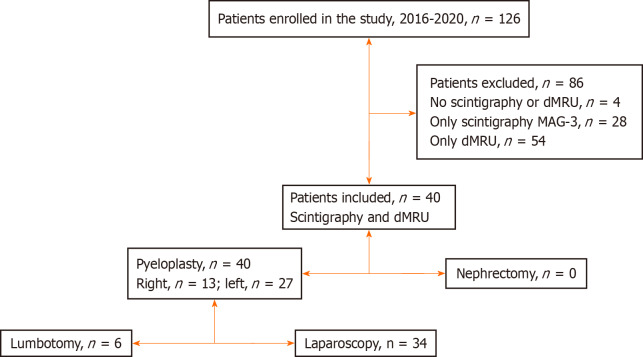
Methods: Between 2016 and 2020, 126 patients with UPJO underwent surgery at Robert Debré Hospital. Of these, 83 received a prenatal diagnosis, and 43 were diagnosed during childhood. Four of the 126 patients underwent surgery based on the clinical situation and postnatal ultrasound findings without undergoing functional imaging evaluation. Split renal function was evaluated preoperatively using scintigraphy MAG-3 (n = 28), dMRU (n = 53), or both (n = 40). In this study, we included patients who underwent surgery for UPJO and scintigraphy MAG-3 + dMRU but excluded those who underwent only scintigraphy MAG-3 or dMRU. The patients were divided into groups A (< 10% discrepancy) and B (> 10% discrepancy). We examined the discrepancy in split renal function between the two modalities and investigated the possible risk factors.
Results: The split renal function between the two kidneys was compared in 40 patients (28 boys and 12 girls) using scintigraphy MAG-3 and dMRU. Differential renal function, as determined using both modalities, showed a difference of < 10% in 31 children and > 10% in 9 children. Calculation of the relative renal function using dMRU revealed an excellent correlation coefficient with renal scintigraphy MAG-3 for both kidneys.
Conclusion: Our findings demonstrated that dMRU is equivalent to scintigraphy MAG-3 for evaluating split renal function in patients with UPJO.
Keywords: Dynamic contrast-enhanced magnetic resonance urography; Magnetic resonance imaging; Scintigraphy 99m-technetium mercaptoacetyltriglycine; Uteropelvic junction obstruction.
©The Author(s) 2024. Published by Baishideng Publishing Group Inc. All rights reserved.
Conflict of interest statement
Conflict-of-interest statement: All the authors report no relevant conflicts of interest for this article.
Figures

References
-
- Groth TW, Mitchell ME. Ureteropelvic junction obstruction. In: Coran A, editor. Paediatric Surgery. 7th ed. Philadelphia: Elsevier, 2012: 1411-1425.
-
- Zderic SA, Lambert SM. Developmental abnormalities of the genitourinary system. In: Gleason CA, Devaskar SU, editor. Avery's Diseases of the Newborn. 9th ed. Philadelphia: Elsevier, 2012: 1191-1204.
-
- Brown T, Mandell J, Lebowitz RL. Neonatal hydronephrosis in the era of sonography. AJR Am J Roentgenol. 1987;148:959–963. - PubMed
-
- Lawler LP, Jarret TW, Corl FM, Fishman EK. Adult ureteropelvic junction obstruction: insights with three-dimensional multi-detector row CT. Radiographics. 2005;25:121–134. - PubMed
-
- González R, Schimke CM. Ureteropelvic junction obstruction in infants and children. Pediatr Clin North Am. 2001;48:1505–1518. - PubMed
LinkOut - more resources
Full Text Sources
Research Materials
