Case Reports
. 2024 Apr 5;19(6):2545-2548.
doi: 10.1016/j.radcr.2024.02.103. eCollection 2024 Jun.

Spinal metastasis of nephroblastoma: Yes it exists

Affiliations
Case Reports

Spinal metastasis of nephroblastoma: Yes it exists

Nourrelhouda Bahlouli et al. Radiol Case Rep. .

Abstract

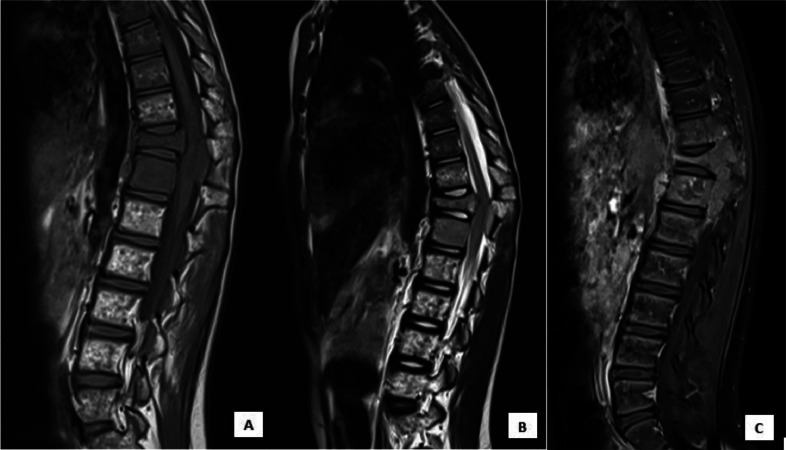
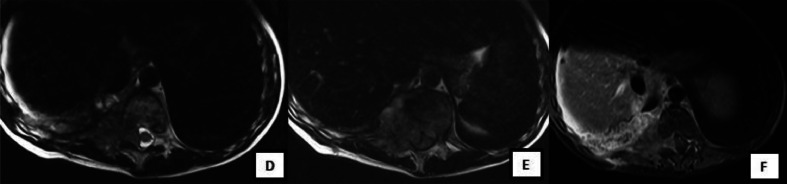
Nephroblastoma or Wilms' tumor is the most common malignant tumor of the kidney in the pediatric population. Metastasis is caused by hematogenous spread. The most common localizations in decreasing order of frequency are lymph nodes, lungs, and liver. The bone is very rarely affected. According to the literature, bone metastases have been described in the iliac bone, skull, and mandible. The vertebral localization was described in 3 cases only, the first 1 in 2009, and the 2 others in 2015 . The goal of our work is to report a very rare case of metastatic vertebral localization of a Wilms' tumor in relapse after treatment; and thus to underline the potential for vertebral and intracanal involvement in nephroblastoma.

Keywords: Adolescent; Intraspinal; Nephroblastoma; Pleural; Spinal metastasis; Wilms tumor.


Figures

Fig 1
Fig. 1
HE , Gx40 : infiltration of pleural and fatty tissue by the tumor.
Fig 2
Fig. 2
HE , Gx20 : blastomatous component of a metastatic nephroblastoma on pleuro-pulmonary.
Fig 3
Fig. 3
MRI images in sagital section: (A) T1 sequence, (B) T2 sequence, (C) injected T1 sequence showing: T1 hyposignal of the vertebral bodies of D11 and D12 which are enhanced after injection of Gadolinium. The latter is the site of 2 nodular signal anomalies enhanced in the periphery. A biconcave compression fracture D11 with posterior wall recession and angulation of the spine.
Fig 4
Fig. 4
MRI images in axial section: (D) T2 sequence, (E) T2 sequence, (F) injected T1 sequence showing: Right posterobasal pleural mass with intermediate T2 signal, T1 hyposignal, enhanced after injection of Gadolinium. This mass: Invades the transverse processes D10-D11-D12 and their foramens with endocanal extension amputating the subarachnoid spaces and compressing the medulla which is displaced to the left without any signal abnormalities in the medulla. It extends to the right paravertebral muscles and the soft tissues in front of it.

Similar articles

References

    1. Hélène S-B, Pierre L, Cecile C, Mathieu V, Cyril L, Nathalie Rocourt, et al. Bone vertebrae metastases with spinal cord compression: a rare event in Wilms tumor. J Pediatr Hematol Oncol. 2015 doi: 10.1097/MPH.0000000000000332. - DOI - PubMed
    1. Watanabe R, Takahashi A, Suzuki M, Toki F, Kanazawa T, Hirato J, et al. Adolescent Wilms tumor with intraspinal and bone metastases a case report and the review of literature. J Pediatr Hematol Oncol. 2006 doi: 10.1097/MPH.0b013e318190d718. - DOI - PubMed
    1. Cohn SL, Hamre M, Kletzel M, Chou P, Radkowski MA. Intraspinal Wilms’ tumor metastasis. Cancer. 1994;73:2444–2449. doi: 10.1002/1097-0142(19940501)73:9<2444::aid-cncr2820730930>3.0.co;2-a. - DOI - PubMed
    1. Ebb DH, Kerasidis H, Vezina G, Packer RJ, Carabell S, Ivy P. Spinal cord compression in widely metastatic Wilms’ tumor. Paraplegia in two children with anaplastic Wilms’ tumor. Cancer. 1992;69:2726–2730. doi: 10.1002/1097-0142(19920601)69:11<2726::aid-cncr2820691116>3.0.co;2-z. - DOI - PubMed
    1. bond JV, martin EC. Bone metastases in wilms' tumour. Clin Radiol. 1975;26:103–106. doi: 10.1016/s0009-9260(75)80023-7. - DOI - PubMed

Publication types

LinkOut - more resources