

A Rare Presentation of Occipital Dermoid Cyst with Intracranial Extension and Secondary Infection: Case Report and Follow-Up
- PMID: 38596231
- PMCID: PMC11001448
- DOI: 10.1055/a-2287-2108
A Rare Presentation of Occipital Dermoid Cyst with Intracranial Extension and Secondary Infection: Case Report and Follow-Up
Abstract
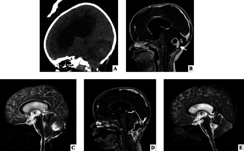
Craniofacial dermoid cysts are congenital anomalies that rarely have intracranial extension and can be associated with other anomalies. Common sites of these lesions are the lateral brow and parietal scalp. Presentation of the dermoid cyst in the occipital region with intracranial extension is extremely rare. We report a 2-year-old female with a presentation of an occipital dermoid cyst with intracranial extension and secondary cerebellar abscess. This case highlights the rarity of the presentation of an occipital dermoid cyst with intracranial extension and secondary infection and the importance of early imaging for suspected dermoid cysts in the occipital region for identification of intracranial extension.
Keywords: case report; intracranial abscess; occipital dermoid cyst; pediatrics.
The Author(s). This is an open access article published by Thieme under the terms of the Creative Commons Attribution-NonDerivative-NonCommercial License, permitting copying and reproduction so long as the original work is given appropriate credit. Contents may not be used for commercial purposes, or adapted, remixed, transformed or built upon. ( https://creativecommons.org/licenses/by-nc-nd/4.0/ ).
Conflict of interest statement
Conflict of Interest None declared.
Figures


References
-
- Orozco-Covarrubias L, Lara-Carpio R, Saez-De-Ocariz M, Duran-McKinster C, Palacios-Lopez C, Ruiz-Maldonado R. Dermoid cysts: a report of 75 pediatric patients. Pediatr Dermatol. 2013;30(06):706–711. - PubMed
-
- Hanikeri M, Waterhouse N, Kirkpatrick N, Peterson D, Macleod I. The management of midline transcranial nasal dermoid sinus cysts. Br J Plast Surg. 2005;58(08):1043–1050. - PubMed
-
- Overland J, Hall C, Holmes A, Burge J. Risk of intracranial extension of craniofacial dermoid cysts. Plast Reconstr Surg. 2020;145(04):779e–787e. - PubMed
-
- Martens F, Ectors P, Noel P, Hanquinet S, Faverly D. Unusual cause of cerebellar abscess: occipital dermal sinus and dermoid cyst. Neuropediatrics. 1987;18(02):107–109. - PubMed
-
- Hayek G, Mercier P, Fournier H D, Menei P, Pouplard F, Guy G.Dermal sinus and dermoid cyst revealed by abscess formation in posterior fossa. Report of 2 pediatric cases and review of the literature [in French] Neurochirurgie 200147(2-3 Pt 1):123–127. - PubMed