. 2024 Feb 22;32(1):200780.
doi: 10.1016/j.omton.2024.200780. eCollection 2024 Mar 21.

Minute virus of mice shows oncolytic activity against pancreatic cancer cells exhibiting a mesenchymal phenotype

Affiliations

Minute virus of mice shows oncolytic activity against pancreatic cancer cells exhibiting a mesenchymal phenotype

Margaux Vienne et al. Mol Ther Oncol. .

Abstract

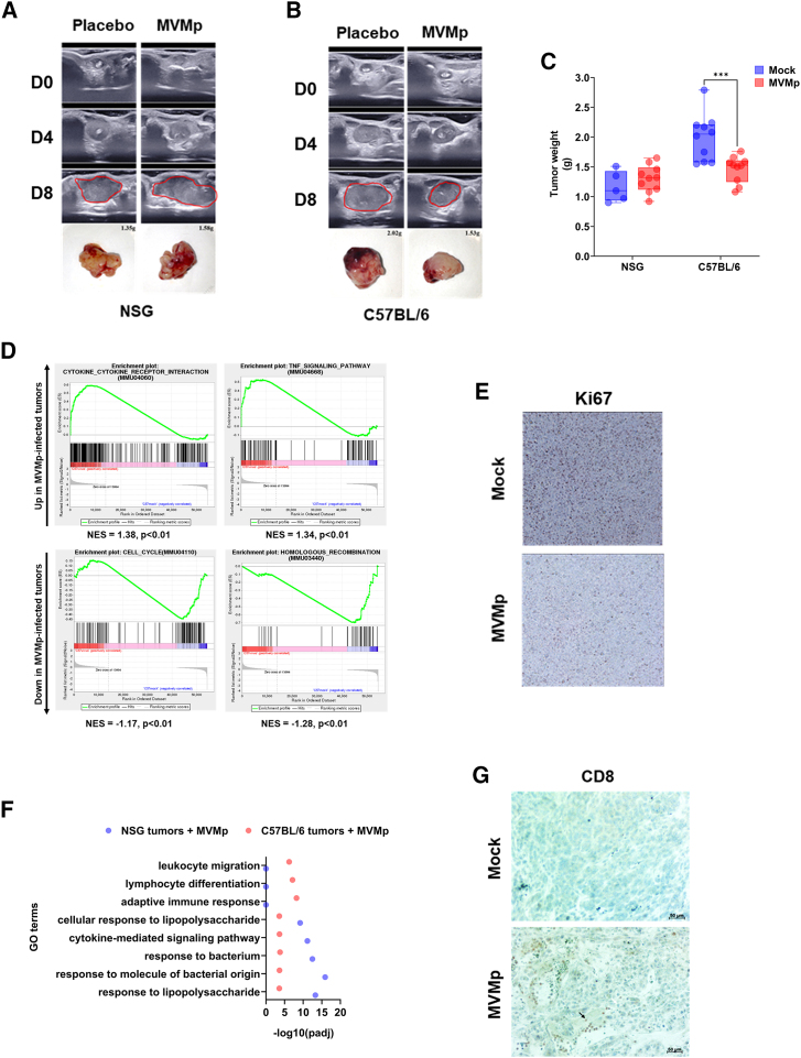
Pancreatic cancer will soon become the second cause of death by cancer in Western countries. The main barrier to increase the survival of patients with this disease requires the development of novel and efficient therapeutic strategies that better consider tumor biology. In this context, oncolytic viruses emerge as promising therapeutics. Among them, the fibrotropic minute virus of mice prototype (MVMp) preferentially infects migrating and undifferentiated cells that highly resemble poorly differentiated, basal-like pancreatic tumors showing the worst clinical outcome. We report here that MVMp specifically infects, replicates in, and kills pancreatic cancer cells from murine and human origin with a mesenchymal, basal-like profile, while sparing cancer cells with an epithelial phenotype. Remarkably, MVMp infection, at a dose that does not provoke tumor growth inhibition in athymic mice, shows significant antitumoral effect in immune-competent models; extended mouse survival; and promoted the massive infiltration of tumors by innate, myeloid, and cytotoxic T cells that exhibit a less terminally exhausted phenotype. Collectively, we demonstrate herein for the first time that MVMp is specific and oncolytic for pancreatic tumors with mesenchymal, basal-like profile, paving the way for precision-medicine opportunities for the management of the most aggressive and lethal form of this disease.

Keywords: MT: Regular Issue; MVMp; immune mobilization; mesenchymal phenotype; oncolytic virus; pancreatic cancer; targeted virotherapy.


Conflict of interest statement

The authors declare no competing interests.

Figures

None
Graphical abstract
Figure 1
Figure 1
Isolation of mesenchymal and epithelial subtypes of murine pancreatic cancer cells R211-E and R211-M cells were isolated by differential adherence to plastic as mentioned in the “materials and methods” section. (A) Representative images of long-term culture of R211 cells (top) and of isolated R211-E (middle) and R211-M (bottom) cells. (B) Western blot analysis for the indicated proteins in LA9, parental R211, R211-E, and R211-M cells. Representative of two independent experiments. Representative images of tumor echography (C) and tumor growth kinetics (D) analyzed by echography of R211-M and R211-E tumors engrafted in NSG mice. N = 3–8 mice per group. (E) Hematoxylin and eosin (H&E) staining of orthotopic R211-E and R211-M tumors engrafted in nude mice at endpoint. Representative of n = 3 mice per group.
Figure 2
Figure 2
MVM is oncolytic in murine and human PDAC cells with mesenchymal phenotype R211-M and R211-E cells were infected with MVMp at MOI = 100. Control cells were incubated with infection medium. LA9 wells were used as positive control. Representative images (A), real-time cell death analysis (B), and western blot for PARP cleavage of cells infected by MVMp or mock-treated cells, up to 72 h (A and B) or 48 h (C) following infection. Results are mean ± SEM or representative of at least three independent experiments performed in duplicate. ∗∗∗∗p < 0.001, multiple unpaired Student’s t test. R211-M and R211-E cells were infected with MVMp at MOI = 100, when LA9 were treated with 0.1 M MVMp. (D) Cellular content in viral genomes was analyzed by qPCR for NS1 viral gene at the time indicated. Results are mean ± SEM of at three independent experiments performed in duplicate. ∗p < 0.05, ∗∗∗:p < 0.005, multiple unpaired Student’s t test. (E) Cell supernatant was collected at the time indicated and analyzed in real time for oncolytic activity on LA9 reporter cells. Results are mean ± SEM of three independent experiments performed in duplicate. ∗p < 0.05, ∗∗p < 0.01, ∗∗∗p < 0.005, ∗∗∗∗p < 0.001, multiple unpaired Student’s t test. (F) Western blot analysis for the indicated proteins in human primary PDAC cells PDAC087T, PDAC015T, PDAC051T, and PDACT1. Representative of two independent experiments. PDAC087T, PDAC015T, PDAC051T, and PDACT1 were infected with MVMp at MOI = 100 when control cultures were incubated with infection medium. E, epithelial; M, mesenchymal. Representative images (G) and live cell death analysis (H) of human primary PDAC cells 72 h following infection by MVMp. Results are mean ± SEM of three independent experiments performed in duplicate. ∗∗p < 0.01, unpaired Student’s t test.
Figure 3
Figure 3
MVM is oncolytic in murine models of PDAC with mesenchymal phenotype R211-M or R211-E tumors were induced in the pancreas of NSG mice as described in the “materials and methods” section. Eleven days later, when tumors reached 100 mm3 in size, mice were randomized and received two sequential i.v. injections of MVMp (10e6 or 10e7 p.f.u.) in two separate injections 4 days apart (D0 = day of the first injection). N = 3 mice were used per group. (A) R211-M tumor volume by echography, 9 days after starting treatment. Results are mean ± SEM or representative of three mice per group. ∗∗∗∗p < 0.001, unpaired Student’s t test. At endpoint, tumors were sampled and tumor viral genome content was quantified by qPCR (B, results are shown as mean of three independent experiments. ∗p < 0.05, ∗∗∗p > 0.005). (C) Total RNA from R211-M tumors was analyzed for the presence of NS1 and viral capsid mRNA by qPCR. RNA-seq followed by gene set enrichment analysis (GSEA, D) and western blot for PARP cleavage (E) were performed. NES, normalized enrichment score. Results are mean ± SEM or representative of three mice per group. ∗p < 0.05, ∗∗∗p < 0.005, unpaired Student’s t test.
Figure 4
Figure 4
MVMp shows increased antitumoral efficacy in immune-competent models of pancreatic cancer R211-M tumors were induced in the pancreas of NSG mice or of C57BL/6 mice as described in the “materials and methods” section. When tumors reached 100 mm3 in size, mice were randomized and received two sequential i.v. injections of MVMp (10e4 or 10e6 p.f.u.) in two separate injections 4 days apart (D0 = day of the first injection). Eight to 10 mice were used per group. Representative echography images and photographs with weight of R211-M tumors grown in NSG (A) or C57Bl6 (B) mice, up to 8 days following initial administration of MVMp. Tumors are circled in red. (C) Tumor weight at endpoint. Results are mean ± SEM or representative of 8–10 mice per group. ∗∗∗p < 0.005, unpaired Student’s t test. At endpoint, tumors were sampled and RNA-seq followed by GSEA (D), immunochemistry for Ki67 (E, R211-M tumors in C57Bl/6 mice only), GO terms analysis (F), and immunochemistry for Ki67 (G, R211-M tumors in C57Bl/6 mice only) were performed in mice receiving 10e6 p.f.u. of the virus. Results are mean ± SEM or representative of 8–10 mice per group.
Figure 5
Figure 5
MVMp infection increases the infiltration of immune cells into experimental pancreatic tumors R211-M tumors were induced in the pancreas of C57BL/6 mice as described in the “materials and methods” section. When tumors reached 100 mm3 in size, mice were randomized and received two sequential i.v. injections of MVMp (10e6 p.f.u.) in two separate injections 4 days apart (D0 = day of the first injection). At endpoint, tumors were resected, minced, and single-cell suspensions were analyzed by cytometry for live cells (A), CD45+ cells (B), neutrophils (C), NKp46+ cells (D), conventional dendritic cells (cDC, E), CCR2+ monocytes (F), CCR2-macrophages (G), CD4+ (H), and CD8+ T cells (I). Results are mean ± SEM of three to six mice per group ∗p < 0.005, unpaired Student’s t test.
Figure 6
Figure 6
MVMp infection reduces the expression of selected immune checkpoint by intratumoral CD8+ T cells R211-M tumors were induced in the pancreas of C57BL/6 mice as described in the “materials and methods” section. When tumors reached 100 mm3 in size, mice were randomized and received two sequential i.v. injections of MVMp (10e6 p.f.u.) in two separate injections 4 days apart (D0 = day of the first injection). Five to eight mice were used per group. At endpoint, tumors were resected, minced, and single-cell suspensions were analyzed by cytometry for CD8+ T cells that were co-stained with PD-1 (A), TIGIT (B), LAG-3 (C), and TIM-3 (D) immune checkpoints. Results are mean ± SEM of three to six mice per group. ∗p < 0.005, Student’s t test. ∗p < 0.005, unpaired Student’s t test.

Similar articles

Cited by

References

    1. Park W., Chawla A., O’Reilly E.M. Pancreatic Cancer: A Review. JAMA. 2021;326:851–862. doi: 10.1001/jama.2021.13027. - DOI - PMC - PubMed
    1. Martens S., Lefesvre P., Nicolle R., Biankin A.V., Puleo F., Van Laethem J.L., Rooman I. Different shades of pancreatic ductal adenocarcinoma, different paths towards precision therapeutic applications. Ann. Oncol. 2019;30:1428–1436. doi: 10.1093/annonc/mdz181. - DOI - PubMed
    1. Moffitt R.A., Marayati R., Flate E.L., Volmar K.E., Loeza S.G.H., Hoadley K.A., Rashid N.U., Williams L.A., Eaton S.C., Chung A.H., et al. Virtual microdissection identifies distinct tumor- and stroma-specific subtypes of pancreatic ductal adenocarcinoma. Nat. Genet. 2015;47:1168–1178. doi: 10.1038/ng.3398. - DOI - PMC - PubMed
    1. Puleo F., Nicolle R., Blum Y., Cros J., Marisa L., Demetter P., Quertinmont E., Svrcek M., Elarouci N., Iovanna J., et al. Stratification of Pancreatic Ductal Adenocarcinomas Based on Tumor and Microenvironment Features. Gastroenterology. 2018;155:1999–2013.e3. doi: 10.1053/j.gastro.2018.08.033. - DOI - PubMed
    1. Nicolle R., Blum Y., Marisa L., Loncle C., Gayet O., Moutardier V., Turrini O., Giovannini M., Bian B., Bigonnet M., et al. Pancreatic Adenocarcinoma Therapeutic Targets Revealed by Tumor-Stroma Cross-Talk Analyses in Patient-Derived Xenografts. Cell Rep. 2017;21:2458–2470. doi: 10.1016/j.celrep.2017.11.003. - DOI - PMC - PubMed

LinkOut - more resources