

A Multifunctional Nanozyme Integrating Antioxidant, Antimicrobial and Pro-Vascularity for Skin Wound Management
- PMID: 38596410
- PMCID: PMC11001553
- DOI: 10.2147/IJN.S452216
A Multifunctional Nanozyme Integrating Antioxidant, Antimicrobial and Pro-Vascularity for Skin Wound Management
Abstract
Background: Skin wounds are a prevalent issue that can have severe health consequences if not treated correctly. Nanozymes offer a promising therapeutic approach for the treatment of skin wounds, owing to their advantages in regulating redox homeostasis to reduce oxidative damage and kill bacteria. These properties make them an effective treatment option for skin wounds. However, most of current nanozymes lack the capability to simultaneously address inflammation, oxidative stress, and bacterial infection during the wound healing process. There is still great potential for nanozymes to increase their therapeutic functional diversity and efficacy.
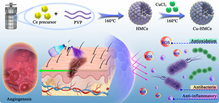
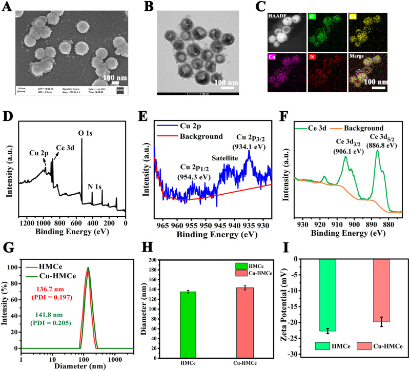
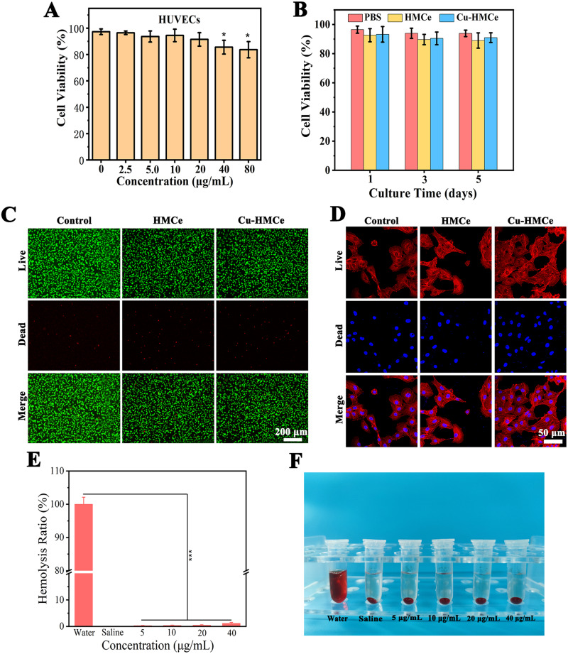
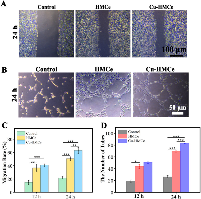
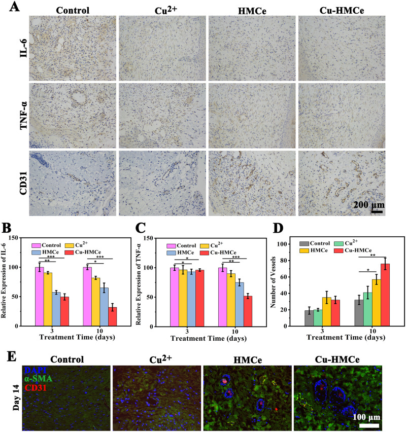
Methods: Herein, copper-doped hollow mesopores cerium oxide (Cu-HMCe) nanozymes with multifunctional of antioxidant, antimicrobial and pro-vascularity is successfully prepared. Cu-HMCe can be efficiently prepared through a simple and rapid solution method and displays sound physiological stability. The biocompatibility, pro-angiogenic, antimicrobial, and antioxidant properties of Cu-HMCe were assessed. Moreover, a full-thickness skin defect infection model was utilized to investigate the wound healing capacity, as well as anti-inflammatory and pro-angiogenic properties of nanozymes in vivo.
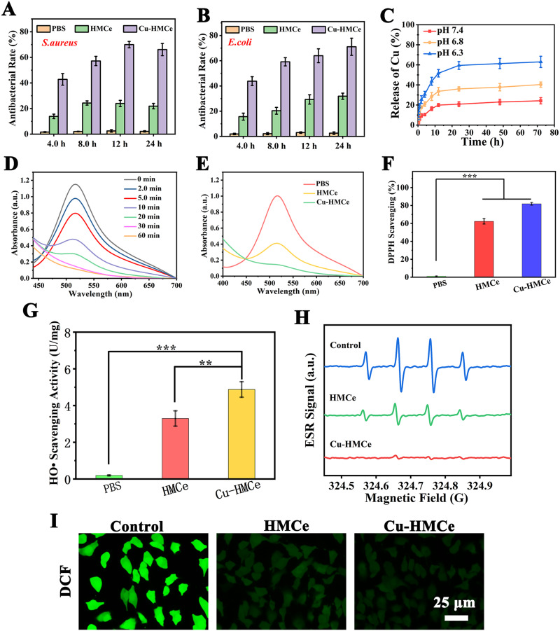
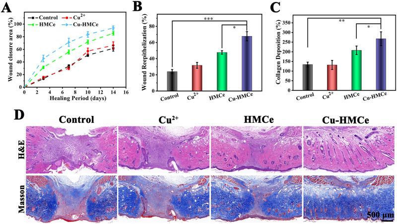
Results: Both in vitro and in vivo experiments have substantiated Cu-HMCe's remarkable biocompatibility. Moreover, Cu-HMCe possesses potent antioxidant enzyme-like catalytic activity, effectively clearing DPPH radicals (with a scavenging rate of 80%), hydroxyl radicals, and reactive oxygen species. Additionally, Cu-HMCe exhibits excellent antimicrobial and pro-angiogenic properties, with over 70% inhibition of both E. coli and S. aureus. These properties collectively promote wound healing, and the wound treated with Cu-HMCe achieved a closure rate of over 90% on the 14th day.
Conclusion: The results indicate that multifunctional Cu-HMCe with antioxidant, antimicrobial, and pro-angiogenic properties was successfully prepared and exhibited remarkable efficacy in promoting wound healing. This nanozymes providing a promising strategy for skin repair.
Keywords: Cu-HMCe nanozyme; antibacterial; antioxidation; vascularization; wound healing.
© 2024 Jiang et al.
Conflict of interest statement
The authors report no conflicts of interest in this work.
Figures
Similar articles
-
Gelatin/Dopamine/Zinc-Doped Ceria/Curcumin nanocomposite hydrogels for repair of chronic refractory wounds.Int J Pharm. 2024 Sep 30;663:124575. doi: 10.1016/j.ijpharm.2024.124575. Epub 2024 Aug 10. Int J Pharm. 2024. PMID: 39134289
-
[Effects and mechanism of copper oxide nanozymes on wound healing of full-thickness skin defects in diabetic mice].Zhonghua Shao Shang Za Zhi. 2020 Dec 20;36(12):1139-1148. doi: 10.3760/cma.j.cn501120-20200929-00426. Zhonghua Shao Shang Za Zhi. 2020. PMID: 33379850 Chinese.
-
Cu-GA-coordination polymer nanozymes with triple enzymatic activity for wound disinfection and accelerated wound healing.Acta Biomater. 2023 Sep 1;167:449-462. doi: 10.1016/j.actbio.2023.05.048. Epub 2023 Jun 2. Acta Biomater. 2023. PMID: 37270076
-
Copper-Based Nanozymes: Potential Therapies for Infectious Wounds.Small. 2025 Feb;21(8):e2407195. doi: 10.1002/smll.202407195. Epub 2025 Jan 5. Small. 2025. PMID: 39757568 Review.
-
Multifunctional and theranostic hydrogels for wound healing acceleration: An emphasis on diabetic-related chronic wounds.Environ Res. 2023 Dec 1;238(Pt 1):117087. doi: 10.1016/j.envres.2023.117087. Epub 2023 Sep 15. Environ Res. 2023. PMID: 37716390 Review.
Cited by
-
Advanced wound healing with Stimuli-Responsive nanozymes: mechanisms, design and applications.J Nanobiotechnology. 2025 Jul 1;23(1):479. doi: 10.1186/s12951-025-03558-w. J Nanobiotechnology. 2025. PMID: 40598229 Free PMC article. Review.
References
-
- Alizadeh S, Seyedalipour B, Shafieyan S, et al. Copper nanoparticles promote rapid wound healing in acute full thickness defect via acceleration of skin cell migration, proliferation, and neovascularization. Biochem Biophys Res Commun. 2019;517(4):684–690. doi:10.1016/j.bbrc.2019.07.110 - DOI - PubMed
MeSH terms
Substances
LinkOut - more resources
Full Text Sources
Medical
