. 2022 Jul 7:1:kvac009.
doi: 10.1093/oons/kvac009. eCollection 2022.

Tissue-Wide Effects Override Cell-Intrinsic Gene Function in Radial Neuron Migration

Affiliations

Tissue-Wide Effects Override Cell-Intrinsic Gene Function in Radial Neuron Migration

Andi H Hansen et al. Oxf Open Neurosci. .

Abstract

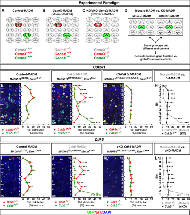
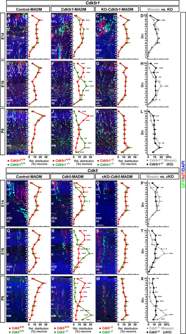
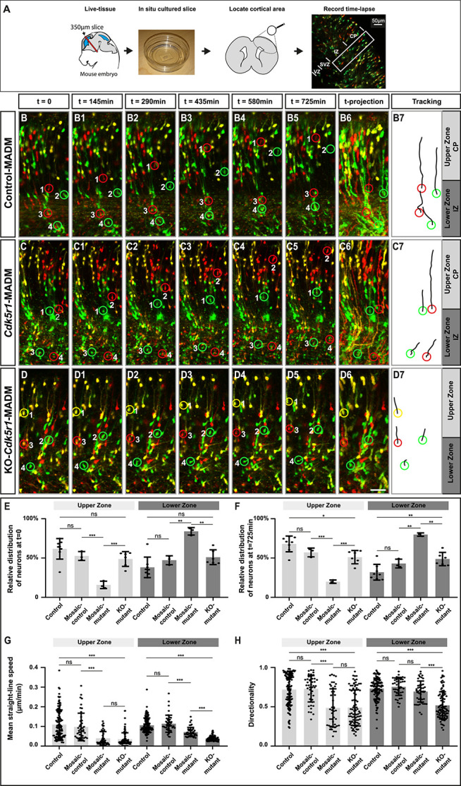
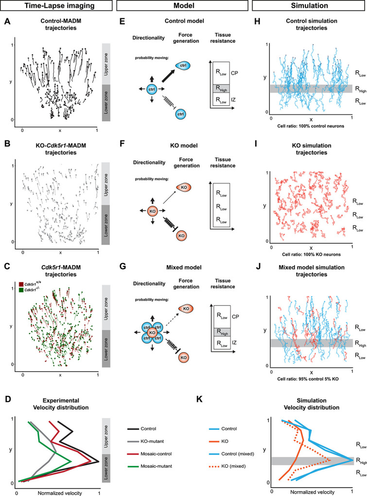
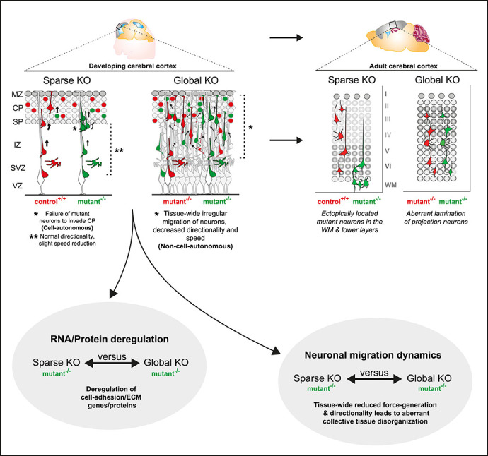
The mammalian neocortex is composed of diverse neuronal and glial cell classes that broadly arrange in six distinct laminae. Cortical layers emerge during development and defects in the developmental programs that orchestrate cortical lamination are associated with neurodevelopmental diseases. The developmental principle of cortical layer formation depends on concerted radial projection neuron migration, from their birthplace to their final target position. Radial migration occurs in defined sequential steps, regulated by a large array of signaling pathways. However, based on genetic loss-of-function experiments, most studies have thus far focused on the role of cell-autonomous gene function. Yet, cortical neuron migration in situ is a complex process and migrating neurons traverse along diverse cellular compartments and environments. The role of tissue-wide properties and genetic state in radial neuron migration is however not clear. Here we utilized mosaic analysis with double markers (MADM) technology to either sparsely or globally delete gene function, followed by quantitative single-cell phenotyping. The MADM-based gene ablation paradigms in combination with computational modeling demonstrated that global tissue-wide effects predominate cell-autonomous gene function albeit in a gene-specific manner. Our results thus suggest that the genetic landscape in a tissue critically affects the overall migration phenotype of individual cortical projection neurons. In a broader context, our findings imply that global tissue-wide effects represent an essential component of the underlying etiology associated with focal malformations of cortical development in particular, and neurological diseases in general.

Keywords: 4D live-imaging; cell-autonomous gene function; cerebral cortex development; mosaic analysis with double markers (MADM); neuronal migration; non-cell-autonomous effects; single-cell genetics.


Conflict of interest statement

All authors declare that they have no conflicts of interest.

Figures

Figure 1
Figure 1
MADM analysis reveals that global tissue-wide effects predominates the cell-autonomous phenotype due to loss of p35/CDK5. (AD) Experimental MADM paradigm to genetically dissect cell-autonomous gene function and non-cell-autonomous effects. (A) Control (control-MADM: all cells GeneX+/+); (B) sparse genetic mosaic (GeneX-MADM: only green cells are GeneX−/− mutant, red cells are GeneX+/+ in an otherwise heterozygous GeneX+/− environment); (C) global/whole-tissue gene knockout (KO/cKO-GeneX-MADM: all cells mutant); (D) direct phenotypic comparison of mutant cells in GeneX-MADM (mosaic MADM) to mutant cells in KO/cKO-gene-MADM (KO/cKO-MADM). Any significant difference in their respective phenotypes implies non-cell-autonomous effects. (E–H) Analysis of green (GFP+) and red (tdT+) MADM-labeled projection neurons in (E) control-MADM (MADM-11GT/TG;Emx1Cre/+); (F) Cdk5r1-MADM (MADM-11GT/TG,Cdk5r1;Emx1Cre/+); and (G) KO-Cdk5r1-MADM (MADM-11GT,Cdk5r1/TG,Cdk5r1;Emx1Cre/+). Relative distribution (%) of MADM-labeled projection neurons is plotted in ten equal zones across the cortical wall. (H) Direct distribution comparison of Cdk5r1−/− mutant cells in Cdk5r1-MADM (grey) versus KO-Cdk5r1-MADM (black) distribution. (I–L) Analysis of green (GFP+) and red (tdT+) MADM-labeled projection neurons in (I) control-MADM (MADM-5GT/TG;Emx1Cre/+); (J) Cdk5-MADM (MADM-5GT/TG,Cdk5;Emx1Cre/+); and (K) KO-Cdk5-MADM (MADM-11GT,Cdk5/TG,Cdk5;Emx1Cre/+). Relative distribution (%) of MADM-labeled projection neurons is plotted in 10 equal zones (1–10) across the cortical wall. (L) Direct distribution comparison of Cdk5−/− mutant cells in Cdk5-MADM (grey) versus KO-Cdk5-MADM (black) distribution. Nuclei were stained using DAPI (blue). N = 3 for each genotype with 10 (MADM-11) and 20 (MADM-5) hemispheres analysed. Data indicate mean ± SD, *P < 0.05, **P < 0.01 and ***P < 0.001. Scale bar: 100 μm. Marginal zone (MZ), cortical layers (II–VI), white matter (WM).
Figure 2
Figure 2
Developmental time course analysis of MADM-based sparse and global KO of p35/CDK5. (A–L) Analysis of green (GFP+) and red (tdT+) MADM-labeled projection neurons in (A, E, I) control-MADM (MADM-11GT/TG;Emx1Cre/+); (B, F, J) Cdk5r1-MADM (MADM-11GT/TG,Cdk5r1;Emx1Cre/+); and (C, G, K) KO-Cdk5r1-MADM (MADM-11GT,Cdk5r1/TG,Cdk5r1; Emx1Cre/+) at E14 (A–D), E16 (E–H) and P0 (I–L). Relative distribution (%) of MADM-labeled projection neurons is plotted in ten equal zones across the developing cortical wall. (D, H, L) Direct distribution comparison of Cdk5r1−/− mutant cells at E14 (D), E16 (H) and P0 (L) in Cdk5r1-MADM (grey) versus KO-Cdk5r1-MADM (black) distribution. (M–X) Analysis of green (GFP+) and red (tdT+) MADM-labeled projection neurons in (M, Q, U) control-MADM (MADM-11GT/TG;Emx1Cre/+); (N, R, V) Cdk5-MADM (MADM-11GT/TG,Cdk5;Emx1Cre/+); and (O, S, W) KO-Cdk5-MADM (MADM-11GT,Cdk5/TG,Cdk5; Emx1Cre/+) at E14 (M–P), E16 (Q–T) and P0 (U–X). Relative distribution (%) of MADM-labeled projection neurons is plotted in 10 equal zones across the developing cortical wall. (P, T, X) Direct distribution comparison of Cdk5−/− mutant cells at E14 (P), E16 (T), and P0 (X) in Cdk5-MADM (grey) versus KO-Cdk5-MADM (black) distribution. Nuclei were stained using DAPI (blue). N = 3 for each genotype with 10 (MADM-11) and 20 (MADM-5) hemispheres analysed. Data indicate mean ± SD, *P < 0.05, **P < 0.01 and ***P < 0.001. Cortical plate (CP), intermediate zone (IZ), subventricular zone/ventricular zone (SVZ/VZ), cortical layers (II–VI), white matter (WM). Scale bars: 100 μm.
Figure 3
Figure 3
Projection neuron migration dynamics upon sparse and global KO of p35/CDK5. (A) Experimental setup for time-lapse imaging of MADM-labeled neurons in developing somatosensory cortex at E16. (B–D) Time-lapse imaging of (B-B5) control-MADM (MADM-11GT/TG;Emx1Cre/+), (C-C5) Cdk5r1-MADM (MADM-11GT/TG,Cdk5r1;Emx1Cre/+) and (D-D5) KO-Cdk5r1-MADM (MADM-11GT,Cdk5r1/TG,Cdk5r1; Emx1Cre/+). (B6, C6, D6) 12 h time projection of sequential images in IZ (lower zone) and emerging CP (upper zone) at 15 min frame rate. (B7, C7, D7) Tracking trajectories of indicated neurons (red and green rings) in control-MADM (B7), Cdk5r1-MADM (C7) and KO-Cdk5r1-MADM (D7). (E) Relative distribution of neurons at the start of the time-lapse t = 0 for each replicate time-lapse per genotype. (F) Relative distribution of neurons at the end of the time-lapse t = 725 min for each replicate time-lapse per genotype. (G) Mean straight-line speed of the top 15 tracks per replicate time-lapse per genotype. (H) Directionality of the top 15 tracks per replicate time-lapse per genotype. N = 3 videos from >2 independent animals. Data indicate mean ± SD, *P < 0.05, **P < 0.01 and ***P < 0.001. Scale bar: 40 μm.
Figure 4
Figure 4
In silico modelling of neuronal migration dynamics upon MADM-based Cdk5r1 ablation. (A–C) Representative migration trajectories in (A) control-MADM, (B) KO-Cdk5r1-MADM and (C) Cdk5r1-MADM. (D) Normalized velocity distributions from experimental data in control-MADM (n = 4), KO-Cdk5r1-MADM (n = 3) and Cdk5r1-MADM (n = 3). (E) Model of neuron migration in control environment with corresponding resistance zones. The thickness of the arrows indicates the probability to move in any direction. The random walk bias of cells in form of directionality and force generation when cells move (arrow) and with force conservation included as a spring constant when cells do not move (spring arrow). The directionality bias is defined as 65% in pial-direction for control. (F) Model of neuron migration in global Cdk5r1 KO environment with a single resistance zone. The thickness of the arrows indicates the probability to move in any direction, here the directionality bias is defined as 51% in pial-direction. The random walk bias of cells in form of directionality and force generation when cells move (arrow) and with force conservation included as a spring constant when cells do not move (spring arrow). (G) Mixed model indicating cross interactions of Cdk5r1−/− mutant and control cells, respectively. The directionality bias is defined in pial-direction as a function of N_ctrl/N_Mut with a minimum at 51% and a maximum at 65%. The thickness of the arrows indicates the probability of the mutant neuron to move in any direction. The random walk bias of cells in form of directionality and force generation when cells move (arrow) and with force conservation included as a spring constant when cells do not move (spring arrow). (H–I) Simulation of migration trajectories in (H) control model, (I) global Cdk5r1 KO model and (J) mixed model (95% Ctrl, 5% KO). Resistant zones are indicated accordingly. (K) Normalized velocity distributions of simulation trajectories.
Figure 5
Figure 5
Gene and protein expression in Cdk5r1−/− mutant cells upon sparse and global KO. (A) Experimental paradigm and pipelines for gene expression profiling in control-MADM (left), Cdk5r1-MADM (middle) and KO-Cdk5r1-MADM (right). (B) Number of differentially expressed genes (DEGs) in Cdk5r1-MADM and KO-Cdk5r1-MADM versus control at E13, E16 and P0. (C) Number of DEGs in KO-Cdk5r1-MADM versus Cdk5r1-MADM at E13, E16 and P0. (D) Percentage of up- and downregulated genes in KO-Cdk5r1-MADM versus Cdk5r1-MADM at E13, E16 and P0. (E) Top GO terms associated with genes in (C and D) at P0. Note that GO term enrichments for upregulated genes are non-significant. (F) Experimental paradigm and pipelines for proteome profiling in control-MADM (left), Cdk5r1-MADM (middle) and KO-Cdk5r1-MADM (right). (G) Volcano plot showing deregulated proteins in control-MADM versus Cdk5r1-MADM comparison at P0. Note that only three proteins were significantly upregulated. (H) Volcano plot showing deregulated proteins KO-Cdk5r1-MADM versus Cdk5r1-MADM comparison at P0. Asterisks indicate that Rab and Atp1a protein groups consist of several isoforms not listed in the figure. (I) Top enriched GO terms associated with genes encoding the proteins as shown in (H). (J) Number of genes associated with differentially expressed proteins in KO-Cdk5r1-MADM versus Cdk5r1-MADM. Note that criteria for significant differential expression were relaxed compared to (H). (K) Venn diagrams indicating the overlap of deregulated genes in transcriptomic and proteomic datasets in KO-Cdk5r1-MADM versus Cdk5r1-MADM. (L) Top 10 GO-terms associated with gene sets that are up- and downregulated in both (transcriptomic and proteomic) data sets (overlap in K).
Figure 6
Figure 6
MADM-based analysis of Dab1 and Cdk5r1/Dab1 epistasis. (A–D) Analysis of green (GFP+) and red (tdT+) MADM-labeled projection neurons in (a) control-MADM (MADM-4GT/TG;Emx1Cre/+); (F) Dab1-MADM (MADM-4GT/TG,Dab1;Emx1Cre/+); and (G) KO-Dab1-MADM (MADM-4GT,Dab1/TG,Dab1;Emx1Cre/+). Relative distribution (%) of MADM-labeled projection neurons is plotted in 10 equal zones across the cortical wall. (H) Direct distribution comparison of Dab1−/− mutant cells in Dab1-MADM (grey) versus KO-Dab1-MADM (black) distribution. (E–H) Cdk5r1/Dab1 epistasis in MADM-11 labeling background. Analysis of green (GFP+) and red (tdT+) MADM-labeled projection neurons in (E) KO-Dab1; MADM-11 (MADM-11GT/TG;Dab1−/−;Emx1Cre/+), (F) Cdk5r1-MADM-11;KO-Dab1 (MADM-11GT/TG,Cdk5r1;Dab1−/−;Emx1Cre/+) and (G) double-KO-Cdk5r1-MADM-11;KO-Dab1 (MADM-11GT,Cdk5r1/TG,Cdk5r1;Dab1−/−;Emx1Cre/+). Relative distribution (%) of MADM-labeled projection neurons is plotted in 10 equal zones across the cortical wall. (H) Direct distribution comparison of Cdk5r1−/− mutant cells upon sparse (grey) and global (black) KO in Dab1−/− mutant background. (I–L) Dab1/Cdk5r1 epistasis in MADM-4 labeling background. Analysis of green (GFP+) and red (tdT+) MADM-labeled projection neurons in (I) KO-Cdk5r1; MADM-4 (MADM-4GT/TG;Cdk5−/−;Emx1Cre/+), (J) Dab1-MADM-4;KO-Cdk5r1 (MADM-4GT/TG,Dab1;Cdk5r1−/−;Emx1Cre/+) and (G) double-KO Dab1-MADM-4;KO-Cdk5r1 (MADM-4GT,Dab1/TG,Dab1;Cdk5r1−/−;Emx1Cre/+). Relative distribution (%) of MADM-labeled projection neurons is plotted in 10 equal zones across the cortical wall. (H) Direct distribution comparison of Dab1−/− mutant cells upon sparse (grey) and global (black) KO in Cdk5r1−/− mutant background. All analysis was carried out at P21. N = 3 for each genotype. From each animal 10 (MADM-11) or 20 (MADM-4) hemispheres were analysed. Nuclei were stained using DAPI (blue). Data indicate mean ± SD, *P < 0.05, **P < 0.01 and ***P < 0.001. Marginal zone (MZ), superplate (SPP), white matter (WM). Scale bar: 100 μm.
Figure 7
Figure 7
Gene expression upon combined global KO of Cdk5r1 and Dab1. (A) Experimental paradigm and pipelines for gene expression profiling in control-MADM (left), KO-Cdk5r1-MADM (middle; KO-Cdk5r1) and MADM;KO-Dab1 (right; KO-Dab1) at P0. (B) Number of differentially expressed genes (DEGs) in KO-Cdk5r1 and KO-Dab1 versus control. (C) Percentage of up- and downregulated genes in KO-Cdk5r1 and KO-Dab1 versus control. (D) Venn diagrams showing common up- and down-regulated genes in KO-Cdk5r1 and KO-Dab1 versus control. (E) Percentage of common up- and downregulated genes in KO-Cdk5r1 and KO-Dab1 versus control. (F) Significance of all pairwise overlaps of DEGs shown in (D). (G) Top 10 GO terms of commonly downregulated genes in KO-Cdkr5r1 and KO-Dab1, according to overlap shown in (D, right). Commonly upregulated genes did not yield any significant GO term enrichment.
Figure 8
Figure 8
Interplay of cell-autonomous and global tissue-wide properties in cortical projection neuron migration. Schematic illustrating the MADM-based subtractive phenotypic analysis of sparse genetic mosaics (control background) and global knockout (cKO/KO) (mutant background), both coupled with fluorescent MADM-labeling of homozygous mutant and control neurons. Such assay enabled the high-resolution analysis of projection neuron migration dynamics in distinct genetic environments with concomitant isolation of genomic and proteomic profiles. In combination with computational modeling, we utilized these experimental paradigms to visualize non-cell-autonomous effects in radial neuron migration at single-cell resolution. In sparse KO, mutant neurons migrated more dynamically and expressed cell adhesion molecules similar like in control. However, in global KO, we observed that cell adhesion molecules were significantly downregulated. Mutant neurons in global KO also showed much more severe migration phenotype resulting in drastic disorganization of the mature cortical wall.

References

    1. Amberg N, Hippenmeyer S. Genetic mosaic dissection of candidate genes in mice using mosaic analysis with double markers. STAR Protoc. 2021;2: 100939 - PMC - PubMed
    1. Angevine JB, Sidman RL. Autoradiographic study of cell migration during histogenesis of cerebral cortex in the mouse. Nature. 1961;192:766–8 - PubMed
    1. Arnaud L, Ballif BA, Cooper JA. Regulation of protein tyrosine kinase signaling by substrate degradation during brain development. Mol. Cell. Biol. 2003;23:9293–302 - PMC - PubMed
    1. Ayala R, Shu T, Tsai LH. Trekking across the brain: the journey of neuronal migration. Cell. 2007;128:29–43 - PubMed
    1. Baek ST, Copeland B, Yun E-J et al. An AKT3-FOXG1-reelin network underlies defective migration in human focal malformations of cortical development. Nat Med. 2015;21:1445–54 - PMC - PubMed