Review
. 2022 May 4:1:kvac004.
doi: 10.1093/oons/kvac004. eCollection 2022.

Non-autonomous regulation of neurogenesis by extrinsic cues: a Drosophila perspective

Affiliations
Review

Non-autonomous regulation of neurogenesis by extrinsic cues: a Drosophila perspective

Phuong-Khanh Nguyen et al. Oxf Open Neurosci. .

Abstract

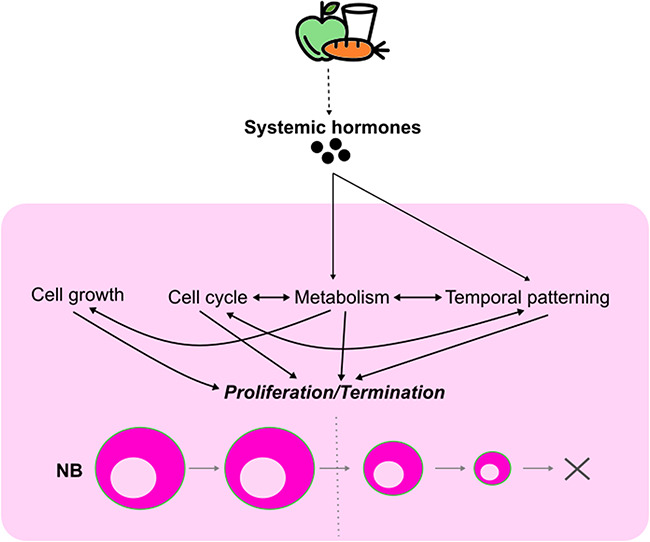
The formation of a functional circuitry in the central nervous system (CNS) requires the correct number and subtypes of neural cells. In the developing brain, neural stem cells (NSCs) self-renew while giving rise to progenitors that in turn generate differentiated progeny. As such, the size and the diversity of cells that make up the functional CNS depend on the proliferative properties of NSCs. In the fruit fly Drosophila, where the process of neurogenesis has been extensively investigated, extrinsic factors such as the microenvironment of NSCs, nutrients, oxygen levels and systemic signals have been identified as regulators of NSC proliferation. Here, we review decades of work that explores how extrinsic signals non-autonomously regulate key NSC characteristics such as quiescence, proliferation and termination in the fly.

Keywords: Drosophila; neuroblast; niche.


Conflict of interest statement

None declared.

Figures

Figure 1
Figure 1
The cell types and their interactions within the developing CNS during early and late neural development. (A) The larval central nervous system (CNS) is divided into three neurogenic regions: the central brain (CB), the ventral nerve cord (VNC) and the optic lobe (OL). (B–C′) The neurogenic niche of neuroblasts (NBs) is composed of the extracellular matrix at the outer most layer of the CNS, different glial cell types (green) and the trachea (blue). The glial niche contains perineural glia (PG), subperineural glia (SPG), cortex glia (CG) and neuropil glia. Neuropil glia is consisted of ensheathing glia and astrocyte-like glia in which only astrocyte-like glia infiltrate the neuropil (yellow). The CNS primary trachea includes the cerebral trachea (CT) that invades the CB as well as the OL, and the ganglion branch (GB) that invades the VNC. Primary branches develop on the surface of the neuropil and are embedded between the neuropil and neuropil glia. From the primary branch, secondary tracheal branches elaborate towards the brain cortex. Dashed line indicates the cross-sections shown in B′ and C′. (B) Dorsal view of the CNS and (B′) cross-section of the VNC at the first-instar larval (L1) stage. At this stage, many NBs of type I (pink) and type II (orange) are in quiescence, each with a primary basal protrusion in contact with the neuropil. (C) Dorsal view of the CNS at the third-instar larval (L3) stage. At this stage, NBs of type I and type II are reactivated to resume postembryonic neurogenesis. In the OL, NBs in the OPC and the IPC are produced from two neuroepithelia. Note that mushroom body NBs (MBNBs, purple) do not become quiescent but continue proliferating throughout the embryonic-larval transition. (C′) Cross-section of the VNC at L3. During larval development, CG undergoes an extensive process of remodelling such that they extend their membrane to encase NBs and their progeny. As a result, CG forms chambers for each individual NB lineage. Concomitantly, the CNS trachea continues to elaborate towards the brain cortex.
Figure 2
Figure 2
Extrinsic signals regulate the balance between neuroblast quiescence and reactivation during early larval development. The entry of neuroblasts (pink) into quiescence during late embryogenesis is partially induced by Dl/Notch signalling mediated by newborn GMCs in the proximity. The quiescent state of neuroblasts was suggested to be maintained by multiple factors derived from the glial niche such as FMRP, Ana, as well as the Ed/Crb-mediated Hippo signalling. Each quiescent neuroblast extends a primary basal protrusion that is in contact with the neuropil and is enriched with DE-cad. DE-cad in turn promotes neuroblast reactivation during early larval development. Neuroblast reactivation is induced by the release of dILP6 from glial cells, especially cortex glia, and non-autonomous upregulation of PI3K signalling in cortex glia and the trachea (blue). The extracellular matrix component Trol can promote neuroblast reactivation by upregulating the Hh and Bnl signalling cascades. Systemic hormones, such as dILP2 and Ecdysone, were also suggested to play roles in facilitating neuroblast reactivation at the early larval stages.
Figure 3
Figure 3
Extrinsic signals regulate neuroblast proliferation during larval development. The glial niche (green) provides multiple factors to regulate neuroblast (pink) proliferation. Perineural glia (PG) produce Dlp, which facilitates the activation of autocrine Gbb-induced TGFβ signalling in the neuroblast to promote proliferation. PG also express Tret-1 that uptakes Trehalose from the haemolymph to process it into lactate and alanine that are supplied to neurons (grey) for energy production. Subperineural glia (SPG) express Scaf while cortex glia (CG) express the amino acid transporters path, Sbm and the chloride channel ClC-a, which all promote neuroblast proliferation. The CG and the neuroblast crosstalk through Pvr/Pvf signalling in which Pvr signalling in the CG can non-autonomously upregulate PI3K signalling and DE-Cad in the neuroblast. Jeb, which can be produced by glial cells, activates Alk/PI3K signalling in the neuroblast to promote proliferation regardless of the organismal nutritional status (dashed line indicate unclear cellular source). At the late larval stages, CG produces Hh that activates Hh signalling in the neuroblasts to suppress their proliferation. CG and SPG also contain lipid droplets (LD) that can non-autonomously affect neuroblast proliferation.
Figure 4
Figure 4
Integration of extrinsic and intrinsic cues to schedule neuroblast termination. nutrient availability induces changes in the synthesis and the secretion of systemic hormones in the animal. This influences multiple intrinsic properties of the neuroblasts, including cell growth, cell cycle controls, metabolism and temporal patterning that govern their proliferation. Additionally, intrinsic mechanisms can interact and cross-regulate each other to stimulate timely neuroblast termination.
Figure 5
Figure 5
The cell types and the niche of the OL OPC in late neural development. Left: The OL during late larval development. Right: Inset showed in the left panel. In the OPC, neuroblasts (NBs, blue) are produced from the neuroepithelium (NE, black) due to a differentiating proneural wave. NE cells initially divide symmetrically to expand the progenitor pool. As the proneural wave travels across the tissue, NE cells at the wave front acquire the epi-neuroblast (epi-NB) state in which they asymmetrically divide to give rise to a NB and a ganglion mother cell (GMC). The GMC then differentiates into two neurons or glial cells of the OL. The OL glial niche (green) lies on top of the OPC, including perineural glia (PG), subperineural glia (SPG) and cortex glia (CG). The proneural wave propagation is driven by Spi secreted by epi-NBs. Spi is also produced by the CG and is directly regulated by miR-8 expression in the CG. In addition, wave propagation was suggested to be non-autonomously regulated ClC-a expression in the CG and the Ser/Cno/Notch complex formed between the SPG and the NE.

Similar articles

References

    1. Penzo-Méndez AI, Stanger BZ. Organ-size regulation in mammals. Cold Spring Harb Perspect Biol. 2015;7:a019240. - PMC - PubMed
    1. Texada MJ, Koyama T, Rewitz K. Regulation of body size and growth control. Genetics. 2020;216:269–313 - PMC - PubMed
    1. Götz M, Huttner WB. The cell biology of neurogenesis. Nat Rev Mol Cell Biol. 2005;6:777–88 - PubMed
    1. Harding K, White K. Drosophila as a model for developmental biology: stem cell-fate decisions in the developing nervous system. J Dev Biol. 2018;6:25. - PMC - PubMed
    1. Li G, Hidalgo A. Adult neurogenesis in the Drosophila brain: the evidence and the void. Int J Mol Sci. 2020;21:6653. - PMC - PubMed

LinkOut - more resources