

Comparative study on the effect of maxillary expansion on the displacement of maxillary alveolar bone before and after alveolar bone graft: a three-dimensional finite element analysis
- PMID: 38596963
- PMCID: PMC9396421
- DOI: 10.7518/hxkq.2022.04.013
Comparative study on the effect of maxillary expansion on the displacement of maxillary alveolar bone before and after alveolar bone graft: a three-dimensional finite element analysis
Abstract
Objectives: This paper aimed to simulate and compare the effect of maxillary expansion on the displacement of maxillary alveolar bone before and after alveolar bone graft.
Methods: On the established finite element model of the maxilla before bone grafting, ANSYS was used to simulate alveolar bone graft to form the model of maxilla after bone grafting. The same expansion force was applied to the two models, and the three-dimensional displacement of alveolar bone was observed and compared between them.
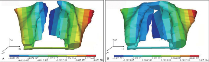
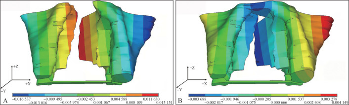
Results: Comparison of the three-dimensional displacement showed that expansion before bone grafting was significantly larger than that after bone grafting (P<0.05). For horizontal displacement, in the expansion group before bone grafting, the displacement was gradually decreased from anterior to posterior alveolar bone. In the expansion group after bone grafting, the displacement was gradually increased from anterior to posterior alveolar bone. Displacement of noncleft side alveolar bone was significantly larger than that of cleft side alveolar bone with maxillary expansion before and after alveolar bone graft (P<0.05). In terms of vertical displacement, the anteromedial alveolar bone moved downward and the posterolateral alveolar bone moved upward with maxillary expansion before and after alveolar bone graft. For sagittal displacement, the anteromedial alveolar bone moved forward and the posterolateral alveolar bone moved backward with maxillary expansion before alveolar bone graft. However, the movement trend was opposite with maxillary expansion after alveolar bone graft.
Conclusions: The three-dimensional movement with maxillary expansion before alveolar bone graft is more obvious than that after bone grafting for patients with unilateral complete cleft lip and palate. Expander should be moved backward properly with maxillary expansion before alveolar bone graft and moved forward properly and cooperated with maxillary protraction with maxillary expansion after alveolar bone graft. Meanwhile, precaution must be taken in terms of asymmetric expansion and anterior open bite in operation.
目的: 模拟比较牙槽突裂植骨前后上颌扩弓对上颌牙槽骨位移影响。方法: 在已建立的植骨前上颌骨有限元模型上,采用ANSYS软件模拟牙槽突裂植骨,形成植骨后上颌骨模型。在2组模型上分别施加相同上颌扩弓力,观察比较牙槽骨区域三维方向位移形变情况。结果: 三维方向位移量比较,植骨前扩弓组均显著大于植骨后扩弓组(P<0.05)。水平向位移:植骨前扩弓,由前向后牙槽骨区域位移量逐渐降低;植骨后扩弓,由前向后牙槽骨区域位移量逐渐升高;植骨前后扩弓健侧牙槽骨位移量均显著大于患侧(P<0.05)。垂直向位移:植骨前后扩弓,牙槽骨前内侧均向下移动,牙槽骨后外侧均向上移动。矢状向位移:植骨前扩弓,牙槽骨前内侧向前移动,后外侧向后移动,植骨后扩弓移动趋势相反。结论: 单侧完全性唇腭裂患者植骨前扩弓三维方向移动均较植骨后明显,植骨前扩弓建议扩弓器适当向后移动,植骨后扩弓建议扩弓器适当向前移动并配合前牵引治疗,同时治疗中需警惕不对称扩弓及前牙开牙合的发生。.
Keywords: alveolar bone graft; finite e-lement analysis; maxillary expansion; three dimensional displacement of alveolar bone.
Conflict of interest statement
利益冲突声明:作者声明本文无利益冲突。
Figures


Similar articles
-
Effects of cleft type, facemask anchorage method, and alveolar bone graft on maxillary protraction: a three-dimensional finite element analysis.Cleft Palate Craniofac J. 2012 Mar;49(2):221-9. doi: 10.1597/10-265. Epub 2011 Jul 8. Cleft Palate Craniofac J. 2012. PMID: 21740169
-
Biomechanical effects on maxillary protraction of the craniofacial skeleton with cleft lip and palate after alveolar bone graft.J Craniofac Surg. 2013 Mar;24(2):446-53. doi: 10.1097/SCS.0b013e31826cfe27. J Craniofac Surg. 2013. PMID: 23524712
-
[Maxillary protraction with and without maxillary expansion on unilateral cleft lip and palate model: a finite element analysis].Shanghai Kou Qiang Yi Xue. 2012 Jun;21(3):287-93. Shanghai Kou Qiang Yi Xue. 2012. PMID: 22885489 Chinese.
-
Orthodontic Preparation for Secondary Alveolar Bone Grafting in Patients with Complete Cleft Lip and Palate.Oral Maxillofac Surg Clin North Am. 2020 May;32(2):205-217. doi: 10.1016/j.coms.2020.01.003. Epub 2020 Feb 22. Oral Maxillofac Surg Clin North Am. 2020. PMID: 32098718 Review.
-
Alveolar bone grafts: the surgical/orthodontic management of the cleft maxilla.Ann Acad Med Singap. 1999 Sep;28(5):721-7. Ann Acad Med Singap. 1999. PMID: 10597360 Review.
References
-
- Ahmed M, Fida M, Jeelani W. Management of an adolescent with complete bilateral cleft lip and palate using fan-shaped expander and secondary alveolar bone graft: a case report[J] Int Orthod. 2020;18(3):593–602. - PubMed
-
- Ayub PV, Janson G, Gribel BF, et al. Analysis of the maxillary dental arch after rapid maxillary expansion in patients with unilateral complete cleft lip and palate[J] Am J Orthod Dentofacial Orthop. 2016;149(5):705–715. - PubMed
-
- Allareddy V, Bruun R, MacLaine J, et al. Orthodontic preparation for secondary alveolar bone grafting in patients with complete cleft lip and palate[J] Oral Maxillofac Surg Clin North Am. 2020;32(2):205–217. - PubMed
-
- Garib D, Miranda F, Sathler R, et al. Rapid maxillary expansion after alveolar bone grafting with rhBMP-2 in UCLP evaluated by means of CBCT[J] Cleft Palate Craniofac J. 2017;54(4):474–480. - PubMed
LinkOut - more resources
Full Text Sources