

Effects of surface nanomorphology on the senescence of periodontal ligament stem cells
- PMID: 38597077
- PMCID: PMC11034406
- DOI: 10.7518/hxkq.2024.2023244
Effects of surface nanomorphology on the senescence of periodontal ligament stem cells
Abstract
Objectives: The effect of TiO2 nanotube morphology on the differentiation potency of senescent periodontal ligament stem cells was investigated.
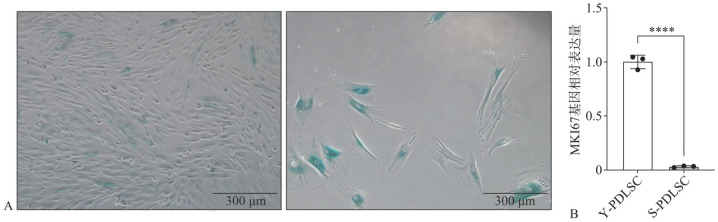
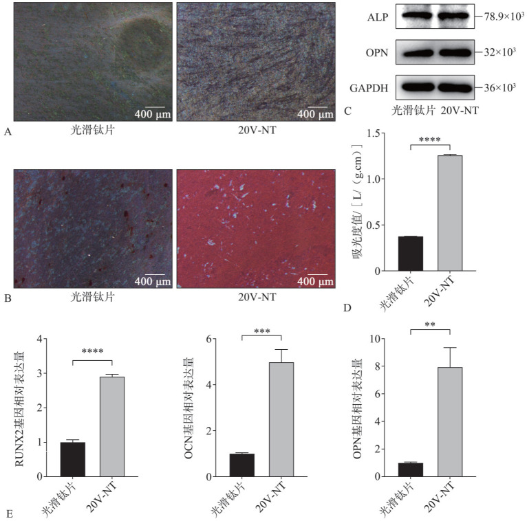
Methods: Two types of titanium sheets with TiO2 nanotube morphology (20V-NT and 70V-NT) were prepared via anodic oxidation at 20 and 70 V separately, and their surface morphology was observed. Young periodontal ligament stem cells were cultivated in an osteogenic induction medium, and the most effective surface morphology in promoting osteogenic differentiation was selected. RO3306 and Nutlin-3a were used to induce the aging of young periodontal ligament stem cells, and senescent periodontal ligament stem cells were obtained. The osteogenic differentiation of senescent periodontal ligament stem cells was induced, and the effect of surface morphology on osteogenic differentiation was observed.
Results: Nanotube morphology was achieved on the surfaces of titanium sheets through anodic oxidation, and the diameters of the nanotubes increased with voltage. A significant difference in the effect of nanotube morphology was found among nanotubes with different diameters in the young periodontal ligament stem cells. The surface nanotube morphology of 20V-NT had a more significant effect that promoted osteogenic differentiation. Compared with a smooth titanium sheet, the surface nanotube morphology of 20V-NT increased the number of alkaline phosphatase-positive senescent periodontal ligament stem cells and promoted calcium deposition and the expression of osteogenic marker genes Runt-related transcription factor 2, osteopontin, and osteocalcin.
Conclusions: A special nanotube morphology enhances the differentiation ability of senescent periodontal ligament stem cells, provides an effective method for periodontal regeneration, and further improves the performance of implants.
目的: 探讨二氧化钛纳米管形貌对衰老牙周膜干细胞分化能力的影响。方法: 利用阳极氧化法,分别于20、70 V电压下制备出2种具有二氧化钛纳米管形貌的钛片(20V-NT、70V-NT),观察其表面形貌特征。在成骨诱导条件下培养年轻牙周膜干细胞,挑选具有促进成骨分化作用的表面形貌。用RO3306和Nutlin-3a诱导年轻牙周膜干细胞衰老,获得衰老的牙周膜干细胞。诱导衰老牙周膜干细胞成骨分化,观察表面形貌对衰老牙周膜干细胞成骨分化的影响。结果: 阳极氧化法可在钛片表面形成纳米管形貌,且纳米管直径随电压的增大而增大;不同直径纳米管形貌对年轻牙周膜干细胞成骨分化的影响存在较大差异,20V-NT表面纳米形貌促成骨分化效果更明显。与光滑钛片相比,20V-NT表面纳米形貌提高了衰老牙周膜干细胞碱性磷酸酶阳性数量,促进了钙沉积以及成骨标志性基因Runt相关转录因子2、骨桥蛋白、骨钙素的表达。结论: 特定的表面纳米形貌能增强衰老牙周膜干细胞的分化能力,为牙周再生和进一步提高种植体的性能提供了一种有效方法。.
Keywords: nanotubes; osteogenesis; periodontal ligament stem cells; senescence; surface morphology; titanium.
Conflict of interest statement
利益冲突声明:作者声明本文无利益冲突。
Figures


References
-
- Long M, Rack HJ. Titanium alloys in total joint replacement—a materials science perspective[J] Biomaterials. 1998;19(18):1621–1639. - PubMed
-
- Rocca M, Fini M, Giavaresi G, et al. Osteointegration of hydroxyapatite-coated and uncoated titanium screws in long-term ovariectomized sheep[J] Biomaterials. 2002;23(4):1017–1023. - PubMed
MeSH terms
Substances
LinkOut - more resources
Full Text Sources
Research Materials