

Verification of the expression trend and interaction prediction of innate immune cells and immune-checkpoint molecules in the process of oral mucosal carcinogenesis
- PMID: 38597079
- PMCID: PMC11034413
- DOI: 10.7518/hxkq.2024.2023280
Verification of the expression trend and interaction prediction of innate immune cells and immune-checkpoint molecules in the process of oral mucosal carcinogenesis
Abstract
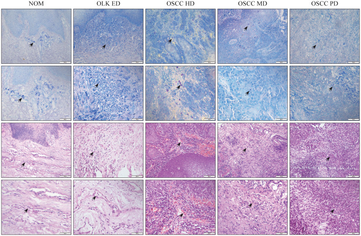
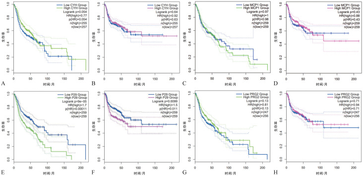
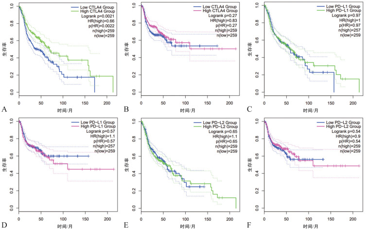
Objectives: This study aimed to explore the expression trends of innate immune cells and immune-checkpoint molecules validated by data calculation in the process of oral mucosal carcinogenesis, as well as to explore methods of suppressing oral mucosal carcinogenesis based on immunotherapy by predicting their interactions. Me-thods 1) The cancer genome atlas (TCGA) database comprehensively scores immune cells and immune-checkpoint molecules in the process of oral mucosal carcinogenesis and screens out intrinsic immune cells and immune-checkpoint molecules that interfere with tumor immune escape. 2) Clinical patient blood routine data were collected for the statistical analysis of peripheral blood immune cells during the progression of oral mucosal carcinogenesis. Immune cells in peripheral blood that may affect the progression of oral mucosal carcinogenesis were screened. 3) Immunohistochemical staining was performed on intrinsic immune cells and immune-checkpoint molecules validated based on data calculation in various stages of oral mucosal carcinogenesis. 4) Special staining was used to identify innate immune cells in various stages of oral mucosal carcinogenesis based on data-calculation verification. 5) Survival analysis was conducted on intrinsic immune cells and immune-checkpoint molecules validated based on data calculation during the process of oral mucosal carcinogenesis. The association of intrinsic immune cells and immune-checkpoint molecules with the prognosis of oral squamous cell carcinoma was verified.
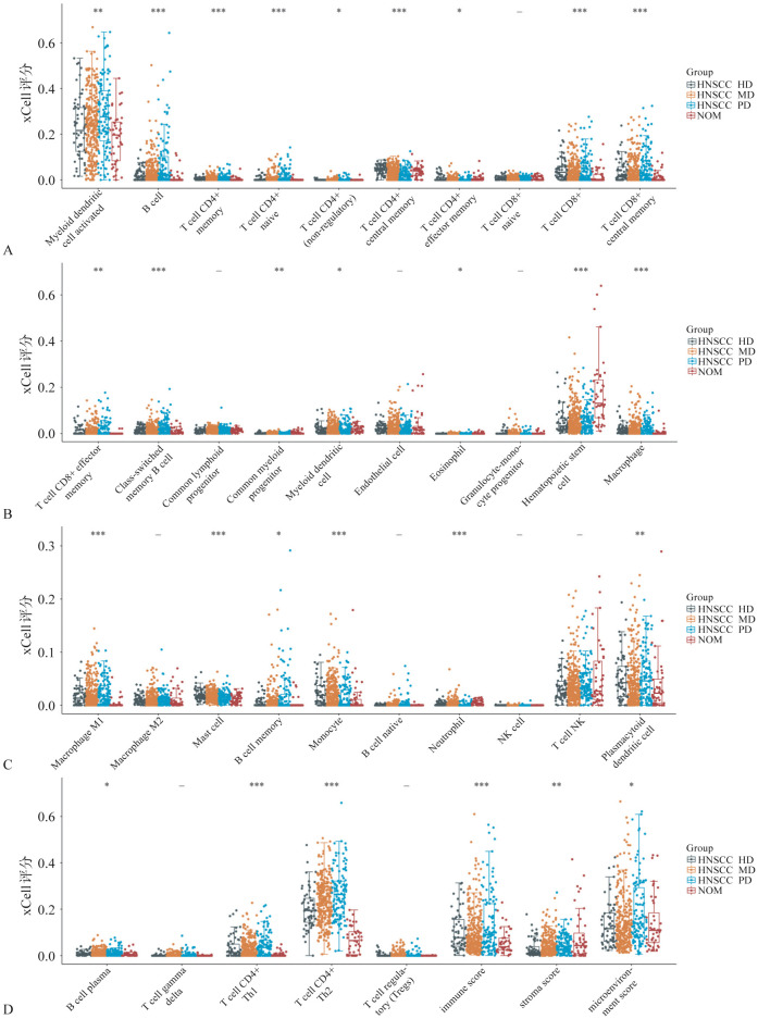
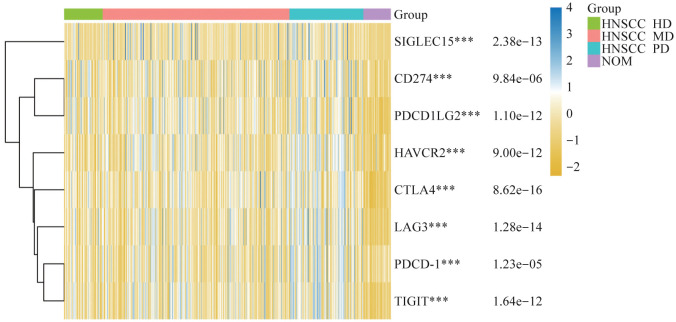
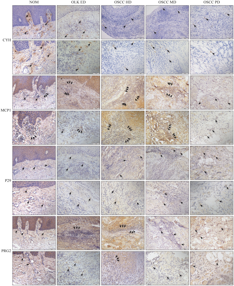
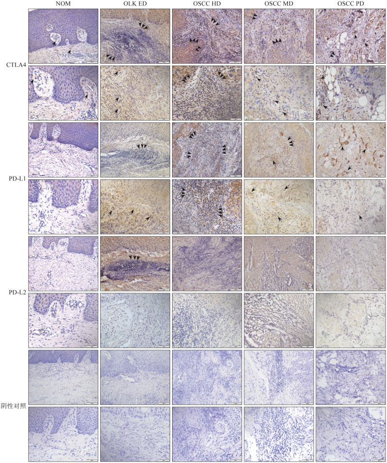
Results: The expression of monocytes and neutrophils increased during the process of oral mucosal carcinogenesis. The expression of eosinophils showed a single peak trend of up and down. The expression of mast cells decreased. In the process of oral mucosal carcinogenesis, the expression of the immune-checkpoint molecules cytotoxic T-lymphocyte-associated protein 4 (CTLA4) and programmed cell death-ligand (PD-L1) increased. The expression trends of monocytes, neutrophils, and eosinophils were positively correlated with those of CTLA4 and PD-L1 immune-checkpoint molecules. The expression trend of mast cells was negatively correlated with the expression of CTLA4 and PD-L1. Monocytes, neutrophils, and eosinophils may promote tumor immune escape mediated by CTLA4 and/or PD-L1, thereby accelerating the progression of oral mucosal carcinogenesis. Mast cells may inhibit tumor immune escape mediated by CTLA4 and/or PD-L1, delaying the progression of oral mucosal carcinogenesis.
Conclusions: Therefore, interference with specific immune cells in innate immunity can regulate the expression of CTLA4 and/or PD-L1 to a certain extent, inhibit tumor immune escape, and delay the progression of oral mucosal carcinogenesis.
目的: 研究口腔黏膜癌变进程中基于数据计算验证的固有免疫细胞和免疫检查点分子的表达趋势,并通过预测其交互作用,探索免疫治疗抑制口腔黏膜癌变进程的方法。方法: 1)利用癌症基因组图谱对口腔黏膜癌变进程中的免疫细胞和免疫检查点分子进行全面评分,筛选出干扰肿瘤细胞免疫逃逸的固有免疫细胞和免疫检查点分子;2)收集血常规资料,对口腔黏膜癌变进程中外周血免疫细胞进行统计学分析,筛选外周血中可能影响口腔黏膜癌变进程的免疫细胞;3)对口腔黏膜癌变进程各阶段中基于数据计算验证的固有免疫细胞和免疫检查点分子进行免疫组织化学染色;4)采用特殊染色鉴定口腔黏膜癌变进程各阶段中基于数据计算验证的固有免疫细胞;5)对口腔黏膜癌变进程中基于数据计算验证的固有免疫细胞和免疫检查点分子进行生存分析,验证固有免疫细胞和免疫检查点分子与口腔鳞状细胞癌预后间的关联。结果: 在口腔黏膜癌变进程中,单核细胞、中性粒细胞表达呈上升趋势;嗜酸性粒细胞表达呈升降单峰趋势;肥大细胞表达呈下降趋势;免疫检查点分子细胞毒性T淋巴细胞相关蛋白4(CTLA4)和细胞程序性死亡-配体1(PD-L1)的表达呈上升趋势。单核细胞、中性粒细胞和嗜酸性粒细胞表达趋势与CTLA4和PD-L1免疫检查点分子的表达趋势正相关;肥大细胞表达趋势与CTLA4和PD-L1免疫检查点分子的表达趋势负相关。单核细胞、中性粒细胞和嗜酸性粒细胞可能促进CTLA4和(或)PD-L1介导的肿瘤细胞免疫逃逸,加速口腔黏膜癌变进程;肥大细胞可能抑制CTLA4和(或)PD-L1介导的肿瘤细胞免疫逃逸,延缓口腔黏膜癌变进程。结论: 干扰固有免疫中特定免疫细胞可在一定程度上调控CTLA4和(或)PD-L1的表达,抑制肿瘤细胞免疫逃逸,延缓口腔黏膜癌变进程。.
Keywords: immune checkpoint molecules; immune escape; immunotherapy; inherent immune cells; malignant transformation of oral mucosa.
Conflict of interest statement
利益冲突声明:作者声明本文无利益冲突。
Figures


Similar articles
-
Correlation between messenger RNA expression and protein expression of immune checkpoint-associated molecules in bladder urothelial carcinoma: A retrospective study.Urol Oncol. 2017 May;35(5):257-263. doi: 10.1016/j.urolonc.2017.01.014. Epub 2017 Mar 11. Urol Oncol. 2017. PMID: 28291636
-
Tumour cell-intrinsic CTLA4 regulates PD-L1 expression in non-small cell lung cancer.J Cell Mol Med. 2019 Jan;23(1):535-542. doi: 10.1111/jcmm.13956. Epub 2018 Oct 30. J Cell Mol Med. 2019. PMID: 30378264 Free PMC article.
-
Immune checkpoints indoleamine 2,3-dioxygenase 1 and programmed death-ligand 1 in oral mucosal dysplasia.J Oral Pathol Med. 2018 Sep;47(8):773-780. doi: 10.1111/jop.12737. Epub 2018 Jun 20. J Oral Pathol Med. 2018. PMID: 29851145
-
T cell checkpoint regulators in the heart.Cardiovasc Res. 2019 Apr 15;115(5):869-877. doi: 10.1093/cvr/cvz025. Cardiovasc Res. 2019. PMID: 30721928 Free PMC article. Review.
-
Regulatory mechanisms of immune checkpoints PD-L1 and CTLA-4 in cancer.J Exp Clin Cancer Res. 2021 Jun 4;40(1):184. doi: 10.1186/s13046-021-01987-7. J Exp Clin Cancer Res. 2021. PMID: 34088360 Free PMC article. Review.
References
-
- 王 海青, 向 婉婷, 卢 俊米. PD-L1在口腔鳞状细胞癌中表达的临床意义[J] 中国卫生标准管理. 2023;14(6):83–87.
- Wang HQ, Xiang WT, Lu JM. Clinical significance of PD-L1 expression in oral squamous cell carcinoma[J] China Health Stand Manag. 2023;14(6):83–87.
MeSH terms
Substances
LinkOut - more resources
Full Text Sources
Medical
Research Materials