Review
. 2024 Jul 1;48(7):777-789.
doi: 10.1097/PAS.0000000000002218. Epub 2024 Apr 10.

TFE3 -Rearranged PEComa/PEComa-like Neoplasms : Report of 25 New Cases Expanding the Clinicopathologic Spectrum and Highlighting its Association With Prior Exposure to Chemotherapy

Affiliations
Review

TFE3 -Rearranged PEComa/PEComa-like Neoplasms : Report of 25 New Cases Expanding the Clinicopathologic Spectrum and Highlighting its Association With Prior Exposure to Chemotherapy

Pedram Argani et al. Am J Surg Pathol. .

Abstract

Since their original description as a distinctive neoplastic entity, ~50 TFE3 -rearranged perivascular epithelioid cell tumors (PEComas) have been reported. We herein report 25 new TFE3 -rearranged PEComas and review the published literature to further investigate their clinicopathologic spectrum. Notably, 5 of the 25 cases were associated with a prior history of chemotherapy treatment for cancer. This is in keeping with prior reports, based mainly on small case series, with overall 11% of TFE3 -rearranged PEComas being diagnosed postchemotherapy. The median age of our cohort was 38 years. Most neoplasms demonstrated characteristic features such as nested architecture, epithelioid cytology, HMB45 positive, and muscle marker negative immunophenotype. SFPQ was the most common TFE3 fusion partner present in half of the cases, followed by ASPSCR1 and NONO genes. Four of 7 cases in our cohort with meaningful follow-up presented with or developed systemic metastasis, while over half of the reported cases either recurred locally, metastasized, or caused patient death. Follow-up for the remaining cases was limited (median 18.5 months), suggesting that the prognosis may be worse. Size, mitotic activity, and necrosis were correlated with aggressive behavior. There is little evidence that treatment with MTOR inhibitors, which are beneficial against TSC -mutated PEComas, is effective against TFE3 -rearranged PEComas: only one of 6 reported cases demonstrated disease stabilization. As co-expression of melanocytic and muscle markers, a hallmark of conventional TSC -mutated PEComa is uncommon in the spectrum of TFE3 -rearranged PEComa, an alternative terminology may be more appropriate, such as " TFE3 -rearranged PEComa-like neoplasms," highlighting their distinctive morphologic features and therapeutic implications.


Conflict of interest statement

Conflicts of Interest and Source of Funding: The authors have disclosed that they have no significant relationships with, or financial interest in, any commercial companies pertaining to this article.

Figures

Figure 1 (case 4):
Figure 1 (case 4):
This bladder biopsy reveals an epithelioid cell lesion with pale eosinophilic cytoplasm arranged in sheets and cords within hyalinized stroma (A). At higher power, the neoplastic cells have bland round nuclei and granular eosinophilic cytoplasm, resembling histiocytes (B). The neoplastic cells demonstrate diffuse cytoplasmic labeling for HMB45 (C) and strong diffuse nuclear labeling for TFE3 (D).
Figure 2 (case 1):
Figure 2 (case 1):
This renal biopsy reveals an epithelioid cell lesion (right) associated with abundant hemorrhage (left). At high power the epithelioid cells have round nuclei and pale granular eosinophilic cytoplasm, resembling histocytes (B). The neoplastic cells label diffusely for HMB45 (C) and strong diffuse nuclear labeling for TFE3 (D).
Figure 3 (case 2):
Figure 3 (case 2):
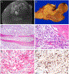
MRI STIR images reveal an aggressive lesion in the proximal femur showing cortical breakthrough and extension into adjacent soft tissue (A). On gross examination, the fleshy tumor involves the femoral neck and breaks through the cortex to extend into soft tissue. Defects consistent with prior rod placement are also evident (B). The epithelioid neoplastic cells are arranged in nests, and permeate into existing lamellar bony trabeculae (C). At high power, the neoplastic cells abut bone lined by osteoblasts. The neoplastic cells have round nuclei with pinpoint nucleoli and granular eosinophilic to pale cytoplasm (D). The neoplastic cells demonstrate diffuse cytoplasmic labeling for HMB45 (red chromogen, E) and diffuse strong nuclear labeling for TFE3 (F).
Figure 4 (case 10):
Figure 4 (case 10):
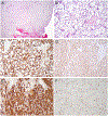
This polypoid nasal lesion undermines the respiratory epithelium (right), which is focally ulcerated (bottom) (A). High power view demonstrates an epithelioid cell lesion with voluminous, water clear cytoplasm and hyalinized stroma (B). The neoplastic cells are negative for melanocytic markers but instead are diffusely immunoreactive for muscle markers desmin (C) and smooth muscle actin (D). They are diffusely immunoreactive for the downstream marker of TFE3 signaling, glycoprotein non-metastatic B (GPNMB) (E) and demonstrate diffuse strong nuclear labeling for TFE3 (F).
Figure 5 (case 22):
Figure 5 (case 22):
This lung nodule is composed of neoplastic spindle cells with moderate atypia associated with a prominent chronic lymphoplasmacytic inflammatory infiltrate, reminiscent of an inflammatory myofibroblastic tumor (A). At higher magnification, some of the neoplastic cells have a more epithelioid appearance with a syncytial growth pattern with associated inflammatory infiltrate (B). The neoplastic cells label diffusely for HMB45, highlighting their spindled morphology (C) and demonstrate diffuse nuclear labeling for TFE3 (D).
Figure 6 (case 20):
Figure 6 (case 20):
This thigh mass demonstrated pleomorphic spindled to epithelioid morphology, with dense collagen (A). The pleomorphic epithelioid cells have voluminous xanthomatous cytoplasm, mimicking lipoblasts (B). The xanthomatous cells have areas of sheet like growth with high mitotic activity including atypical forms (C). The neoplastic cells demonstrate diffuse cytoplasmic labeling for cathepsin K (D) but an absence of melanocytic or muscle marker expression.

References

    1. Ladanyi M, Lui MY, Antonescu CR, et al. The der(17)t(X;17)(p11;q25) of human alveolar soft part sarcoma fuses the TFE3 transcription factor gene to ASPL, a novel gene at 17q25. Oncogene. 2001;20:48–57. - PubMed
    1. Argani P Antonescu CR, Illei PB, et al. Primary renal neoplasms with ASPL-TFE3 gene fusion of alveolar soft part sarcoma: a distinctive tumor entity previously included among renal cell carcinomas of children and adolescents. Am J Pathol. 2001;159:179–192. - PMC - PubMed
    1. Argani P, Antonescu CR, Couturier J, et al. PRCC-TFE3 renal carcinomas: morphologic, immunohistochemical, ultrastructural, and molecular analysis of an entity associated with the t(X;1)(p11.2;q21). Am J Surg Pathol. 2002;26:1553–1566. - PubMed
    1. Clark J, Lu YJ, Sidhar SK, et al. Fusion of splicing factor genes PSF and NonO (p54nrb) to the TFE3 gene in papillary renal cell carcinoma. Oncogene. 1997;15:2233–2239. - PubMed
    1. Argani P, Zhang L, Reuter VE, et al. RBM10-TFE3 Renal Cell Carcinoma: A Potential Diagnostic Pitfall Due to Cryptic Intrachromosomal Xp11.2 Inversion Resulting in False-negative TFE3 FISH. Am J Surg Pathol. 2017;41:655–662. - PMC - PubMed

MeSH terms

Substances