

[PI3K/Akt/Erk signaling pathway mediates neuroprotection of CaMKⅡγ and CaMKⅡδ against ischemic reperfusion injury in mice]
- PMID: 38597448
- PMCID: PMC11006692
- DOI: 10.12122/j.issn.1673-4254.2024.03.18
[PI3K/Akt/Erk signaling pathway mediates neuroprotection of CaMKⅡγ and CaMKⅡδ against ischemic reperfusion injury in mice]
Abstract
Objective: To observe neuroprotective effects of Ca2+/calmodulin-dependent kinase Ⅱ (CaMK Ⅱ)γ and CaMkII δ against acute neuronal ischemic reperfusion injury in mice and explore the underlying mechanism.
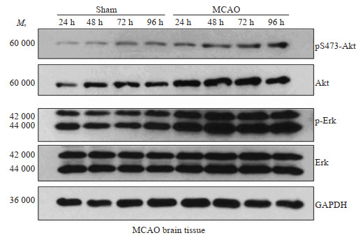
Methods: Primary cultures of brain neurons isolated from fetal mice (gestational age of 18 days) were transfected with two specific siRNAs (si-CAMK2G and si-CAMK2D) or a control sequence (si-NT). After the transfection, the cells were exposed to oxygen-glucose deprivation/reperfusion (OGD/R) conditions for 1 h followed by routine culture. The expressions of phosphatidylinositol-3-kinase/extracellular signal-regulated kinase (PI3K/Akt/Erk) signaling pathway components in the neurons were detected using immunoblotting. The expressions of the PI3K/Akt/Erk signaling pathway proteins were also detected in the brain tissues of mice receiving middle cerebral artery occlusion (MCAO) or sham operation.
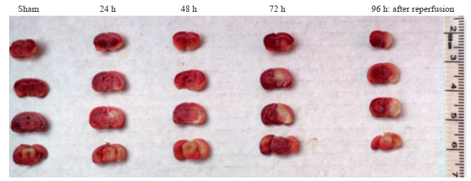
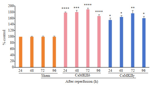
Results: The neuronal cells transfected with siCAMK2G showed significantly lower survival rates than those with si-NT transfection at 12, 24, 48, and 72 h after OGD/R (P < 0.01), and si-CAMK2G transfection inhibited OGD/R-induced upregulation of CaMKⅡγ expression. Compared to si-NT, transfection with si-CAMK2G and si-CAMK2D both significantly inhibited the expressions of PI3K/Akt/Erk signaling pathway components (P < 0.01). In the mouse models of MCAO, the expressions of CaMKⅡδ and CaMKⅡγ were significantly increased in the brain, where activation of the PI3K/Akt/Erk signaling pathway was detected. The expression levels of CaMKⅡδ, CaMKⅡγ, Erk, phosphorylated Erk, Akt, and phosphorylated Akt were all significantly higher in MCAO mice than in the sham-operated mice at 24, 48, 72, and 96 h after reperfusion (P < 0.05).
Conclusion: The neuroprotective effects of CaMKⅡδ and CaMKⅡγ against acute neuronal ischemic reperfusion injury are mediated probably by the PI3K/Akt/Erk pathway.
目的: 观察钙/钙调蛋白依赖性激酶Ⅱ(CaMKⅡ)的同工型CaMKⅡγ和CaMKⅡδ对鼠神经元细胞缺血缺氧再灌注损伤的影响,并探究其作用机制。
方法: 分离胚胎期第18天胎鼠大脑用于提取原代神经元,在5% CO2、37 ℃条件下培养,分为常规培养的对照组、空白对照组(si-NT)、CaMKⅡγ敲除组(si-CAMK2G)和CaMKⅡδ敲除组(si-CAMK2D)。研究组神经元转染处理后,更换为无糖培养基,并将其置于缺氧环境中模拟氧糖剥夺(OGD/R)条件,持续1 h,随后复原标准培养环境。通过对神经元裂解物行免疫蛋白印迹检测磷脂酰肌醇-3-激酶/细胞外信号调节激酶(PI3K/Akt/Erk)信号通路构件的表达量,并建立小鼠大脑中动脉闭塞(MCAO)模型,通过对比假手术组(Sham)(n=25)和MCAO组(n=25)PI3K/Akt/Erk信号通路表达来进行验证。
结果: si-CAMK2G组的胎鼠神经元细胞在OGD/R 12、24、48、72 h的生存率明显低于si-NT组(P < 0.01或0.001),并可逆转OGD/R介导的胎鼠神经元细胞CaMKⅡγ表达上调。si-CAMK2G组和si-CAMK2D组与si-NT组相比,PI3K/Akt/Erk信号通路表达受到明显抑制(P < 0.01)。在MCAO模型中,MCAO组小鼠脑CaMKⅡδ和CaMKⅡγ的表达显著增加并激活了PI3K /Akt/Erk信号通路,CaMKⅡδ和CaMKⅡγ,Erk、磷酸化Erk、Akt和磷酸化Akt在MCAO造模成功再灌注后24、48、72和96 h的表达显著高于Sham组再灌注24 h(P < 0.05,0.01或0.0001)。
结论: CaMKⅡγ和CaMKⅡδ在神经元细胞发生缺血缺氧损伤时的神经保护作用可能是通过PI3K/Akt/Erk信号通路介导的。
Keywords: Ca2+/calmodulin-dependent kinase Ⅱ; cerebral ischemia/reperfusion injury; extracellular signal-regulated kinase; neuroprotection; phosphatidylinositol-3-kinase; signaling pathway.
Figures


References
Publication types
MeSH terms
Substances
LinkOut - more resources
Full Text Sources
Molecular Biology Databases
Research Materials
Miscellaneous