. 2024 Apr 10;81(1):172.
doi: 10.1007/s00018-023-05057-3.

Bioengineered MSCCxcr2 transdifferentiated keratinocyte-like cell-derived organoid potentiates skin regeneration through ERK1/2 and STAT3 signaling in diabetic wound

Affiliations

Bioengineered MSCCxcr2 transdifferentiated keratinocyte-like cell-derived organoid potentiates skin regeneration through ERK1/2 and STAT3 signaling in diabetic wound

Subholakshmi Choudhury et al. Cell Mol Life Sci. .

Abstract

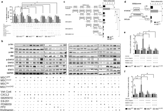
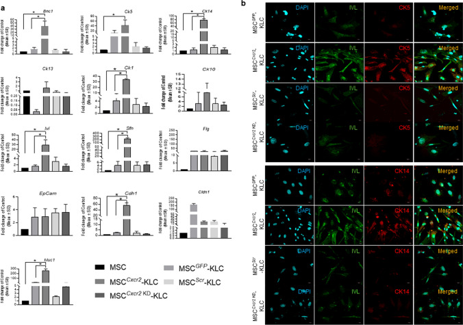
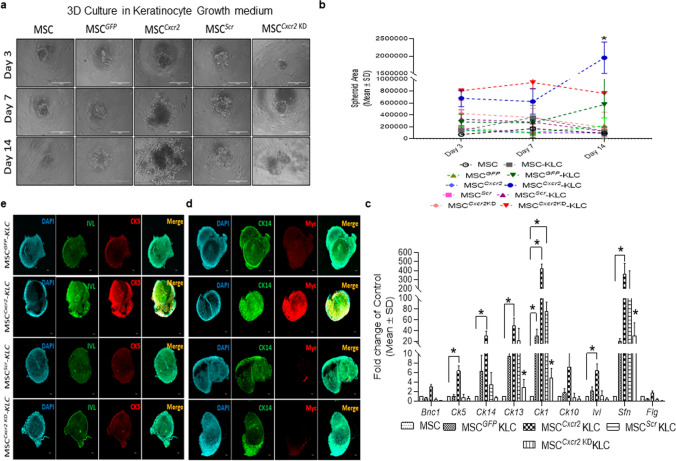
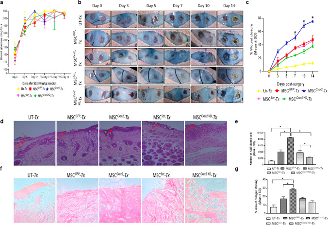
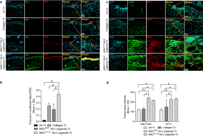
Skin regeneration is severely compromised in diabetic foot ulcers. Allogeneic mesenchymal stem cell (MSC) transplantation is limited due to the poor engraftment, mitogenic, and differentiation potential in the harsh wound microenvironment. Thus, to improve the efficacy of cell therapy, the chemokine receptor Cxcr2 was overexpressed in MSCs (MSCCxcr2). CXCL2/CXCR2 axis induction led to the enhanced proliferation of MSCs through the activation of STAT3 and ERK1/2 signaling. Transcriptional upregulation of FGFR2IIIb (KGF Receptor) promoter by the activated STAT3 and ERK1/2 suggested trans-differentiation of MSCs into keratinocytes. These stable MSCCxcr2 in 2D and 3D (spheroid) cell cultures efficiently transdifferentiated into keratinocyte-like cells (KLCs). An in vivo therapeutic potential of MSCCxcr2 transplantation and its keratinocyte-specific cell fate was observed by accelerated skin tissue regeneration in an excisional splinting wound healing murine model of streptozotocin-induced type 1 diabetes. Finally, 3D skin organoids generated using MSCCxcr2-derived KLCs upon grafting in a relatively avascular and non-healing wounds of type 2 diabetic db/db transgenic old mice resulted in a significant enhancement in the rate of wound closure by increased epithelialization (epidermal layer) and endothelialization (dermal layer). Our findings emphasize the therapeutic role of the CXCL2/CXCR2 axis in inducing trans-differentiation of the MSCs toward KLCs through the activation of ERK1/2 and STAT3 signaling and enhanced skin regeneration potential of 3D organoids grafting in chronic diabetic wounds.

Keywords: 3D skin organoids; Bioengineered MSCs; Cell transplantation; Chronic non-healing wounds; Keratinocytes; Organoid grafting; Tissue regeneration.


Conflict of interest statement

The authors have no financial or non-financial interests to disclose.

Figures

Fig. 1
Fig. 1
Molecular signaling of Cxcr2-mediated transcriptional regulation of FGFR2IIIb in MSCs a Graph depicting significantly increased proliferation of mouse bone marrow-derived MSCs with Cxcr2 overexpression that was further increased with CXCL2, which was abrogated in the presence of CXCR2 inhibitor SB2656101 and signaling molecule inhibitors PP2 (Src), Wortmannin (Akt), PD98059 (ERK), and S31-201 (STAT3). b Immunoblot analysis showed increased expression of p-ERK1/2, and p-STAT3 with Cxcr2 overexpression in MSCs which was reverted in the presence of the respective inhibitors, PD98059, and S3I-201. Cxcr2 silencing in MSCs differentially downregulated the activation of these mediators. c In silico analysis of the FGFR2IIIb promoter showing the presence of four proximal SP1 (p-ERK1/2)-binding sites. Luciferase promoter–reporter assay with Cxcr2-bioengineered MSCs transfected with the promoter–reporter constructs revealed that SP1 BS4 is essential for FGFR2IIIb promoter activity. d Similarly, in silico analysis of the FGFR2IIIb promoter showing the presence of one STAT3-binding site and promoter–reporter assay depicting the positive role of STAT3 BS in FGFR2IIIb promoter activity. Graph representing CXCL2-mediated increase in the luciferase activity in e SP1 (ERK1/2) WT construct and f STAT3 WT construct of FGFR2IIIb promoter in MSCCxcr2 which was abrogated by SB265610 and PD98059. (n = 3, *p < 0.05 as compared with MSCGFP, MSCCxcr2 + CXCL2 in proliferation assay; FGFR2IIIb SP1 (ERK1/2) WT/FGFR2IIIb STAT3 WT construct in MSCGFP, FGFR2IIIb SP1 (ERK1/2) WT/FGFR2IIIb STAT3 WT construct in MSCCxcr2; MSCGFP Vehicle Control, MSCCxcr2, and MSCCxcr2 + CXCL2 in Luciferase-reporter assay)
Fig. 2
Fig. 2
Cxcr2-mediated increased transdifferentiation potential of MSCs toward keratinocyte-like cells a Differential expression analysis of keratinocyte and epithelial markers in MSCGFP, MSCCxcr2, MSCScr, and MSCCxcr2 KD-derived KLCs revealed a significant increase in the expression of Basonuclin (Bnc1), Cytokeratin 5, 14, 1, Involucrin (Ivl), Stratifin (Sfn), E-Cadherin (Cdh1), and Mucin-1 (Muc1) in MSCCxcr2-KLCs. b Confocal microscopy images of MSCGFP, MSCCxcr2, MSCScr, and MSCCxcr2 KD-derived KLCs immuno-stained with Involucrin and CK5 and c Involucrin and CK14 showing a marked increase in the expression of Involucrin, CK5, and CK14 in MSCCxcr2-derived KLCs. (n = 3, *p < 0.05 as compared with MSC and MSCGFP-KLC)
Fig. 3
Fig. 3
Enhanced spheroid forming efficiency of MSCCxcr2-KLCs. a Representative bright-field microscopy images of Cxcr2-modulated MSCs in spheroid culture assay at days 3, 7, and 14. b Graph depicting a significant increase in MSCCxcr2-KLC spheroid area (lower panel depicting the graph of linear regression analysis). c Differential expression analysis of keratinocyte markers in spheroids depicting a significant increase in the expression of Ck5, Ck14, Ck13, Sfn, and Ivl. Confocal microscopy images of spheroids immuno-stained with d CK14, and Myc, e Involucrin, and CK5, depicting increased expression in MSCCxcr2-KLC spheroids. (n = 3, *p < 0.05 as compared with MSC-KLC/MSCGFP-KLC/MSCScr-KLC/MSCCxcr2KD-KLC; day 4/8 in spheroid area; and MSC in gene expression analysis)
Fig. 4
Fig. 4
Accelerated wound closure with MSCCxcr2 transplantation in type 1 diabetic mice a Graph depicting blood glucose levels in type 1 diabetic mice. b Representative images depicting morphometric analysis of type 1 diabetic wounds from post-surgery day 0 to day 14. c Graph depicting a significant increase in wound closure in the MSCCxcr2 Tx group from day 7 to day 14. d Representative images of hematoxylin and eosin (H&E) staining in regenerated type 1 diabetic mice wound tissue indicating epithelial thickness (marked by red arrow) and e quantitation of H&E-stained cells indicating increased granulation in the MSCCxcr2 transplanted group. Similarly, representative images of f Sirius red staining and g Quantitative analysis depicting a significant increase in the percent area of collagen-stained tissue in the MSCCxcr2 Tx group, whereas no significant change was depicted in the control and negative control groups (UT-Untransplanted, n = 3, *p < 0.05 as compared with UT/MSCGFP/MSCScr/MSCCxcr2 KD in morphometric analysis; and UT/MSCGFP/MSCScr/MSCCxcr2 in histology quantitation)
Fig. 5
Fig. 5
Enhanced re-epithelialization with MSCCxcr2 transplantation at type 1 diabetic wound bed a Representative confocal microscopy images of regenerated type 1 diabetic mice wound tissue co-immunostained with CXCR2 and Myc tag, b showing significantly increased Pearson’s correlation coefficient in MSCCxcr2 Tx group. c Representative confocal microscopy images of type 1 diabetic mice wound tissue co-immunostained with the keratinocyte markers Involucrin and CK14, d showing significantly increased Pearson’s correlation coefficient in MSCCxcr2 Tx group (n = 3, *p < 0.05 as compared with Un-Tx/MSCGFP Tx/MSCScr Tx/MSCCxcr2 KD Tx) (Scale bar: 10×–100 µm, 60×–20 µm)
Fig. 6
Fig. 6
Generation of bi-layered 3D skin organoids with fibroblasts and bioengineered MSC-KLCs a Schematic representation of the organoid culture system. b Differential expression analysis of keratinocyte and epithelial markers in MSCGFP-KLC (control) organoids and MSCCxcr2-KLC organoids depicting a significantly high expression of the keratinocyte markers Ck1 and Flg, and epithelial marker Cldn1 in MSCCxcr2-KLC organoids. c Representative H&E-stained images of the skin organoids. d Representative confocal microscopy images of whole mount MSCGFP-KLC and/or MSCCxcr2-KLC organoids, co-immunostained with Myc and CK5 (upper panel), and Involucrin and CK14 (lower panel). e Quantification of Pearson’s correlation coefficient depicting significant co-localization of Myc and CK5 (left), and Involucrin and CK14 (right) in MSCCxcr2-KLC organoids (n = 3, *p < 0.05 as compared with MSCGFP-KLC organoids) (Scale bar: 60×–20 µm)
Fig. 7
Fig. 7
Accelerated wound closure with MSCCxcr2-KLC organoid grafting in type 2 diabetic (db/db) transgenic mice a Morphometric analysis of representative images of db/db type 2 diabetic wounds from post-surgery day 0 to day 14. b Graph depicting a significant increase in wound closure in the MSCCxcr2-KLC organoid grafted group from day 7 to day 14. c Representative images of hematoxylin and eosin (H&E) staining (upper panel) and Sirius red staining (lower panel) in regenerated db/db type 2 diabetic mice wound tissue (red arrow indicating the epidermal region of the regenerated skin tissue sections). d Quantitation of H&E-stained cells (left panel) indicating increased granulation in MSCCxcr2-KLC organoid grafted group, and Sirius red staining (right panel) depicting a significant increase in percent area of collagen-stained tissue in MSCCxcr2-KLC organoid grafted group. (n = 3, *p < 0.05 as compared with Un-Tx/Collagen implanted/Tx group/MSCGFP-KLC organoid in morphometric analysis and histology quantification)
Fig. 8
Fig. 8
Enhanced re-epithelialization and vascularisation at type 2 diabetic mice wound bed a Representative confocal microscopy images and b quantification of Pearson’s correlation coefficient of db/db type 2 diabetic mice wound tissue immunostained with Myc tag and CK5 depicting significantly higher colocalization in MSCCxcr2-KLC organoid grafted group. c Representative confocal microscopy images and d quantification of fluorescence intensity of db/db type 2 diabetic regenerated tissue sections immunostained with α-SMA and CD31 depicting significantly increased staining of these markers in both MSCGFP-KLC and MSCCxcr2-KLC organoid grafted groups. (n = 3, *p < 0.05 as compared with UT/Collagen implanted/Tx/MSCGFP-KLC for re-epithelialization and Un-Tx/Collagen implantation for vascularization) (Scale bar: 10×–100 µm, 60×–20 µm)

Similar articles

Cited by

References

    1. Aitcheson SM, Frentiu FD, Hurn SE, Edwards K, Murray RZ. Skin wound healing: normal macrophage function and macrophage dysfunction in diabetic wounds. Molecules. 2021;26(16):4917. doi: 10.3390/molecules26164917. - DOI - PMC - PubMed
    1. Yang S, Xu M, Meng G, Lu Y. SIRT3 deficiency delays diabetic skin wound healing via oxidative stress and necroptosis enhancement. J Cell Mol Med. 2020;24(8):4415–4427. doi: 10.1111/jcmm.15100. - DOI - PMC - PubMed
    1. Furue K, Ulzii D, Tanaka Y, Ito T, Tsuji G, Kido-Nakahara M, Nakahara T, Furue M. Pathogenic implication of epidermal scratch injury in psoriasis and atopic dermatitis. J Dermatol. 2020;47(9):979–988. doi: 10.1111/1346-8138.15507. - DOI - PubMed
    1. Bieber T. Atopic dermatitis: an expanding therapeutic pipeline for a complex disease. Nat Rev Drug Discov. 2022;21(1):21–40. doi: 10.1038/s41573-021-00266-6. - DOI - PMC - PubMed
    1. Yao D, Ye S, He Z, Huang Y, Deng J, Wen Z, Chen X, Li H, Han Q, Deng H, Zhao RC. Adipose-derived mesenchymal stem cells (AD-MSCs) in the treatment for psoriasis: results of a single-arm pilot trial. Ann Transl Med. 2021;9(22):1653. doi: 10.21037/atm-21-5028. - DOI - PMC - PubMed