. 2024 Apr 10;81(1):171.
doi: 10.1007/s00018-024-05165-8.

The E3 ubiquitin ligase ITCH negatively regulates intercellular communication via gap junctions by targeting connexin43 for lysosomal degradation

Affiliations

The E3 ubiquitin ligase ITCH negatively regulates intercellular communication via gap junctions by targeting connexin43 for lysosomal degradation

Max Zachrisson Totland et al. Cell Mol Life Sci. .

Abstract

Intercellular communication via gap junctions has a fundamental role in regulating cell growth and tissue homeostasis, and its dysregulation may be involved in cancer development and radio- and chemotherapy resistance. Connexin43 (Cx43) is the most ubiquitously expressed gap junction channel protein in human tissues. Emerging evidence indicates that dysregulation of the sorting of Cx43 to lysosomes is important in mediating the loss of Cx43-based gap junctions in cancer cells. However, the molecular basis underlying this process is currently poorly understood. Here, we identified the E3 ubiquitin ligase ITCH as a novel regulator of intercellular communication via gap junctions. We demonstrate that ITCH promotes loss of gap junctions in cervical cancer cells, which is associated with increased degradation of Cx43 in lysosomes. The data further indicate that ITCH interacts with and regulates Cx43 ubiquitination and that the ITCH-induced loss of Cx43-based gap junctions requires its catalytic HECT (homologous to E6-AP C-terminus) domain. The data also suggest that the ability of ITCH to efficiently promote loss of Cx43-based gap junctions and degradation of Cx43 depends on a functional PY (PPXY) motif in the C-terminal tail of Cx43. Together, these data provide new insights into the molecular basis underlying the degradation of Cx43 and have implications for the understanding of how intercellular communication via gap junctions is lost during cancer development.

Keywords: Cervix; Connexin; Deubiquitination; Endosome; NEDD4.


Conflict of interest statement

The authors declare no competing interests concerning this study.

Figures
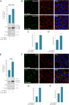
Fig. 1
Fig. 1
Role of ITCH in regulating the Cx43 protein level and gap junction size in HeLa-Cx43 and C33A cells. HeLa-Cx43 (A) or C33A (E) cells were transfected with control siRNA or with an siRNA sequence against ITCH (corresponding to ITCH #1 in Suppl. Figs. S1 and S2). Cell lysates were prepared 96 h after transfection, and equal amounts of total cell protein lysates were subjected to SDS-PAGE. ITCH, Cx43, and β-actin were detected by western blotting. The intensities of the Cx43 bands on western blots were quantified and normalized to the β-actin level. Values shown are the mean ± S.E.M. of four independent experiments. *p < 0.0005. Approximate molecular mass in kDa is indicated. HeLa-Cx43 (B) or C33A (F) cells were transfected with control siRNA or with an siRNA sequence against ITCH (corresponding to ITCH #1 in Suppl. Figs. S1 and S2) for 96 h. The cells were fixed and stained with anti-Cx43 (green) antibodies followed by Alexa488-conjugated secondary antibodies. The plasma membrane was stained with Alexa555-conjugated WGA (red). Nuclei were stained with Hoechst 33342 (blue). The cells were visualized by confocal fluorescence microscopy. Scale bar, 10 μm, applies to all images. (C, D, G, H) Quantification of the area of Cx43-based gap junctions per cell (C,G) and number of Cx43-based gap junctions per cell (D,H) based on confocal fluorescence microscopy images in B and F. Values shown are the means ± S.E.M. of three independent experiments. *p < 0.05. n.s., not significant
Fig. 2
Fig. 2
Role of ITCH in regulating gap junction intercellular communication in HeLa-Cx43 cells. (A) HeLa-Cx43 cells were transfected with control siRNA or with an siRNA sequence against ITCH (corresponding to siRNA #1 in Suppl. Fig. S1 A,B), as indicated, for 96 h. Gap junction intercellular communication was assessed by performing a scrape loading-dye transfer assay. Scale bar, 100 μm, applies to all images. LY, Lucifer Yellow; TMRD, tetramethylrhodamine-conjugated Dextran; PC, phase contrast. (B) Quantification of gap junction intercellular communication by performing the scrape loading-dye transfer assay experiment shown in A. Values shown are the means ± S.E.M. of four independent experiments. *p < 0.0005
Fig. 3
Fig. 3
ITCH interacts with Cx43 and regulates its ubiquitination status. (A) HeLa-Cx43 cells were transfected with control siRNA or with an siRNA sequence against ITCH, as indicated, for 96 h. Cell lysates were then subjected to immunoprecipitation with anti-Cx43 antibodies or with rabbit IgG as negative control. HeLa cells that do not express Cx43 were included as a negative control. Equal amounts of immunoprecipitates were subjected to SDS-PAGE. Co-immunoprecipitated ITCH was detected with western blotting by using anti-ITCH antibodies (upper panel) and immunoprecipitated Cx43 was detected by using anti-Cx43 antibodies (lower panel). Also shown is the relative expression of Cx43, ITCH, and β-actin in cell lysates prior to immunoprecipitation (Input). Molecular mass in kDa is indicated. (B) HeLa-Cx43 cells were transfected with control siRNA or with an siRNA sequence against ITCH (corresponding to siRNA #1 in Suppl. Figure 1 A,B), as indicated, for 96 h. Cell lysates were then subjected to immunoprecipitation by using anti-Cx43 antibodies or with rabbit IgG as a negative control, and equal amounts of immunoprecipitates were subjected to SDS-PAGE. Ubiquitinated Cx43 was detected with western blotting by using anti-ubiquitin antibodies (upper panel). The blot was stripped and reprobed with anti-Cx43 antibodies (lower panel). Also shown is the relative expression of Cx43, ITCH, and β-actin in cell lysates prior to immunoprecipitation (Input). Molecular mass in kDa is indicated. (C) Quantification of ubiquitinated Cx43 based on the data obtained in B. For each lane, the level of ubiquitin immunoreactivity was normalized to the level of Cx43 immunoreactivity in the immunoprecipitates. Values shown are the means ± S.E.M. of 12 independent experiments. *p < 0.05
Fig. 4
Fig. 4
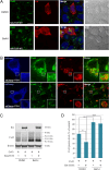
Effect of ectopic expression of ITCH on the level of Cx43-based gap junctions and the cellular Cx43 protein level. (A) HeLa-Cx43 cells were transfected with HA-ITCH-WT or HA-ITCH-C830A for 48 h, as indicated. The cells were fixed and stained with anti-Cx43 (green) and anti-HA (grey) antibodies followed by Alexa488- and Alexa647-conjugated secondary antibodies. The plasma membrane was stained with Alexa555-conjugated WGA (red). Nuclei were stained with Hoechst 33342 (blue). The cells were visualized by confocal fluorescence microscopy. Scale bar, 10 μm, applies for all images. Arrows indicate examples of Cx43-based gap junctions formed between adjacent cells that do not express HA-ITCH-WT or HA-ITCH-C830A. Paired arrows indicate an example of a region of the plasma membrane between two adjacent cells that both express HA-ITCH-WT, where Cx43-based gap junctions are not formed. Arrowheads indicate examples of regions of the plasma membrane between two adjacent cells, one of which express HA-ITCH-WT and the other not, where Cx43-based gap junctions are not formed. Two-headed arrows indicate examples of Cx43-based gap junctions formed in the plasma membrane of cells expressing HA-ITCH-C830A. Double arrowhead indicate an example of Cx43 staining in vesicular structures localized in the perinuclear area that does not appear to be affected by ectopic overexpression of HA-ITCH-WT. (B) Quantification of the area of Cx43-based gap junctions per cell based on confocal fluorescence microscopy images in A. Values shown are the means ± S.E.M. of three independent experiments. *p < 0.0005. (C) HeLa cells negative for Cx43 were co-transfected with Cx43 and empty vector, Cx43 and HA-ITCH-WT, or Cx43 and HA-ITCH-C830A, as indicated, for 48 h. Cell lysates were then prepared and equal amounts of total cell protein were subjected to SDS-PAGE. Cx43 and β-actin were detected by western blotting. Molecular mass in kDa is indicated. (D) The intensities of the Cx43 bands on western blots shown in C were quantified and normalized to the level of β-actin. Values shown are the means ± S.E.M. of three independent experiments. *p < 0.05
Fig. 5
Fig. 5
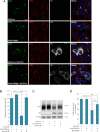
Effect of bafilomycin A1 on the ITCH-induced loss of Cx43 protein. (A) C33A cells were transfected with HA-ITCH-WT for 48 h. The cells were treated with DMSO (vector) or bafilomycin A1 (BafA1; 200 nM) for the last 18 h of the transfection. The cells were fixed and stained with anti-Cx43 (green) and anti-HA (red) antibodies followed by Alexa488- and Alexa594-conjugated secondary antibodies. Nuclei were stained with Hoechst 33342 (blue). The cells were visualized by confocal fluorescence microscopy. Scale bars, 10 μm. (B) C33A cells were transfected with mCherry-ITCH-WT for 48 h. The cells were treated with bafilomycin A1 for 18 h (upper panel) or 5 h (lower panel). Cx43 (green) and LAMP1 (red) were visualized by confocal fluorescence microscopy, as indicated. Scale bars, 10 μm. LAMP1 was stained by using Alexa405-conjugated secondary antibodies, and, following image capture, the LAMP1 signal was converted to red for better visualization of possible colocalization between LAMP1 and Cx43, and the mCherry-ITCH signal was converted to blue. (C) HeLa cells negative for Cx43 were co-transfected with Cx43 and empty vector or with Cx43 and HA-ITCH-WT, as indicated, for 48 h. For the last 18 h of the transfection, the cells were treated with DMSO (vector) or bafilomycin A1 (BafA1; 200 nM). Cell lysates were then prepared and equal amounts of total cell protein were subjected to SDS-PAGE. Cx43 and β-actin were detected by western blotting. Molecular mass in kDa is indicated. (D) The intensities of the Cx43 bands on western blots shown in C were quantified and normalized to the level of β-actin. Values shown are the means ± S.E.M. of four independent experiments. *p < 0.05, **p < 0.005, ***p < 0.001
Fig. 6
Fig. 6
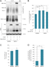
Effect of mutating the PY motif on the interaction between ITCH and Cx43 and on Cx43 ubiquitination. (A) HeLa cells negative for Cx43 were co-transfected with with either Cx43-WT or Cx43-P283A and HA-ITCH-WT or empty plasmid, as indicated, for 48 h. Cell lysates were then subjected to immunoprecipitation with anti-Cx43 antibodies or with rabbit IgG as control, and equal amounts of immunoprecipitates were subjected to SDS-PAGE. HA-ITCH-WT in the immunoprecipitates was detected with western blotting by using anti-HA antibodies and ubiquitinated Cx43 was detected by using anti-ubiquitin antibodies, as indicated (two upper panels, respectively). The blot was stripped and reprobed with anti-Cx43 antibodies, as indicated (lower panel). Also shown is the relative expression of Cx43, HA-ITCH-WT, and β-actin in cell lysates prior to immunoprecipitation (Input). Molecular mass in kDa is indicated. (B) Quantification of the relative level of HA-ITCH that co-immunoprecipitates with Cx43-WT and Cx43-P283A based on the data obtained in A. For each lane, the level of HA-ITCH in the immunoprecipitate was normalized to the level of Cx43 in the immunoprecipitate. Values shown are the means ± S.E.M. of four independent experiments. *p < 0.05. (C) Quantification of the relative level of Cx43 ubiquitination in the various samples based on the data obtained in A. For each lane, the level of ubiquitin immunoreactivity in the immunoprecipitate was normalized to the level of Cx43 in the immunoprecipitate. Values shown are the means ± S.E.M. of four independent experiments. *p < 0.05, **p < 0.005, ***p < 0.001
Fig. 7
Fig. 7
Effect of mutating the PY motif on the ability of ITCH to promote loss of gap junctions and degradation of Cx43. HeLa cells negative for Cx43 were co-transfected with either Cx43-WT or Cx43-P283A and empty plasmid, Cx43-WT and HA-ITCH, or Cx43-P283A and HA-ITCH, as indicated, for 48 h. (A) The cells were fixed and stained with anti-Cx43 (green) and anti-HA (grey) antibodies followed by Alexa488- and Alexa647-conjugated secondary antibodies. The plasma membrane was stained with Alexa555-conjugated WGA (red). Nuclei were stained with Hoechst 33342 (blue). The cells were visualized by confocal fluorescence microscopy. Scale bars, 10 μm. Arrows indicate examples of Cx43-based gap junctions formed between adjacent cells expressing only Cx43-WT or Cx43-P283A and not HA-ITCH-WT. Arrowheads indicate examples of regions of the plasma membrane of cells co-expressing Cx43-WT and HA-ITCH-WT, where Cx43-based gap junctions are not formed. Two-headed arrows indicate examples of Cx43-based gap junctions formed in the plasma membrane of cells co-expressing Cx43-P283A and HA-ITCH-WT. (B) Quantification of the area of Cx43-based gap junctions per cell based on confocal fluorescence microscopy images in A. Values shown are the means ± S.E.M. of three independent experiments. *p < 0.001. n.s., not significant. (C) Cell lysates were prepared and equal amounts of total cell protein were subjected to SDS-PAGE. Cx43 and β-actin were detected by western blotting. Molecular mass in kDa is indicated. (D) The intensities of the Cx43 bands on western blots shown in C were quantified and normalized to the level of β-actin. Values shown are the means ± S.E.M. of six independent experiments. ***p < 0.001
Fig. 8
Fig. 8
Effect of bafilomycin A1 on the interaction between ITCH and Cx43. (A) HeLa cells negative for Cx43 were co-transfected with Cx43 and empty vector or with Cx43 and HA-ITCH, as indicated, for 48 h. For the last 18 h of the transfection, the cells were treated with DMSO (vector) or bafilomycin A1 (BafA1; 200 nM), as indicated. Cell lysates were then subjected to immunoprecipitation with anti-Cx43 antibodies or with rabbit IgG as negative control, and equal amounts of the immunoprecipitates were subjected to SDS-PAGE. HA-ITCH in the immunoprecipitates was detected with western blotting by using anti-HA antibodies and ubiquitinated Cx43 was detected by using anti-ubiquitin antibodies, as indicated (two upper panels, respectively). The blot was stripped and reprobed with anti-Cx43 antibodies, as indicated (lower panel). Also shown is the relative expression of Cx43, HA-ITCH, and β-actin in cell lysates prior to immunoprecipitation (Input). Molecular mass in kDa is indicated. (B) Quantification of the relative level of Cx43 ubiquitination in the various samples based on the data obtained in A. For each lane, the level of ubiquitin immunoreactivity in the immunoprecipitate was normalized to the level of Cx43 in the immunoprecipitate. Values shown are the means ± S.E.M. of five independent experiments. n.s., not significant. (C) Quantification of the relative level of HA-ITCH in the Input samples shown in A. For each lane, the level of HA-ITCH in the lysates was normalized to the level of β-actin. Values shown are the means ± S.E.M. of five independent experiments. n.s., not significant. (D) Quantification of the relative level of HA-ITCH that co-immunoprecipitated with Cx43 based on the data obtained in A. For each lane, the level of HA-ITCH in the immunoprecipitate was normalized to the level of Cx43 in the immunoprecipitate. Values shown are the means ± S.E.M. of five independent experiments. *p < 0.05

Similar articles

Cited by

References

    1. Beyer EC, Berthoud VM. Gap junction gene and protein families: connexins, innexins, and pannexins. Biochim Biophys Acta. 2018;1860:5–8. doi: 10.1016/j.bbamem.2017.05.016. - DOI - PMC - PubMed
    1. Goodenough DA, Paul DL. Gap junctions. Cold Spring Harb Perspect Biol. 2009;1:a002576. doi: 10.1101/cshperspect.a002576. - DOI - PMC - PubMed
    1. Aasen T, Leithe E, Graham SV, Kameritsch P, Mayan MD, Mesnil M, Pogoda K, Tabernero A. Connexins in cancer: bridging the gap to the clinic. Oncogene. 2019;38:4429–4451. doi: 10.1038/s41388-019-0741-6. - DOI - PMC - PubMed
    1. Mesnil M, Aasen T, Boucher J, Chepied A, Cronier L, Defamie N, Kameritsch P, Laird DW, Lampe PD, Lathia JD, Leithe E, Mehta PP, Monvoisin A, Pogoda K, Sin WC, Tabernero A, Yamasaki H, Yeh ES, Dagli MLZ, Naus CC. An update on minding the gap in cancer. Biochim Biophys Acta. 2018;1860:237–243. doi: 10.1016/j.bbamem.2017.06.015. - DOI - PubMed
    1. Sinyuk M, Mulkearns-Hubert EE, Reizes O, Lathia J. Cancer connectors: Connexins, Gap junctions, and communication. Front Oncol. 2018;8:646. doi: 10.3389/fonc.2018.00646. - DOI - PMC - PubMed