. 2024 Jun 1;35(6):ar76.
doi: 10.1091/mbc.E24-01-0043. Epub 2024 Apr 10.

N-terminal signals in the SNX-BAR paralogs Vps5 and Vin1 guide endosomal coat complex formation

Affiliations

N-terminal signals in the SNX-BAR paralogs Vps5 and Vin1 guide endosomal coat complex formation

Shawn P Shortill et al. Mol Biol Cell. .

Abstract

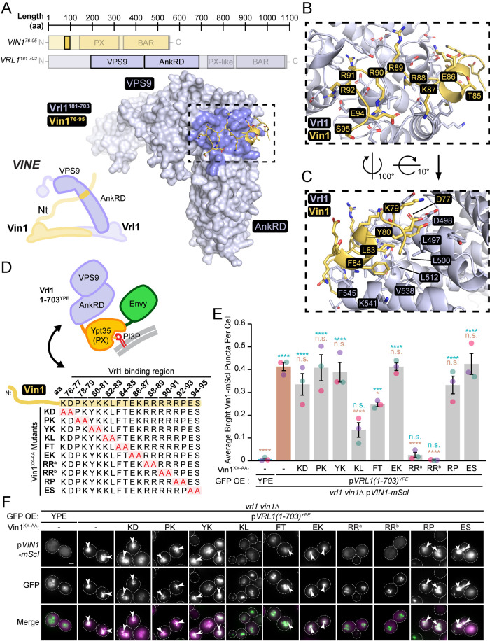
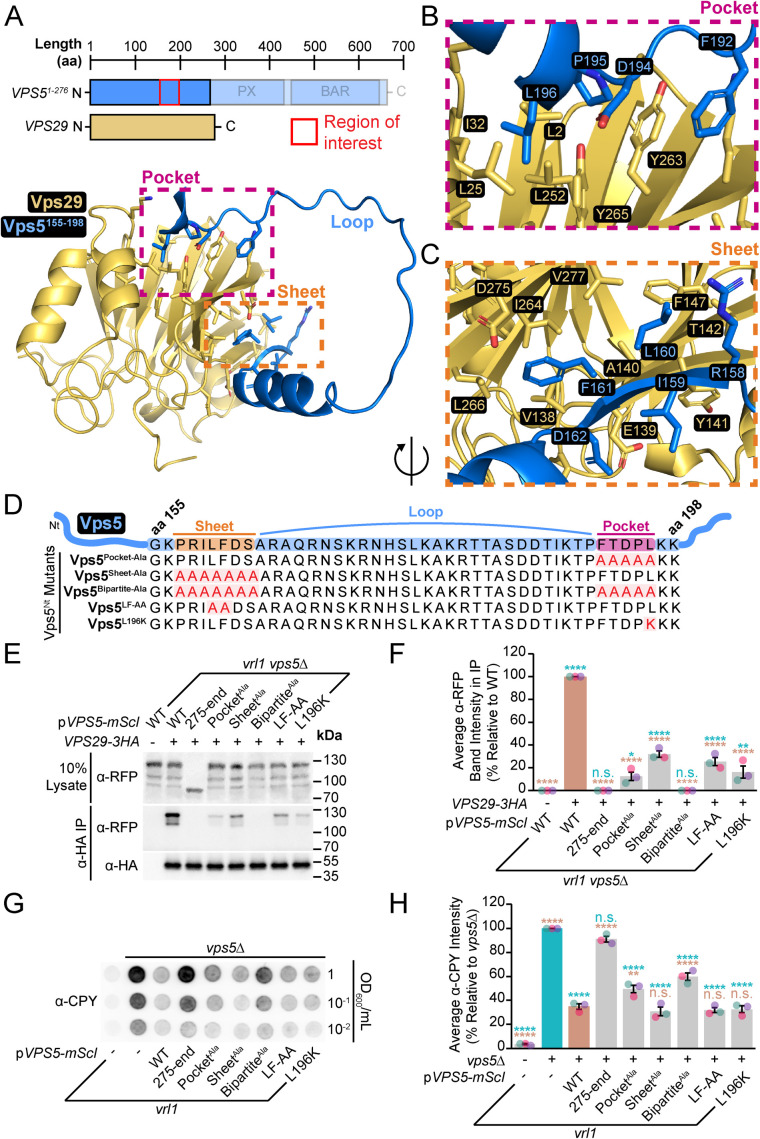
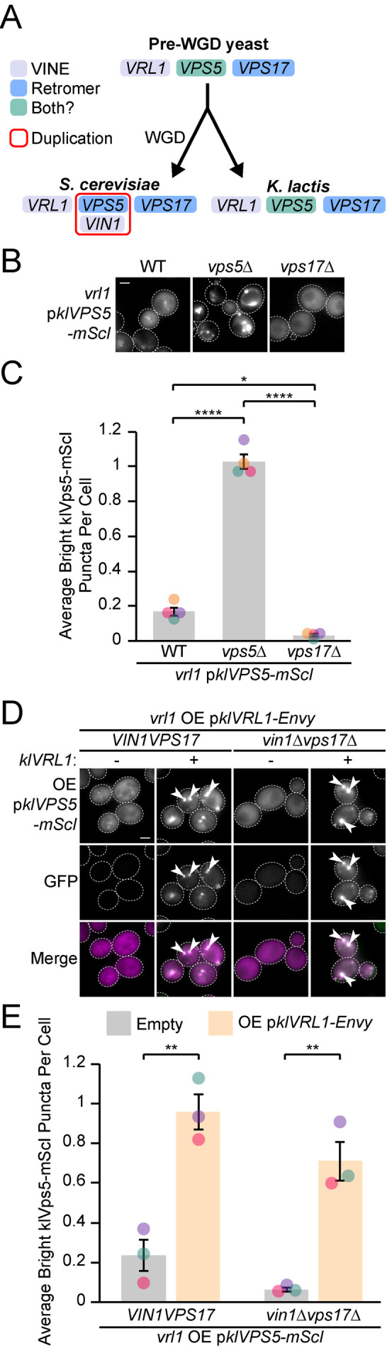
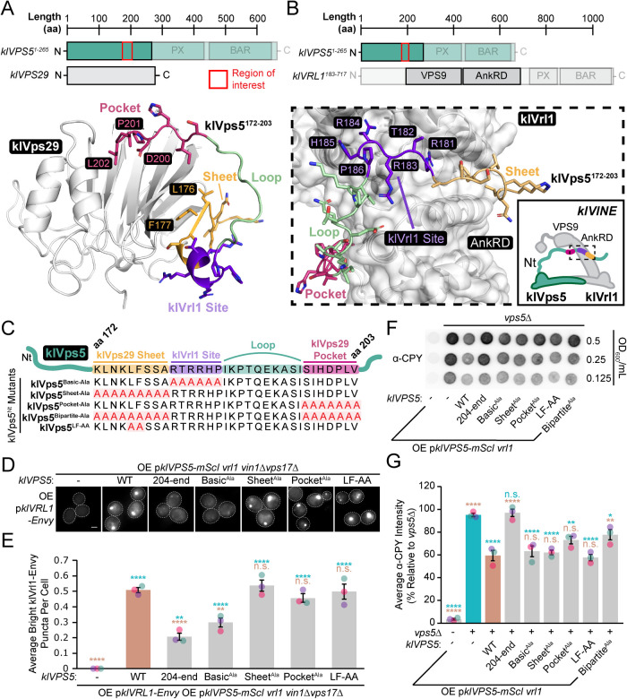
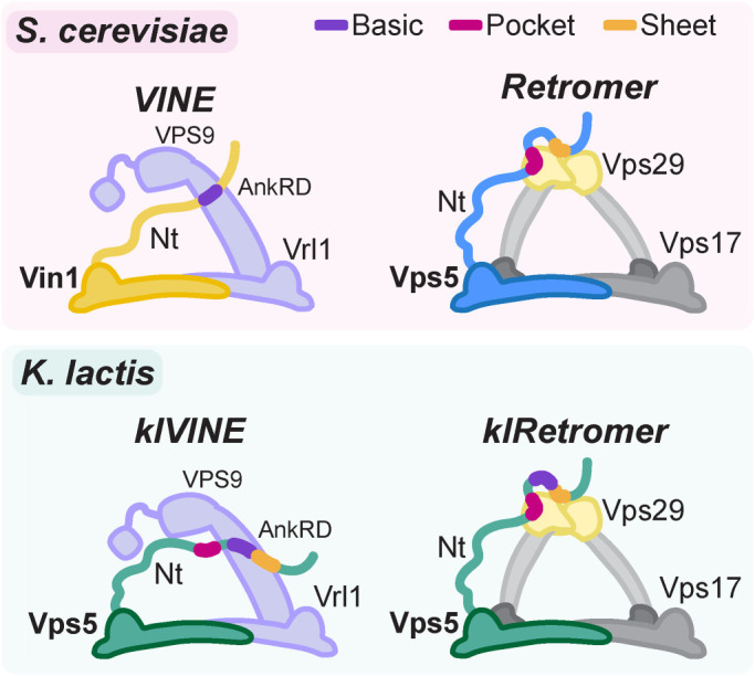
Endosomal coats incorporate membrane-binding subunits such as sorting nexin (SNX) proteins. The Saccharomyces cerevisiae SNX-BAR paralogs Vin1 and Vps5 are respective subunits of the endosomal VINE and retromer complexes whose dimerizing BAR domains are required for complex assembly and membrane association. However, a degree of promiscuity is predicted for yeast BAR-BAR pairings, and recent work has implicated the unstructured N-terminal domains of Vin1 and Vps5 in coat formation. Here, we map N-terminal signals in both SNX-BAR paralogs that contribute to the assembly and function of two distinct endosomal coats in vivo. Whereas Vin1 leverages a polybasic region and adjacent hydrophobic motif to bind Vrl1 and form VINE, the N-terminus of Vps5 interacts with the retromer subunit Vps29 at two sites, including a conserved hydrophobic pocket in Vps29 that engages other accessory proteins in humans. We also examined the sole isoform of Vps5 from the milk yeast Kluyveromyces lactis and found that ancestral yeasts may have used a nested N-terminal signal to form both VINE and retromer. Our results suggest that the specific assembly of Vps5-family SNX-BAR coats depends on inputs from unique N-terminal sequence features in addition to BAR domain coupling, expanding our understanding of endosomal coat biology.


Figures

FIGURE 1:
FIGURE 1:
Mapping features in the Vin1 N-terminus that are required for recognition by Vrl1. (A) AlphaFold2-predicted interaction between Vrl1181-703 and the Vrl1-binding portion of the Vin1 N-terminus (Vin176-95). Vrl1 residues predicted to be within four angstroms of Vin1 are shown in darker color. (B) Basic residues within Vin176-95 are predicted to associate with Vrl1. (C) Nonpolar residues in Vin176-95 are predicted to associate at an adjacent site on the Vrl1 AnkRD. (D) Diagram of assay used to test recruitment by chimeric Vrl1 construct (top) of the indicated pairwise alanine substitution mutants of the Vin1 N-terminus (bottom). (E) Quantification of RFP puncta per cell for Vin1 mutants. One-way ANOVA with Dunnett’s multiple comparison tests; n = 3, cells/strain/replicate ≥ 878; not significant, n.s. = p > 0.05, * = p < 0.05, *** = p < 0.001, **** = p < 0.0001. Blue statistical significance labels correspond to a Dunnett-corrected ANOVA performed against YPE-containing bait while brown labels correspond to a Dunnett-corrected ANOVA performed against wild-type Vin1. (F) Differential effects of pairwise alanine substitution in the Vin1 N-terminus on recruitment by the AnkRD-containing Vrl1(1-703)YPE chimera. Representative images quantified in E. Scale bars, 2 µm. Error bars report SEM. aa, amino acids. OE, over-expressed. Nt, N-terminus. mScI, mScarletI. RRa, R88A R89A. RRb, R90A R91A. YPE, Ypt35(PX)-Envy.
FIGURE 2:
FIGURE 2:
An N-terminal Vin1 Leu-Phe motif is important for recognition by Vrl1 and formation of VINE. (A) Pairwise alanine substitution mutants of Vin1 KL, LF and FT N-terminal residue pairs tested in the chimeric Vrl1 recruitment assay. (B) The Vin1 N-terminal LF-AA mutant displays a severe defect in recruitment by the AnkRD-containing Vrl1(1-703)YPE chimera. (C) Quantification of RFP puncta per cell in B. One-way ANOVA with Dunnett’s multiple comparison tests; n = 3, cells/strain/replicate ≥ 676; not significant, n.s. = p > 0.05, * = p < 0.05, ** = p < 0.01, *** = p < 0.001, **** = p < 0.0001. Blue statistical significance labels correspond to a Dunnett-corrected ANOVA performed against YPE-containing bait while brown labels correspond to a Dunnett-corrected ANOVA performed against wild-type Vin1. (D) Addition of a charged residue to the Vrl1 hydrophobic patch predicted to interface with the Vin1 N-terminal Leu-Phe motif disrupts recruitment of the Vin1 N-terminus by the AnkRD-containing Vrl1(1-703)YPE chimera. (E) Quantification of RFP puncta per cell in D. One-way ANOVA with Dunnett’s multiple comparison tests; n = 3, cells/strain/replicate ≥ 863; ** = p < 0.01, **** = p < 0.0001. Blue statistical significance labels correspond to a Dunnett-corrected ANOVA performed against YPE-containing bait while brown labels correspond to a Dunnett-corrected ANOVA performed against wild-type Vrl1. (F) Pairwise substitution of Vin1-mScI L83A F84A or R90A R91A inhibits formation of Vrl1-Envy puncta in vin1∆ cells. (G) Quantification of GFP puncta per cell in F. One-way ANOVA with Dunnett’s multiple comparison tests; n = 3, cells/strain/replicate ≥ 1108; not significant, n.s. = p > 0.05, * = p < 0.05, **** = p < 0.0001. Blue statistical significance labels correspond to a Dunnett-corrected ANOVA performed against the empty vector vin1∆ strain while brown labels correspond to a Dunnett-corrected ANOVA performed against wild-type Vin1-mScI. Scale bars, 2 µm. Error bars report SEM. aa, amino acid. OE, over-expressed. Nt, N-terminus. mScI, mScarletI. RRb, R90A R91A. YPE, Ypt35(PX)-Envy.
FIGURE 3:
FIGURE 3:
Vps29 binds a bipartite signal in the Vps5 N-terminus. (A) AlphaFold2-predicted interaction of the Vps5 N-terminus (Vps51-276) with Vps29 highlights a bipartite motif between Vps5 aa 155–198. (B) A Vps5 N-terminal FTDPL motif is predicted to interact with Vps29 at a conserved hydrophobic pocket. (C) A Vps5 N-terminal PRILFDS motif is predicted to form a β-strand and extend a Vps29 β-sheet. (D) Diagram of Vps5 substitution mutations in the predicted N-terminal pocket- and sheet-binding sites generated to test retromer assembly and function. (E) Mutagenesis of the Vps5-mScI N-terminus disrupts coIP with Vps29-3HA. Disruption of either the pocket- or sheet-binding site separately causes a strong binding defect, while combined mutation of both (bipartite-ala) completely blocks binding. (F) Quantification of copurified Vps5-mScI levels in E by densitometry. One-way ANOVA with Dunnett’s multiple comparison tests; n = 3; not significant, n.s. = p > 0.05, * = p < 0.05, ** = p < 0.01, **** = p < 0.0001. Blue statistical significance labels correspond to a Dunnett-corrected ANOVA performed against untagged Vps29 while brown labels correspond to a Dunnett-corrected ANOVA performed against 3HA-tagged Vps29 with wild-type Vps5-mScI. (G) Disruption of the Vps5 N-terminal pocket- or sheet-binding sites cause CPY secretion. Paired disruption of the pocket- and sheet-binding sites (bipartite-ala) caused the most severe secretion phenotype. (H) Quantification of secreted CPY by densitometry and normalized to measured secreted CPY from the shown vps5∆ strain. One-way ANOVA with Dunnett’s multiple comparison tests; n = 3; not significant, n.s. = p > 0.05, ** = p < 0.01, **** = p < 0.0001. Blue statistical significance labels correspond to a Dunnett-corrected ANOVA performed against a vps5∆ strain while brown labels correspond to a Dunnett-corrected ANOVA performed against a vps5∆ strain with plasmid-expressed wild-type Vps5-mScI. Error bars report SEM. aa, amino acid. mScI, mScarletI. Nt, N-terminus.
FIGURE 4:
FIGURE 4:
The single K. lactis Vps5 isoform forms both retromer and VINE. (A) Diagram of Vps5 duplication status in S. cerevisiae and K. lactis resulting from an ancient whole genomic duplication event. The single copy of Vps5 in K. lactis may bind both Vrl1 and Vps17 to form VINE and retromer, respectively. (B) Ectopically expressed K. lactis Vps5 (klVps5-mScI) localizes to puncta in S. cerevisiae. Localization depends on Vps17 and is enhanced in a vps5∆ strain. (C) Quantification of RFP puncta per cell in B. One-way ANOVA with Tukey’s multiple comparison tests; n = 4, cells/strain/replicate ≥ 1544; * = p < 0.05, **** = p < 0.0001. (D) Expression of K. lactis Vrl1 (klVrl1-Envy) from the RPL18B promoter enhances klVps5-mScI localization in wild-type cells and induces Vps17-independent localization. (E) Quantification of RFP puncta per cell in D. Two tailed equal variance t tests; n = 3, cells/strain/replicate ≥ 144; ** = p < 0.01. Scale bars, 2 µm. mScI, mScarletI. OE, overexpressed. mScI, mScarletI. WGD.
FIGURE 5:
FIGURE 5:
Nested motifs in the klVps5 N-terminus guide selection for retromer and VINE. (A) AlphaFold2-predicted interaction between K. lactis (kl) Vps29 (white) and the klVps5 N-terminus (klVps51-265). klVps5 is predicted to bind klVps29 through a bipartite motif between N-terminal residues aa 172–203 that involves a hydrophobic pocket site and a β-sheet site. (B) AlphaFold2-predicted interaction between klVrl1183-717 (white) and the klVps5 N-terminus (klVps51-265). klVps5 is predicted to interact with klVrl1 in a similar manner to Vin1 and Vrl1 in S. cerevisiae–through a basic region involving the residues RTRRHP. (C) Diagram of klVps5 substitution mutations in the predicted N-terminal klVrl1-binding and klVps29 pocket- and sheet-binding sites generated to test VINE and retromer assembly. (D) RPL18Bpr-driven expression of klVps5-mScI N-terminal mutants has differential effects on klVrl1-Envy localization in vin1vps17∆ cells. Whereas deletion of the N-terminus (klVps5204-end) and alanine substitution of the polybasic predicted klVrl1-binding site causes loss of klVrl1 localization, disruption of either predicted klVps29 pocket- or sheet-binding sites have no effect. (E) Quantification of GFP puncta per cell in D. One-way ANOVA with Dunnett’s multiple comparison tests; n = 3, cells/strain/replicate ≥ 515; not significant, n.s. = p > 0.05, ** = p < 0.01, **** = p < 0.0001. Blue statistical significance labels correspond to a Dunnett-corrected ANOVA performed on the empty vector vin1vps17∆ strain while brown labels correspond to a Dunnett-corrected ANOVA performed against wild-type klVps5-mScI. (F) Disruption of the klVps5 N-terminus either by complete deletion or alanine substitution of the pocket- and sheet-binding sites causes CPY secretion. Alanine substitution of the predicted klVrl1-binding polybasic site in the klVps5 N-terminus does not create a significant CPY secretion phenotype. (G) Quantification of secreted CPY by densitometry in F. Normalized to measured secreted CPY from the vps5∆ strain. One-way ANOVA with Dunnett’s multiple comparison tests; n = 3; not significant, n.s. = p > 0.05, * = p < 0.05, ** = p < 0.01, **** = p < 0.0001. Blue statistical significance labels correspond to a Dunnett-corrected ANOVA performed against a vps5∆ strain while brown labels correspond to a Dunnett-corrected ANOVA performed against a vps5∆ strain with plasmid-expressed wild-type klVps5-mScI. Error bars report SEM. aa, amino acid. mScI, mScarletI. Nt, N-terminus. OE, overexpressed.
FIGURE 6:
FIGURE 6:
Vps5-family SNX-BAR coat assembly in S. cerevisiae and K. lactis. Model for VINE and retromer assembly with N-terminal binding sites highlighted for Vin1 and Vps5, respectively. A polybasic region unique to Vin1 drives interaction with the Vrl1 AnkRD, while a bipartite motif in the Vps5 N-terminus associates at two distinct sites in Vps29–a conserved hydrophobic pocket and a β-sheet on the opposite surface of the protein. The sole Vps5 isoform in K. lactis possesses all three of these N-terminal motifs and can form both VINE and retromer. Nt, N-terminus.

Similar articles

Cited by

References

    1. Arighi CN, Harmell LM, Aguilar RC, Haft CR, Bonifacino JS (2004). Role of the mammalian retromer in sorting of the cation-independent mannose 6-phosphate receptor. J Cell Biol 165, 123–133. - PMC - PubMed
    1. Baños-Mateos S, Rojas AL, Hierro A (2019). VPS29, a tweak tool of endosomal recycling. Curr Opin Cell Biol 59, 81–87. 10.1016/j.ceb.2019.03.010 - DOI - PubMed
    1. Bean BDM, Davey M, Conibear E (2017). Cargo selectivity of yeast sorting nexins. Traffic 18, 110–122. - PubMed
    1. Burd C, Cullen PJ (2014). Retromer: A master conductor of endosome sorting. Cold Spring Harb Perspect Biol 6, a016774. 10.1101/cshperspect.a016774 - DOI - PMC - PubMed
    1. Byrne K, Wolfe K (2005). The yeast gene order browser: Combining curated homology and syntenic context reveals gene fate in polyploid species. Genome Res 15, 1456–1461. - PMC - PubMed

Publication types

MeSH terms

Substances

LinkOut - more resources