. 2024 Apr 25;67(8):6610-6623.
doi: 10.1021/acs.jmedchem.4c00037. Epub 2024 Apr 10.

Potentiating Activity of GmhA Inhibitors on Gram-Negative Bacteria

Affiliations

Potentiating Activity of GmhA Inhibitors on Gram-Negative Bacteria

François Moreau et al. J Med Chem. .

Abstract

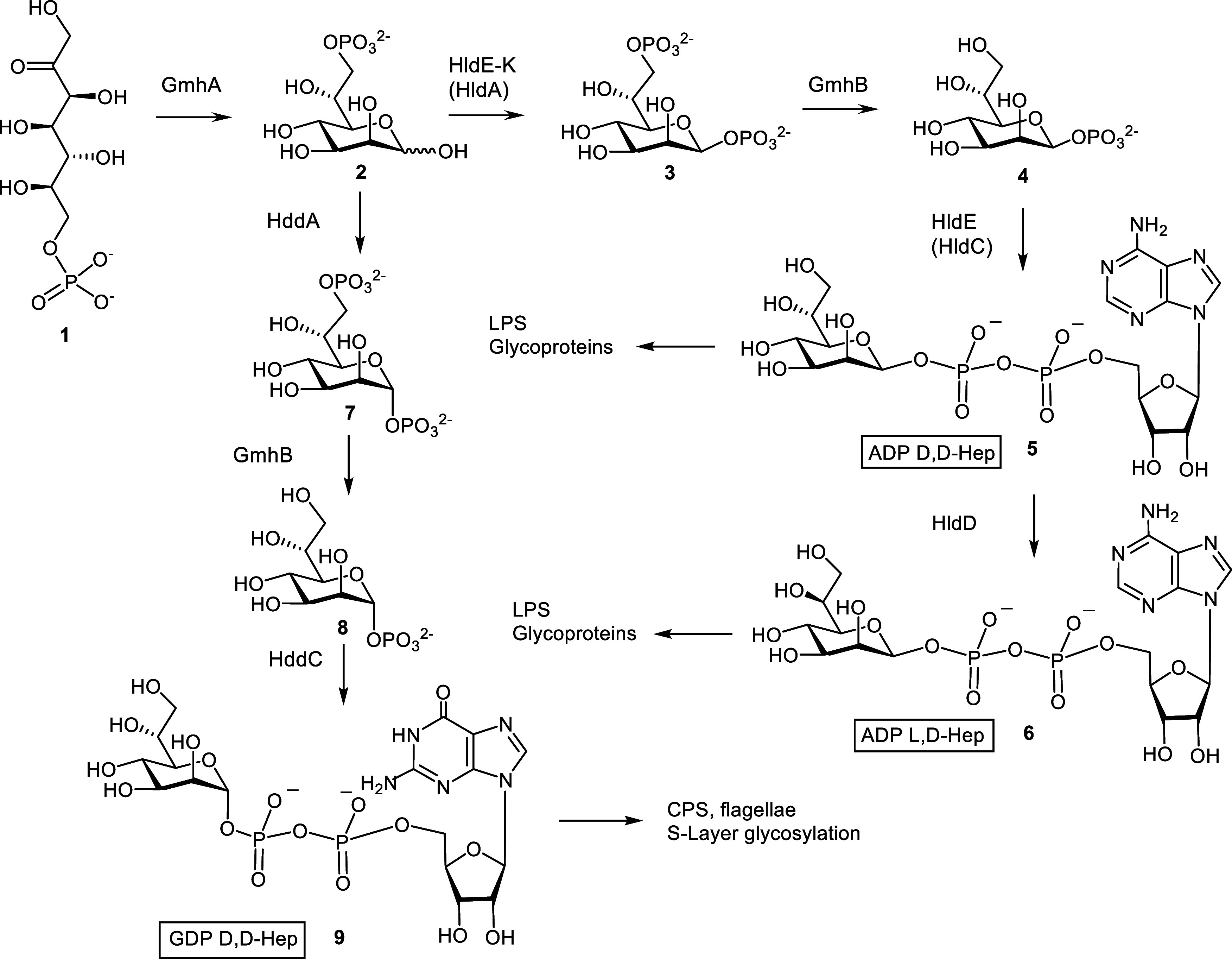
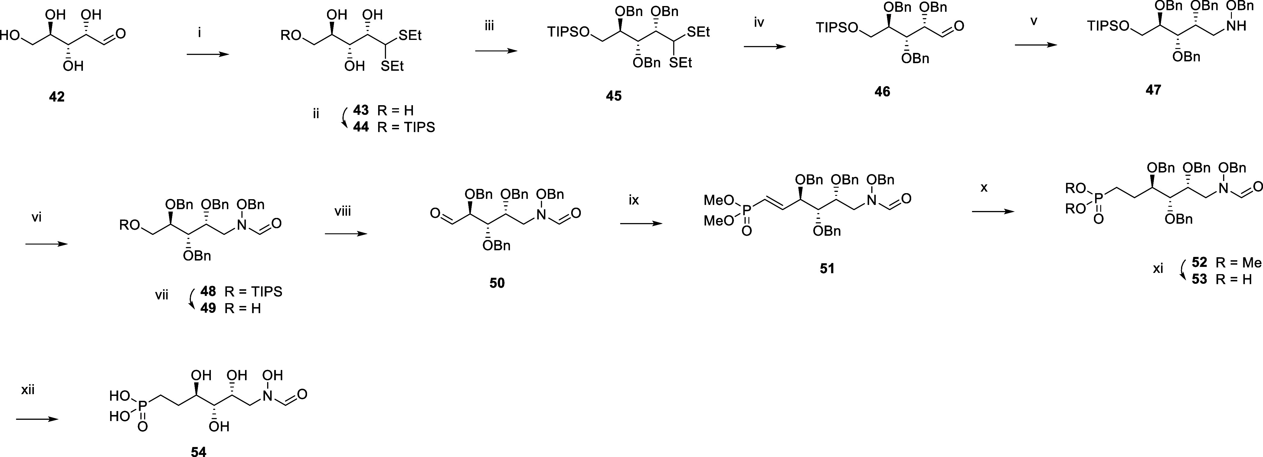
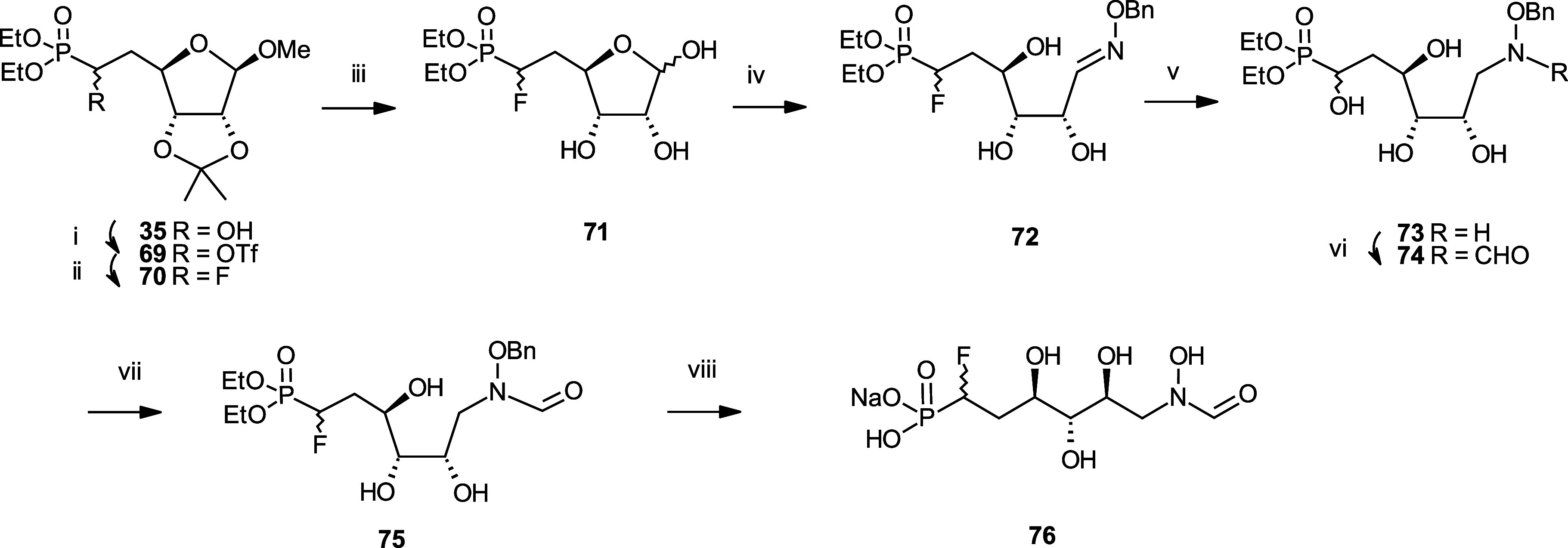
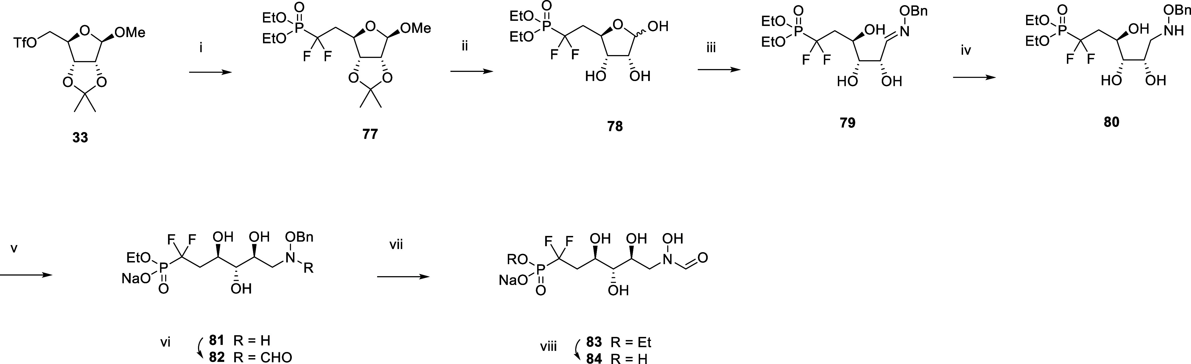
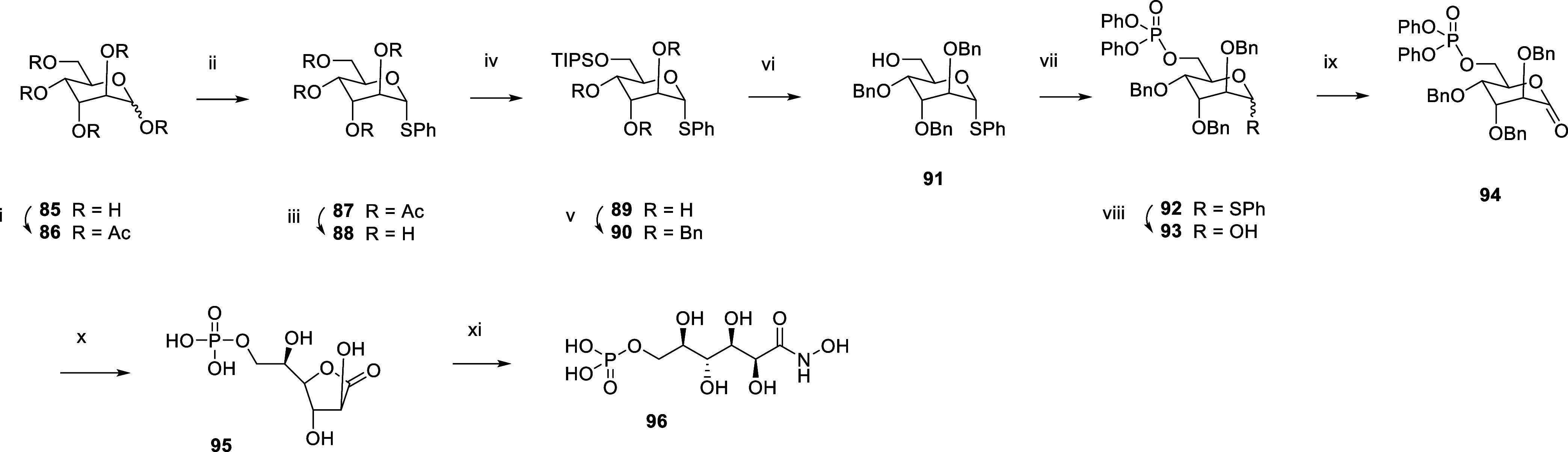
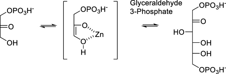
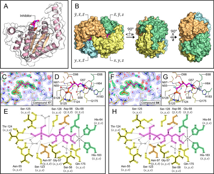
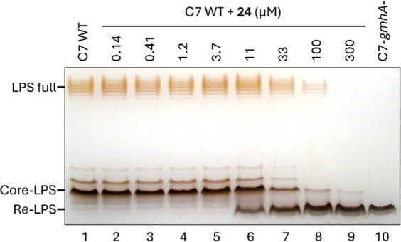
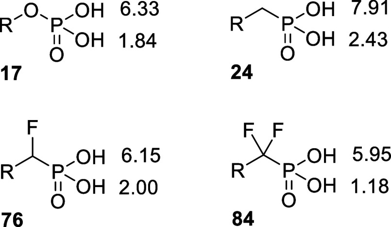
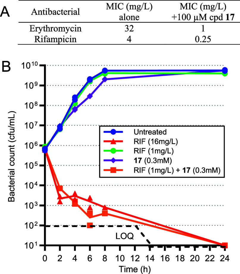
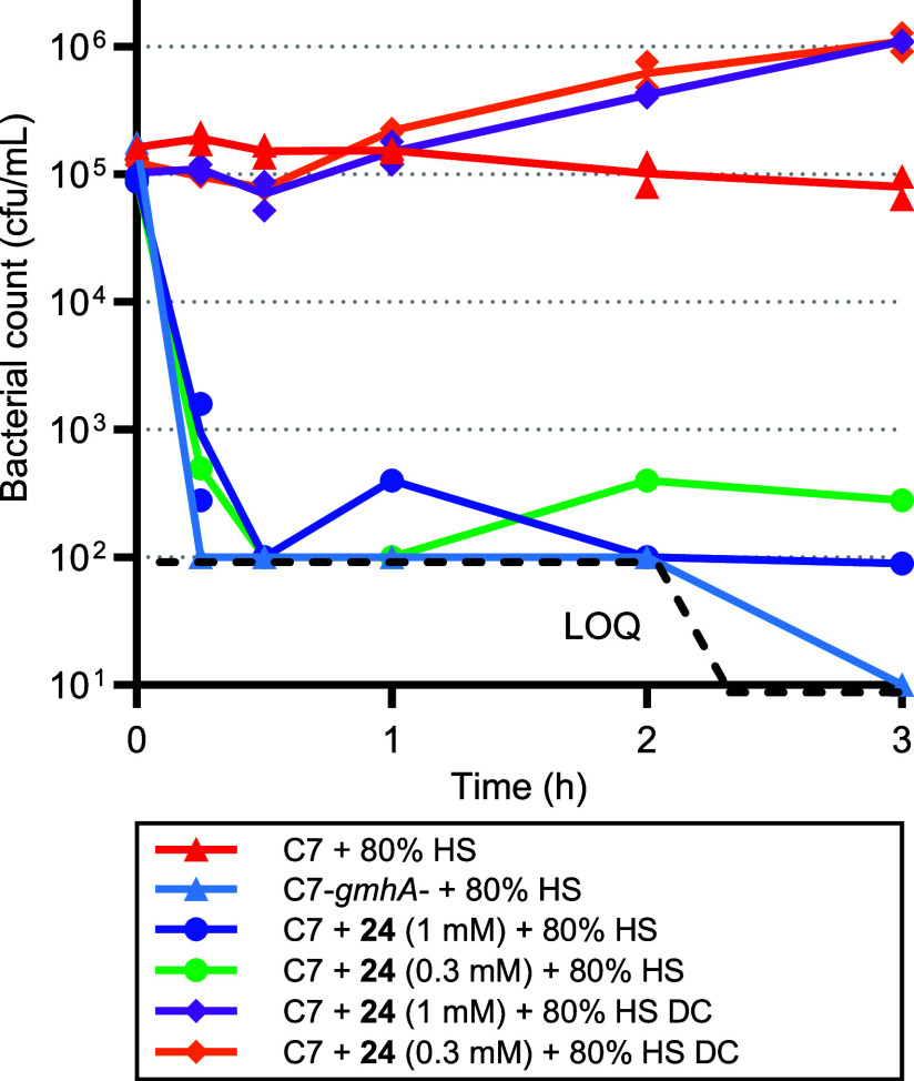
Inhibition of the biosynthesis of bacterial heptoses opens novel perspectives for antimicrobial therapies. The enzyme GmhA responsible for the first committed biosynthetic step catalyzes the conversion of sedoheptulose 7-phosphate into d-glycero-d-manno-heptose 7-phosphate and harbors a Zn2+ ion in the active site. A series of phosphoryl- and phosphonyl-substituted derivatives featuring a hydroxamate moiety were designed and prepared from suitably protected ribose or hexose derivatives. High-resolution crystal structures of GmhA complexed to two N-formyl hydroxamate inhibitors confirmed the binding interactions to a central Zn2+ ion coordination site. Some of these compounds were found to be nanomolar inhibitors of GmhA. While devoid of HepG2 cytotoxicity and antibacterial activity of their own, they demonstrated in vitro lipopolysaccharide heptosylation inhibition in Enterobacteriaceae as well as the potentiation of erythromycin and rifampicin in a wild-type Escherichia coli strain. These inhibitors pave the way for a novel treatment of Gram-negative infections.


Conflict of interest statement

The authors declare the following competing financial interest(s): When this study was performed F.M., D.A., S.F., D.B., and V.G. were Mutabilis full-time employees; M.Bl., M.Ba., M.H., and N.M.X. were full-time employees of the University of Natural Resources and Life Sciences, Vienna, Austria (funded by MUTABILIS, project no. 8389); J.M., E.A., and V.H. were Activation full-time employees, and F.L. was a full-time employee of Carbosynth.

Figures

Scheme 1
Scheme 1. Biosynthetic Pathways toward Nucleotide Activated Heptoses That Would Be Affected by Inhibiting the GmhA Reaction
Scheme 2
Scheme 2. Synthesis of the N-formyl-N-hydroxy Inhibitors 17 and 20
Reagents and Conditions: (i) TIPSCl, DMAP, pyr, 93%; (ii) NBS, aq. Me2CO, 97%; (iii) 1. H2NOBn, pyr., MeOH 2. NaCNBH3, AcOH, 75% over two steps; (iv) CDI, HCOOH, THF, 0 °C, 94%; (v) TBAF, THF, 87%; (vi) 1. 1H-tetrazole, phosphoramidite, rt. 2. mCPBA, TEA, −78 °C, 93%; (vii) Pd/C, H2, aq. THF-HOAc rt, 30%; (viii) I2, Imidazole, P(Ph)3, 66%; (ix) P(OEt)3, 44%; (x) 1. TMSBr, pyr. 2. Pd(OH)2/C, H2, aq. THF, 1% AcOH, 23%.
Scheme 3
Scheme 3. Synthesis of the N-formyl-N-hydroxy Inhibitors 24, 25, 27, and 31
Reagents and Conditions: (i) Dess-Martin ox, 100%; (ii) 22, BuLi, THF, 76%; (iii) Pd(OH)2, aq. THF, MeOH, 16%; (iv) SnCl4, 54%; (v) NMO, K2OsO4, 79%; (vi) Pd(OH)2, aq. THF, 24%; (vii) 28, BuLi, THF, −78 °C, 22%; (viii) CDI, 73%; (ix) Pd(OH)2, aq. THF, 9%.
Scheme 4
Scheme 4. Synthesis of the N-formyl-N-hydroxy Inhibitor 41
Reagents and Conditions: (i) Tf2O, DCM, −10 °C, 86%; (ii) Diethyl [(benzyloxy)methyl]Phosphonate, nBuLi, diisopropylamine, THF, −78 °C, 61%; (iii) H2, 10 bar, Pd/C, EtOH, 50 °C, 95%; (iv) Amberlyst 15, water, 60 °C; (v) Benzylhydroxylamine, NaHCO3, MeOH, water, 60 °C, 56% over two steps; (vi) 1. BH3.THF, THF 0 °C. 2. MeOH, 60 °C, 78%; (vii) Trifluoroethyl formate, THF, 60 °C, 65%; (viii) H2, 1 bar, Pd/C, MeOH, rt, 97%; (ix) 1. TMSBr, pyr, DCM, 5 °C. 2. NaHCO3, MeOH, water, 45%.
Scheme 5
Scheme 5. Synthesis of the N-formyl-N-hydroxy Inhibitor 54
Reagents and Conditions: (i) EtSH, HCl, 0 °C, 83%; (ii) TIPSCl, DMAP, pyr, rt, 87%; (iii) Benzyl bromide, NaH, DMF, 0 °C to rt, 51%; (iv) NBS, acetone, water, 0 °C; (v) 1. H2NOBn.HCl, pyr, MeOH, 60 °C. 2. NaCNBH3, AcOH, rt, 60% over two steps; (vi) CDI, HCO2H, TEA, THF, 0 °C to rt, 77%; (vii) TBAF, THF, rt, 91%; (viii) DMP, DCM, 0 °C to rt, 48%; (ix) Tetramethyl methylenediphosphonate, NaH, Et2O, 0 °C, 55%; (x) H2, 1 bar, Pt/C, EtOAc, rt, 85%; (xi) TMSBr, pyr, DCM, 0 °C to rt, 78%; (xii) H2, 1 bar, Pd(OH)2, THF, AcOH, water, rt, 7%.
Scheme 6
Scheme 6. Synthesis of the N-formyl-N-hydroxy Inhibitor 68
Reagents and Conditions: (i) NaH, CS2, MeI, imidazole, THF, 0 °C to rt; (ii) Bu3SnH, AIBN, toluene, reflux, 76% over two steps; (iii) aq. H2SO4, MeOH, 0 °C, 60%; (iv) NaIO4, MeOH, rt; (v) Tetramethyl methylenediphosphonate, NaH, Et2O, 0 °C to rt, 60% over two steps; (vi) H2, 1 bar, Pd/C, MeOH, rt; (vii) aq. AcOH, 80 °C, 40% over two steps; (viii) H2NOBn.HCl, pyr, MeOH, 60 °C, 67%; (ix) NaCNBH3, AcOH, 0 °C to rt, 88%; (x) Trifluoroethyl formate, THF, 65 °C, 51%; (xi) H2, 1 bar, Pd(OH)2, THF, AcOH, water, rt; (xii) TMSBr, pyr, DCM, 0 °C, 12% over two steps.
Scheme 7
Scheme 7. Synthesis of the N-formyl-N-hydroxy Inhibitor 76
Reagents and Conditions: (i) Triflic anhydride, pyr, DCM, −10 °C; (ii) CsF, tBuOH, 40 °C, 58% over two steps; (iii) Amberlyst 15, water, 60 °C; (iv) H2NOBn.HCl, NaHCO3, EtOH, water, 60 °C, 81% over two steps; (v) 1. BH3.THF, THF, 0 °C. 2. MeOH, 60 °C, 71%; (vi) Trifluoroethyl formate, THF, 60 °C, 63%; (vii) H2, 1 bar, Pd/C, MeOH, rt, 90%; (viii) TMSBr, pyr, DCM, 0 °C, 40%.
Scheme 8
Scheme 8. Synthesis of the N-formyl-N-hydroxy Inhibitor 84
Reagents and conditions: (i) nBuLi, diisopropylamine, tris(N,N-tetramethylene)phosphoric acid triamine, diethyl(difluoromethyl)phosphonate, THF −78 °C, 18%; (ii) Amberlyst 15, water, 60 °C; (iii) H2NOBn.HCl, NaHCO3, water, EtOH, 60 °C, 73% over two steps; (iv) 1. BH3.THF, THF, 0 °C. 2. MeOH, 60 °C, 71%; (v) aq. NaOH, water, rt, 84%; (vi) Trifluoroethyl formate, THF, rt, 29%; (vii) H2, 1 bar, Pd/C, MeOH, rt; (viii) TMSBr, pyr, DCM, 0 °C, 66% over two steps.
Scheme 9
Scheme 9. Synthesis of the Hydroxamic Acid Inhibitor 96
Reagents and Conditions: (i) Ac2O, pyr, DMAP, rt; (ii) PhSH, BF3. Et2O, DCM, 0 °C to rt, 82% over two steps; (iii) TEA, water, MeOH, rt, 91%; (iv) TIPSCl, DMAP, rt, 89%; (v) BnBr, NaH, DMF, rt, 96%; (vi) TBAF, THF, rt, 64%; (vii) Diphenyl phosphochloridate, TEA, DMAP, DCM, rt, 97%; (viii) NBS, acetone, water, −11 to 0 °C, 98%; (ix) PCC, DCM, rt, 65%; (x) 1. H2, 1 bar, Pd/C, THF, rt. 2. H2, 1 bar, PtO2, THF, rt, 90%; (xi) aq. NH2OH, rt, 88%.
Scheme 10
Scheme 10. Mechanism of Class II Fbas
Figure 1
Figure 1
Crystal structures of GmhA in complex with different orthosteric inhibitors. (A) ASU of a GmhA-inhibitor cocrystal structure. The ASU contains one copy of GmhA and one copy of inhibitor. (B) GmhA tetramer assembled from 4 symmetry related ASUs. Each of the 4 active sites contains residues from 3 separate chains of GmhA. (C–E) Co-crystal structure of GmhA with compound 17 deposited under PDB accession code 8V4J. (F–G) Co-crystal structure of GmhA with compound 84 deposited under PDB accession code 8V2T. (C,F) OMIT map verifying the presence of inhibitor in the crystal structure. Observed electron density is represented by a blue volume, while positive and negative features of the OMIT map are shown on green and red meshes, respectively. (D,G) Hydrogen-bonding and zinc-chelating interactions that stabilize the inhibitors in the GmhA active site. (E,H) 2D representation of the same interaction network. GmhA residues are colored according to the ASU, while the inhibitor molecule is labeled in magenta.
Figure 2
Figure 2
Silver-stained SDS-PAGE of the LPS of E. coli C7 grown with 0 to 300 μM compound 24 (columns 1 to 9) and control heptose-deficient Re-LPS of gmhA-deleted strain (column 10).
Figure 3
Figure 3
Predicted pKa of phosphate 17, phosphonate 24, monofluorophosphonate 76, and difluorophosphonate 84 according to ACD Laboratories V12.5.
Figure 4
Figure 4
Combination of compound 17 with antibacterials. (A): E. coli C7 MIC of Erythromycin and Rifampicin alone or combined with 100 μM 17 (n = 1). (B): Time-kill kinetics of E. coli C7 with Rifampicin (RIF) alone or combined with compound 17. Bacterial enumeration replicates are represented in the plot. LOQ: limit of quantification.
Figure 5
Figure 5
Time-kill kinetics of E. coli C7 wild type or gmhA-deleted strain in the presence of 80% human serum untreated or heated (DC). Bacteria are pregrown in LB broth for 5 h in absence or presence of compound 24 at 0.3 and 1 mM. Bacterial enumeration replicates are represented on the plot. LOQ: limit of quantification.

Similar articles

References

    1. Fischbach M. A.; Walsh C. T. Antibiotics for emerging pathogens. Science 2009, 325, 1089–1093. 10.1126/science.1176667. - DOI - PMC - PubMed
    2. Chellat M. F.; Raguž L.; Riedl R. Targeting antibiotic resistance. Angew. Chem., Int. Ed. 2016, 55, 6600–6626. 10.1002/anie.201506818. - DOI - PMC - PubMed
    3. Culyba M. J.; Mo C. Y.; Kohli R. M. Targets for combating the evolution of acquired antibiotic resistance. Biochemistry 2015, 54, 3573–3582. 10.1021/acs.biochem.5b00109. - DOI - PMC - PubMed
    4. Brown E. D.; Wright G. D. Antibacterial drug discovery in the resistance era. Nature 2016, 529, 336–343. 10.1038/nature17042. - DOI - PubMed
    5. Taylor P. L.; Wright G. D. Novel approaches to discovery of antibacterial agents. Anim. Health Res. Rev. 2008, 9, 237–246. 10.1017/S1466252308001527. - DOI - PubMed
    6. Beyer P.; Paulin S. The antibacterial research and development pipeline needs urgent solutions. ACS Infect. Dis. 2020, 6, 1289–1291. 10.1021/acsinfecdis.0c00044. - DOI
    1. Raetz C. R.; Whitfield C. Lipopolysaccharide endotoxins. Annu. Rev. Biochem. 2002, 71, 635–700. 10.1146/annurev.biochem.71.110601.135414. - DOI - PMC - PubMed
    2. Holst O.Structure of the lipopolysaccharide core region. In Bacterial lipopolysaccharides; Knirel Y. A., Valvano M., Eds.; Springer: Wien, NY, 2011, pp 21–39.
    1. Brooke J. S.; Valvano M. A. Molecular cloning of the Haemophilus influenzae gmhA (lpcA) gene encoding a phosphoheptose isomerase required for lipopolysaccharide biosynthesis. J. Bacteriol. 1996, 178, 3339–3341. 10.1128/jb.178.11.3339-3341.1996. - DOI - PMC - PubMed
    2. Loutet S. A.; Flannagan R. S.; Kooi C.; Sokol P. A.; Valvano M. A. A complete lipopolysaccharide inner core oligosaccharide is required for resistance of Burkholderia cenocepacia to antimicrobial peptides and bacterial survival in vivo. J. Bacteriol. 2006, 188, 2073–2080. 10.1128/jb.188.6.2073-2080.2006. - DOI - PMC - PubMed
    3. Vaara M. Antibiotic-supersusceptible mutants of Escherichia coli and Salmonella typhimurium. Antimicrob. Agents Chemother. 1993, 37, 2255–2260. 10.1128/AAC.37.11.2255. - DOI - PMC - PubMed
    1. Escaich S. Antivirulence as a new antibacterial approach for chemotherapy. Curr. Opin. Chem. Biol. 2008, 12, 400–408. 10.1016/j.cbpa.2008.06.022. - DOI - PubMed
    2. Kuo C. J.; Chen J. W.; Chiu H. C.; Teng C. H.; Hsu T. I.; Lu P. J.; Syu W. Jr.; Wang S. T.; Chou T. C.; Chen C. S. Mutation of the enterohemorrhagic Escherichia coli core LPS biosynthesis enzyme rfaD confers hypersusceptibility to host intestinal innate immunity in vivo. Frontiers Cell. Infect. Microbiol. 2016, 6, 82.10.3389/fcimb.2016.00082. - DOI - PMC - PubMed
    3. Clatworthy A. E.; Pierson E.; Hung D. T. Targeting virulence: a new paradigm for antimicrobial therapy. Nat. Chem. Biol. 2007, 3, 541–548. 10.1038/nchembio.2007.24. - DOI - PubMed
    1. Eidels L.; Osborn M. J. Lipopolysaccharide and aldoheptose biosynthesis in transketolase mutants of Salmonella typhimurium. Proc. Natl. Acad. Sci. U.S.A. 1971, 68, 1673–1677. 10.1073/pnas.68.8.1673. - DOI - PMC - PubMed
    2. Chiu S.-F.; Teng K.-W.; Wang P.-C.; Chung H.-Y.; Wang C.-J.; Cheng H.-C.; Kao M.-C. Helicobacter pylori GmhB enzyme involved in ADP-heptose biosynthesis pathway is essential for lipopolysaccharide biosynthesis and bacterial virulence. Virulence 2021, 12, 1610–1628. 10.1080/21505594.2021.1938449. - DOI - PMC - PubMed

Publication types

MeSH terms

Grants and funding