. 2024 Apr 10;20(4):e1011234.
doi: 10.1371/journal.pgen.1011234. eCollection 2024 Apr.

Genetic interaction mapping reveals functional relationships between peptidoglycan endopeptidases and carboxypeptidases

Affiliations

Genetic interaction mapping reveals functional relationships between peptidoglycan endopeptidases and carboxypeptidases

Manuela Alvarado Obando et al. PLoS Genet. .

Abstract

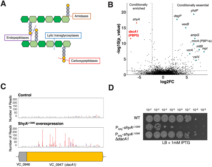
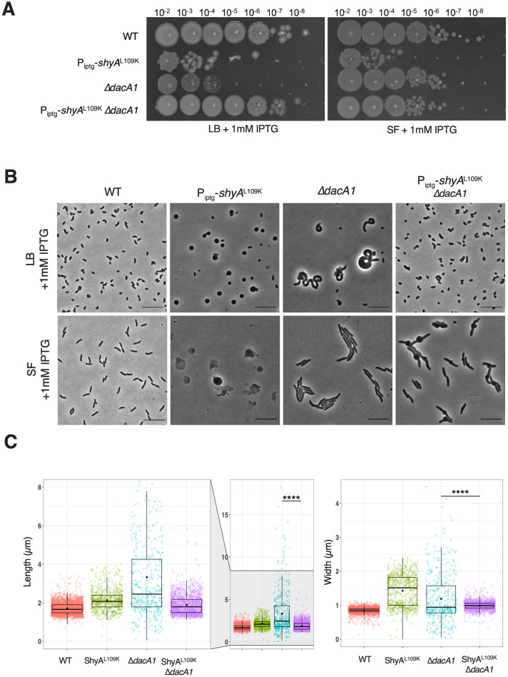
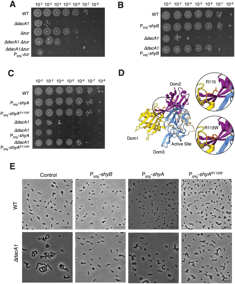
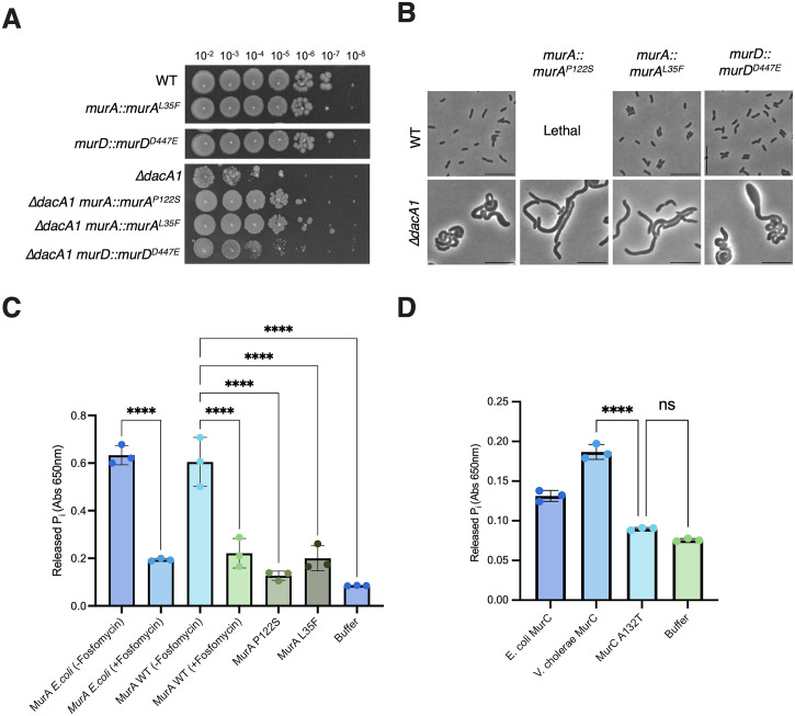
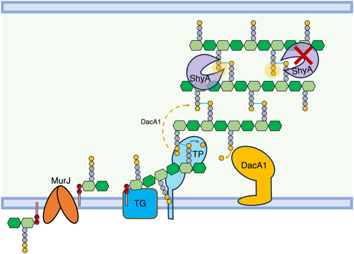
Peptidoglycan (PG) is the main component of the bacterial cell wall; it maintains cell shape while protecting the cell from internal osmotic pressure and external environmental challenges. PG synthesis is essential for bacterial growth and survival, and a series of PG modifications are required to allow expansion of the sacculus. Endopeptidases (EPs), for example, cleave the crosslinks between adjacent PG strands to allow the incorporation of newly synthesized PG. EPs are collectively essential for bacterial growth and must likely be carefully regulated to prevent sacculus degradation and cell death. However, EP regulation mechanisms are poorly understood. Here, we used TnSeq to uncover novel EP regulators in Vibrio cholerae. This screen revealed that the carboxypeptidase DacA1 (PBP5) alleviates EP toxicity. dacA1 is essential for viability on LB medium, and this essentiality was suppressed by EP overexpression, revealing that EP toxicity both mitigates, and is mitigated by, a defect in dacA1. A subsequent suppressor screen to restore viability of ΔdacA1 in LB medium identified hypomorphic mutants in the PG synthesis pathway, as well as mutations that promote EP activation. Our data thus reveal a more complex role of DacA1 in maintaining PG homeostasis than previously assumed.


Conflict of interest statement

The authors have declared that no competing interests exist.

Figures

Fig 1
Fig 1. A screen for EP regulators identifies a genetic relationship between ShyA and PBP5.
(A) Schematic representation of autolysin cleavage sites in the cell wall. (B) Volcano plot of the change in relative abundance of insertion mutants between the control condition (WT) and the experimental condition (ShyAL109K overexpression) (X-axis) versus p-value (Y-axis). The dashed line indicates the cutoff criterion (>2-fold fitness change, P <0.005) for identification of genes that modulate ShyAL109K toxicity. (C) dacA1 was identified as conditionally enriched upon ShyAL109K overexpression using CON-ARTIST, red bars represent the number of reads from transposon insertions found in the forward orientation and blue bars represent insertions found in the reverse orientation. The black dots indicate every TA site available for transposon insertion (D) Overnight cultures of WT, Piptg-shyAL109K and Piptg-shyAL109K ΔdacA1 where plated on LB medium with 1mM IPTG and incubated overnight at 30°C.
Fig 2
Fig 2. ΔdacA1 mutant PG is less susceptible to cleavage by ShyA.
(A) Schematic representation of how DacA1 absence affects the pentapeptide content in the PG sacculus and potentially ShyA activity. (B) Schematic of experimental procedure. The PG sacculi from WT and ΔdacA1 cells were purified using the SDS precipitation method [52]. Purified samples were then stained with Remazol Brilliant Blue and used in sedimentation assays where PG fragmented by hydrolases remains soluble in the supernatant upon centrifugation, providing a quantitative readout of EP activity. Figure was made with Biorender (C) RBB stained sacculi were treated as described in (B) with the addition of proteins indicated on the x-axis. Undigested material was removed by centrifugation and the supernatant was used to measure OD585. Error bars represent standard deviation of two independent experiments. Significance was determined using a two-way ANOVA, **** corresponds to a P-value of <0.0001.
Fig 3
Fig 3. ShyAL109K overexpression restores normal cell growth and morphology in ΔdacA1 cells grown on LB medium.
(A) Overnight cultures were grown on LB (WT background) and salt-free LB (ΔdacA1 background) and then spot-titered on either LB or salt-free LB (SF) with 1mM IPTG. (B) Overnight cultures were sub-cultured 1:100 in either LB or SF at 37°C until they reached exponential phase, followed by addition of inducer (1mM IPTG). Phase contrast images were taken after 2 hours of induction. Scale bar: 10 μm (C) Cells were segmented using Omnipose, and area, width and length were calculated with MicrobeJ. Statistical significance was determined with Welch’s two sample t-test. ****, P < 0.0001. Shown are boxplots with raw data points.
Fig 4
Fig 4. Mutations that enhance endopeptidase activity suppress the ΔdacA1 phenotype in LB.
(A) Overnight cultures were grown on LB (WT background) and salt-free LB (ΔdacA1 background) and then spot-titered on LB medium with 1mM IPTG and incubated at 30°C overnight. (B,C) Overnight cultures of the specified strains were diluted 1:100 in LB medium and grown at 37°C until reaching exponential phase. Following this, 1mM IPTG was added to induce gene expression, and the cultures were further incubated at 37°C for 3 hours. (D) Crystal structure of ShyA showing residue R115 in the interphase between Domain 1 and Domain 3, and prediction of the R115W mutation. (E) Overnight cultures were sub-cultured 1:100 in LB at 37°C until they reached exponential phase, followed by addition of inducer (1mM IPTG). Phase contrast images were taken after 3 hours of induction. Scale bar: 10 μm.
Fig 5
Fig 5. Hypomorphic mutants in the PG precursor synthesis pathway rescue ΔdacA1 growth but not morphology.
(A) Indicated strains are chromosomal replacements of murA and murD with murAP122S, murAL35F and murDD447E, respectively. Overnight cultures were plated on LB medium by spot- titering and incubated at 30°C overnight. (B) Overnight cultures of the indicated strains were sub cultured 1:100 and grown at 37°C in LB medium for 1 hour, then back diluted 1:100 in LB and incubated at 37°C for an additional 2 hours before imaging (C) MurA as well as the different mutants were purified, and their activity was measured in vitro by measuring the inorganic phosphate released from the MurA enzymatic reaction. (D) MurC and MurCA132T were purified and their enzymatic activity was quantified in vitro by measuring the inorganic phosphate released from the MurC enzymatic reaction. (C-D) Statistical significance was determined by one-way ANOVA, error bars represent the standard deviation of three replicates (raw data points also shown).
Fig 6
Fig 6. PG precursor recycling and synthesis modulate dacA1 mutant fitness.
(A) Overnight cultures of the indicated mutants were grown on LB or salt-free LB (ΔdacA1 background) and plated on LB medium via spot-titering and incubated at 30°C overnight. (B) Overnight cultures of WT, ΔdacA1 and both backgrounds harbouring an IPTG inducible copy of murA were plated on salt-free LB with increasing concentrations of NaCl (120mM, 140mM and 160mM) and incubated at 30°C overnight.
Fig 7
Fig 7. ShyA overexpression rescues morphological defects associated with carboxypeptidase insufficiency in E. coli.
(A) E. coli strains CS109 and Δ4 (CS109 ΔdacA ΔdacB ΔdacC ΔpbpG) containing either an empty vector (pHL100) or a vector containing an IPTG-inducible copy of ShyA (pHL100-ShyA) were incubated overnight at 37°C in LB medium with 0.2% glucose, sub-cultured 1:100 in LB and incubated for 2 hours, then back-diluted 1:100 in LB and grown for an additional 2 hours. 1 mM IPTG or 0.2% glucose was added to the cultures, phase contrast images were taken 3 hours after induction. (B) Microscopy images were segmented with Omnipose and analyzed for area, length, and width with MicrobeJ. Statistical significance was assessed via Welch’s t-test. ****, P < 0.0001.
Fig 8
Fig 8. Model of DacA1’s role in balancing PG synthesis and degradation.
DacA1 removes the 5th D-ala residue of the PG side stem and determines which side stem can be used as donor for transpeptidation. By removing the 5th D-ala residue, DacA1 also allows ShyA to cleave crosslinks between strands by producing its preferred substrate, tetrapeptide-containing side stems. In the absence of DacA1, there is an increased amount of pentapeptides in the PG sacculus, hindering ShyA’s ability to cleave crosslinks. This reduction in endopeptidase activity disrupts the balance between PG synthesis and degradation.

Similar articles

Cited by

References

    1. Cho H, Wivagg CN, Kapoor M, Barry Z, Rohs PDA, Suh H, et al.. Bacterial cell wall biogenesis is mediated by SEDS and PBP polymerase families functioning semi-autonomously. Nat Microbiol. 2016. Sep 19;1(10):1–8. doi: 10.1038/nmicrobiol.2016.172 - DOI - PMC - PubMed
    1. Mett H, Bracha R, Mirelman D. Soluble nascent peptidoglycan in growing Escherichia coli cells. J Biol Chem. 1980. Oct 25;255(20):9884–90. - PubMed
    1. Mueller EA, Levin PA. Bacterial Cell Wall Quality Control during Environmental Stress. mBio. 2020;11(5). doi: 10.1128/mBio.02456-20 - DOI - PMC - PubMed
    1. Nelson DE, Young KD. Penicillin Binding Protein 5 Affects Cell Diameter, Contour, and Morphology of Escherichia coli. J Bacteriol. 2000. Mar 15;182(6):1714–21. doi: 10.1128/JB.182.6.1714-1721.2000 - DOI - PMC - PubMed
    1. Ghosh AS, Chowdhury C, Nelson DE. Physiological functions of D-alanine carboxypeptidases in Escherichia coli. Trends Microbiol. 2008. Jul;16(7):309–17. doi: 10.1016/j.tim.2008.04.006 - DOI - PubMed

Publication types

MeSH terms