Review
. 2024 May;38(2):101943.
doi: 10.1016/j.berh.2024.101943. Epub 2024 Apr 9.

Immune checkpoints in autoimmune vasculitis

Affiliations
Review

Immune checkpoints in autoimmune vasculitis

Yuki Sato et al. Best Pract Res Clin Rheumatol. 2024 May.

Abstract

Giant cell arteritis (GCA) is a prototypic autoimmune disease with a highly selective tissue tropism for medium and large arteries. Extravascular GCA manifests with intense systemic inflammation and polymyalgia rheumatica; vascular GCA results in vessel wall damage and stenosis, causing tissue ischemia. Typical granulomatous infiltrates in affected arteries are composed of CD4+ T cells and hyperactivated macrophages, signifying the involvement of the innate and adaptive immune system. Lesional CD4+ T cells undergo antigen-dependent clonal expansion, but antigen-nonspecific pathways ultimately control the intensity and duration of pathogenic immunity. Patient-derived CD4+ T cells receive strong co-stimulatory signals through the NOTCH1 receptor and the CD28/CD80-CD86 pathway. In parallel, co-inhibitory signals, designed to dampen overshooting T cell immunity, are defective, leaving CD4+ T cells unopposed and capable of supporting long-lasting and inappropriate immune responses. Based on recent data, two inhibitory checkpoints are defective in GCA: the Programmed death-1 (PD-1)/Programmed cell death ligand 1 (PD-L1) checkpoint and the CD96/CD155 checkpoint, giving rise to the "lost inhibition concept". Subcellular and molecular analysis has demonstrated trapping of the checkpoint ligands in the endoplasmic reticulum, creating PD-L1low CD155low antigen-presenting cells. Uninhibited CD4+ T cells expand, release copious amounts of the cytokine Interleukin (IL)-9, and differentiate into long-lived effector memory cells. These data place GCA and cancer on opposite ends of the co-inhibition spectrum, with cancer patients developing immune paralysis due to excessive inhibitory checkpoints and GCA patients developing autoimmunity due to nonfunctional inhibitory checkpoints.

Keywords: Antigen-presenting cell; CD155; CD96; Co-inhibition; Costimulation; PD-1; PD-L1; T cells.


Conflict of interest statement

Declaration of competing interest The authors declare that they have no known competing financial interests or personal relationships that could have appeared to influence the work reported in this paper.

Figures

Figure 1.
Figure 1.. Disease Model of Giant Cell Arteritis.
The disease process of GCA begins decades before the onset of clinically overt vasculitis. Genetic risk factors have been mapped to the HLA class II region. At-risk individuals have a metabolic phenotype, characterized by low body mass index (BMI). Following the loss of self-tolerance, patients enter the phase of Pre-GCA, clinically presenting with polymyalgia rheumatica (PMR). Immunologically, they have aberrant expression of NOTCH family receptors on T cells, providing inappropriate immune stimulation. Progression to Phase II involves the transition of peripheral immune cells (monocytes, T cells) into the perivascular space of vasa vasora, where they establish organized lymphoid structures and built in situ supply stations for inflammatory effector cells. The loss of tissue tolerance involves inappropriate signaling of the NOTCH pathway and endothelial cell-T cell interactions. Clinically overt vasculitis (Phase III) requires monocyte and T cell recruitment to the adventitia of the vessel wall. Pathogenic immune cells take residence, mature into effector cells, invade into the medial and intimal layer, injure the vessel wall structures, and elicit a maladaptive tissue repair process (vessel remodeling leading to luminal occlusion). Two pathways critically drive vasculitis: the loss of inhibitory checkpoint function and the in-situ generation of effector T cells from stem-like CD4+ T cells. PD-L1lowCD155low APCs dismantle tissue protection provided by the PD1/PDL1 and the CD96/CD155 checkpoints. CD4+ T effector cells are locally produced from stem-like precursors stationed in adventitial TLS, rendering the disease process autonomous.
Figure 2.
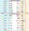
Figure 2.. Expression of immune checkpoint receptors on T cells and their respective ligands on APCs.
T-cell activation is triggered by the recognition of antigen-HLA complexes, but the intensity and duration of T cell responses remains under the control of co-signaling molecules that shape T cell responses through co-stimulation or co-inhibition (31, 83, 84). Well-established co-stimulatory receptors include CD28, inducible costimulatory molecule (ICOS), glucocorticoid-induced TNF receptor family-related protein (GITR), TNF receptor superfamily member 4 (OX40), and TNF receptor superfamily member 9 (4-1BB). T cells receive inhibitory instructions through Cytotoxic T-lymphocyte-associated antigen-4 (CTLA-4) that competitively binds to CD80/86 and lymphocyte activation gene-3 (LAG-3) that competitively binds to major histocompatibility complex II (MHC II). Several co-inhibitory receptors finetune T-cell activation, expansion, and function (e.g. IFN-γ secretion) and include T cell Immunoglobulin and ITIM domain (TIGIT) binding to CD122 or CD155 and CD96 binding to CD155 or CD111. Inhibitory receptors appearing late during T cell stimulation have been implicated in T-cell exhaustion: T cell immunoglobulin and mucin domain 3 (TIM-3) binds to carcino-embryonic antigen cell adhesion molecule 1 (CEACAM-1) or Galectin-9; B and T Lymphocyte Attenuator (BTLA) binds to Herpesvirus entry mediator (HVEM); and programmed cell death protein-1 (PD-1) recognizes PD-L1 and PD-L2.
Figure 3.
Figure 3.. Disease relevant co-signaling molecules in Giant Cell Arteritis.
Tissue destructive granulomas are typically formed by hyper-activated macrophages, receiving instructions from surrounding T cells. The outcome of T cell-macrophage interaction is controlled by antigen recognition and by co-stimulatory and co- inhibitory signals that ultimately determine the lineage commitment, intensity, and duration of T cell immunity. GCA CD4+ T cells are overly sensitive to co-stimulation derived from CD28-CD80/86 interaction. GCA macrophages have a defect in expressing the co-inhibitory ligand PD-L1 and CD155. Consequently, GCA CD4+ T cells are insufficiently restrained and provide long-lasting, pro-inflammatory immunity. The molecular defect leading to PD-L1low CD155low macrophages has been mapped to the endoplasmic reticulum (ER) and to faulty protein trafficking from the ER to the plasma membrane.

References

    1. Jennette JC, Falk RJ, Bacon PA, Basu N, Cid MC, Ferrario F, et al. 2012 revised International Chapel Hill Consensus Conference Nomenclature of Vasculitides. Arthritis Rheum. 2013;65(1):1–11. - PubMed
    1. Watanabe R, Berry GJ, Liang DH, Goronzy JJ, Weyand CM. Pathogenesis of Giant Cell Arteritis and Takayasu Arteritis-Similarities and Differences. Curr Rheumatol Rep. 2020;22(10):68. - PMC - PubMed
    1. McCrindle BW, Rowley AH, Newburger JW, Burns JC, Bolger AF, Gewitz M, et al. Diagnosis, Treatment, and Long-Term Management of Kawasaki Disease: A Scientific Statement for Health Professionals From the American Heart Association. Circulation. 2017;135(17):e927–e99. - PubMed
    1. Bilginer Y, Ozen S. Polyarteritis nodosa. Curr Opin Pediatr. 2022;34(2):229–33. - PubMed
    1. Kitching AR, Anders HJ, Basu N, Brouwer E, Gordon J, Jayne DR, et al. ANCA-associated vasculitis. Nat Rev Dis Primers. 2020;6(1):71. - PubMed

Publication types

MeSH terms

Substances