. 2024 Aug;30(4):362-379.
doi: 10.1007/s13365-024-01204-z. Epub 2024 Apr 10.

Modeling HIV-1 infection and NeuroHIV in hiPSCs-derived cerebral organoid cultures

Affiliations

Modeling HIV-1 infection and NeuroHIV in hiPSCs-derived cerebral organoid cultures

Martina Donadoni et al. J Neurovirol. 2024 Aug.

Abstract

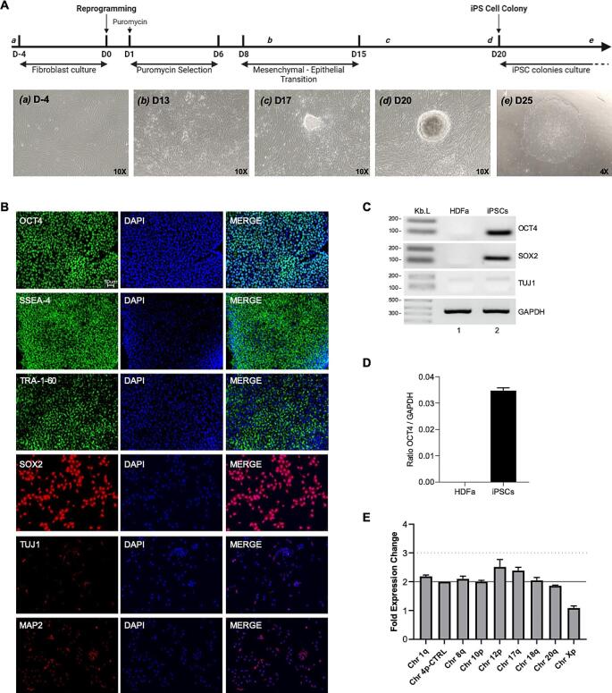
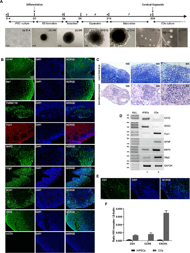
The human immunodeficiency virus (HIV) epidemic is an ongoing global health problem affecting 38 million people worldwide with nearly 1.6 million new infections every year. Despite the advent of combined antiretroviral therapy (cART), a large percentage of people with HIV (PWH) still develop neurological deficits, grouped into the term of HIV-associated neurocognitive disorders (HAND). Investigating the neuropathology of HIV is important for understanding mechanisms associated with cognitive impairment seen in PWH. The major obstacle for studying neuroHIV is the lack of suitable in vitro human culture models that could shed light into the HIV-CNS interactions. Recent advances in induced pluripotent stem cell (iPSC) culture and 3D brain organoid systems have allowed the generation of 2D and 3D culture methods that possess a potential to serve as a model of neurotropic viral diseases, including HIV. In this study, we first generated and characterized several hiPSC lines from healthy human donor skin fibroblast cells. hiPSCs were then used for the generation of microglia-containing human cerebral organoids (hCOs). Once fully characterized, hCOs were infected with HIV-1 in the presence and absence of cART regimens and viral infection was studied by cellular, molecular/biochemical, and virological assays. Our results revealed that hCOs were productively infected with HIV-1 as evident by viral p24-ELISA in culture media, RT-qPCR and RNAscope analysis of viral RNA, as well as ddPCR analysis of proviral HIV-1 in genomic DNA samples. More interestingly, replication and gene expression of HIV-1 were also greatly suppressed by cART in hCOs as early as 7 days post-infections. Our results suggest that hCOs derived from hiPSCs support HIV-1 replication and gene expression and may serve as a unique platform to better understand neuropathology of HIV infection in the brain.

Keywords: Astrocytes; Cerebral organoids; HIV-1; Microglia; NeuroHIV; cART; hiPSCs.


Conflict of interest statement

The authors declare no competing interests.

Figures

Fig. 1
Fig. 1
Generation and characterization of human induced pluripotent stem cells (hiPSCs) from human dermal fibroblast, adult (HDFa). A: Schematic timeline of generation of hiPSCs from HDFa. Representative images of reprogramming of HDFa to generate hiPSC single-cell colony cultures. B: Immunohistochemistry of hiPSCs for pluripotency markers, OCT4, SSEA-4, TRA-1-60 and SOX2, and neuronal differentiation markers, TUJ1 and MAP2. C. RT-PCR to confirm mRNA expression of pluripotency markers, OCT4 and SOX2, and neuronal marker TUJ1. GAPDH was also amplified from same samples as control. Line 1 is HDFa. Line 2 is hiPSC. D. hiPSC Genetic Analysis was performed for any possible karyotypic abnormalities reported for human iPSCs lines
Fig. 2
Fig. 2
Generation and characterization of human cerebral organoids (hCOs) from hiPSCs. A: Schematic timeline of generation of hCOs from hiPSCs. Representative images of generation of hCOs showing Embryoid bodies (EB) formation (b), induction (c), expansion (d) and maturation steps (e-f). Fully mature hCOs representative image at day 50 (g) B: Immunohistochemistry (IHC) of hCOs for astrocytic structural marker, GFAP, microglial marker, Iba-1 and TMEM119, neuronal differentiation markers, TUJ1 and MAP2, oligodendroglia marker, Olig2, neural progenitor markers SOX1 and PAX6, and pluripotency marker, OCT4. C. Luxol Fast Blue for myelin staining and hematoxylin and eosin staining of mature hCOs. D. RT-PCR for mRNA expression of pluripotency markers, OCT4 and SOX2, neuronal markers, TUJ1 and MAP2, astrocytic structural marker, GFAP, and oligodendroglia marker, Olig2. GAPDH was also amplified from same samples. Line 1 is hiPSCs. Line 2 is hCOs. E. Immunohistochemistry (IHC) of hCOs for proliferation marker Ki67. F. RT-qPCR for mRNA expression of HIV-1 receptor, CD4, and co-receptors, CCR5 and CXCR4, in hiPSCs and hCOs. Three different lines of hiPSCs and three different hCOs were used. (Shown as mean ± SEM).
Fig. 3
Fig. 3
HIV-1 infection characterization in human cerebral organoids (hCOs). hCOs were infected with 300 ng of HIV-1 Bal-GFP virus. 4 days post-infection, hCOs were collected and fixed in 4% PFA for 16 h, after which they were cryo-embedded and prepared for staining. (A) Representative images of hCOs stained for GFP (green) and DAPI (blue). Images were taken at 10X magnification. (B) Representative images of hCOs stained for HIV-1 Nef (green) and DAPI (blue). Images were taken at 10X and details at 20X magnification. (C) Dual IHC-RNA Scope. Representative images of co-staining of HIV RNA (red) and cell type specific markers, Iba1, GFAP and MAP2 (brown). Images were taken at 20X and 40X magnifications. N = 3 hCOs.
Fig. 4
Fig. 4
Characterization of HIV-1 infection in hCOs in the presence and absence of cART regimens. A: Schematic representation of experimental outline. hCOs were infected with HIV-1 Bal-GFP virus and treated with cART (Raltegravir, Tenofovir disoproxil fumarate, Emtricitabine) as outlined. B. RT-qPCR for mRNA expression of HIV-1 receptor, CD4, and co-receptors, CCR5 and CXCR4, in uninfected, HIV+, HIV + ART + hCOs. Three different hCOs per condition were used. C: Gag p24 ELISA on supernatant from hCOs uninfected, HIV + hCOs and HIV + ART + hCOs at day 1 (inoculum), day 4 and day 7 post-infections. Fourteen different hCOs per condition were used. D-E: gDNA from uninfected, HIV + and HIV + ART + hCOs was extracted and processed for ddPCR to detect Ψ and Human TERT (D) and data were shown as bar graphs per one million of cells (E). Four different hCOs per condition were used. F-H: RNA from uninfected, HIV + and HIV + ART + hCOs was extracted and processed for RT-qPCR for Ψ (F), GagD (G) and Pol (H) genes. Data were shown as bar graphs normalized to β-Actin. Three different hCOs per condition were used. I: hCOs were collected and fixed in 4% PFA for 16 h, after which they were cryo-embedded and processed for RNAScope to detect HIV-1 RNA (red). Three different hCOs per condition were used. J. hCOs were collected and fixed in 4% PFA for 16 h, after which they were cryo-embedded and processed for IHC for cleaved caspase-3. Three different hCOs per condition were used. (ELISA n = 14, RNA n = 3, gDNA n = 4. Shown as mean ± SEM, * p < 0.05, *** p < 0.001)

References

    1. Adamson DC, Wildemann B, Sasaki M et al (1996) Immunologic NO synthase: elevation in severe AIDS dementia and induction by HIV-1 gp41. Science 274:1917–1921. 10.1126/science.274.5294.1917 - PubMed
    1. Agrawal L, Louboutin J-P, Strayer DS (2007) Preventing HIV-1 tat-induced neuronal apoptosis using antioxidant enzymes: mechanistic and therapeutic implications. Virology 363:462–472. 10.1016/j.virol.2007.02.004 - PubMed
    1. Albright AV, Shieh JT, O’Connor MJ, González-Scarano F (2000) Characterization of cultured microglia that can be infected by HIV-1. J Neurovirol 6(Suppl 1):S53–60 - PubMed
    1. Antinori A, Arendt G, Becker JT et al (2007) Updated research nosology for HIV-associated neurocognitive disorders. Neurology 69:1789–1799. 10.1212/01.WNL.0000287431.88658.8b - PMC - PubMed
    1. Barber TJ, Imaz A, Boffito M et al (2018) CSF inflammatory markers and neurocognitive function after addition of maraviroc to monotherapy darunavir/ritonavir in stable HIV patients: the CINAMMON study. J Neurovirol 24:98–105. 10.1007/s13365-017-0600-6 - PubMed

Publication types

LinkOut - more resources