Case Reports
. 2024 Apr 11;18(1):170.
doi: 10.1186/s13256-024-04499-y.

Paratesticular cellular angiofibroma: a case report

Affiliations
Case Reports

Paratesticular cellular angiofibroma: a case report

Takaya Murashima et al. J Med Case Rep. .

Abstract

Introduction: Paratesticular cellular angiofibroma is a rare benign mesenchymal tumor. The optimal management is surgical resection due to the difficulty of preoperative accurate diagnosis.

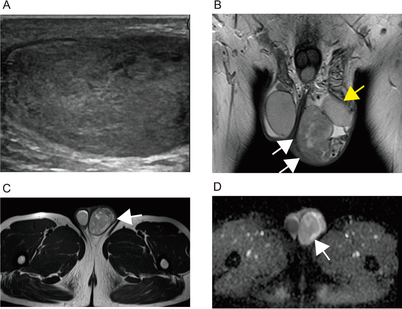
Case presentation: A 51-year-old Japanese male visited our hospital complaining of asymptomatic left scrotal swelling. Physical examination revealed a nontender elastic paratesticular mass (5.5 cm in diameter). Although testicular germ cell tumor was ruled out clinically, the possibility of malignant potential remained for the tumor. Since the patient consented to complete resection, a transinguinal radical orchiectomy was performed. The pathological diagnosis revealed cellular angiofibroma. The patient recovered without perioperative complications, and no apparent recurrence was observed at 5 years after surgery.

Conclusion: The pathological findings were compatible for cellular angiofibroma. The tumor was successfully resected, and no apparent recurrence was observed at 5 years after surgery.

Keywords: Cellular angiofibroma; Orchiectomy; Paratesticular region.


Conflict of interest statement

The authors declare that they have no competing interests.

Figures

Fig. 1
Fig. 1
Ultrasound and magnetic resonance imaging. A Ultrasonography showed internally hypoechoic heterogeneous mass, 5.5-cm-long diameter in size. The tumor showed a clearly demarcated solid mass. B, C T2 weighted images by magnetic resonance imaging examination revealed heterogeneous low intensity mass (white arrows) including focal high intensity areas (B coronal imaging, C axial imaging). Left testis was confirmed (yellow arrow). D Diffusion-weighted imaging shows no apparent decreasing ADC value in the internal area of the mass; however, this is focally observed in peripheral area (white arrow)
Fig. 2
Fig. 2
Gross and histopathological findings of the tumor. A Gross findings. The paratesticular tumor (white arrows) is well-circumscribed, detached from the testis (A). Cut surface shows heterogeneous greyish-white in color. B Microscopically, tumor lesion shows proliferation of short spindle cells in a diffuse or perivascular pattern, accompanied by collagenous stromal fibers and dilated vascular channels. No nuclear atypia or mitotic figure is shown. C Myoid stromal areas are focally seen with hypocellularity. DH Immunohistochemical findings. Short spindle cells are positive for alpha-smooth muscle actin (D), CD34 (E), estrogen receptor (F), and progesterone receptor (G) and negative for desmin (H). Vascular walls and endothelial cells are also positive for alpha smooth muscle actin and CD34 and desmin, respectively

References

    1. Nucci MR, Granter SR, Fletcher CD. Cellular angiofibroma: a benign neoplasm distinct from angiomyofibroblastoma and spindle cell lipoma. Am J Surg Pathol. 1997;21(6):636–644. doi: 10.1097/00000478-199706000-00002. - DOI - PubMed
    1. Iwasa Y, Fletcher CD. Cellular angiofibroma: clinicopathologic and immunohistochemical analysis of 51 cases. Am J Surg Pathol. 2004;28(11):1426–2135. doi: 10.1097/01.pas.0000138002.46650.95. - DOI - PubMed
    1. WHO Classification of Tumours Editorial Board; WHO classification of tumours, urinary and male genital tumours, 5th Edition. - PubMed
    1. Samaratunga H, Fitzpatrick P. Cellular angiofibroma of the scrotum. Pathology. 2008;40(3):330–333. doi: 10.1080/00313020701716409. - DOI - PubMed
    1. Arakaki K, Chinen K, Kamiya M, Tanabe Y, Tawata N, Ikehara F, Uehara K, Shimabukuro H, Kinjo T. Evidence for an association between increased oxidative stress and derangement of FOXO1 signaling in tumorigenesis of a cellular angiofibroma with monoallelic 13q14: a case report. Int J Clin Exp Pathol. 2014;7:8972–8979. - PMC - PubMed

Publication types