. 2024 Mar 29;13(3):603-611.
doi: 10.21037/tlcr-24-25. Epub 2024 Mar 27.

A simple and safe surgical technique for nonpalpable lung tumors: One-stop Solution for a nonpalpable lung tumor, Marking, Resection, and Confirmation of the surgical margin in a Hybrid operating room (OS-MRCH)

Affiliations

A simple and safe surgical technique for nonpalpable lung tumors: One-stop Solution for a nonpalpable lung tumor, Marking, Resection, and Confirmation of the surgical margin in a Hybrid operating room (OS-MRCH)

Aki Fujiwara-Kuroda et al. Transl Lung Cancer Res. .

Abstract

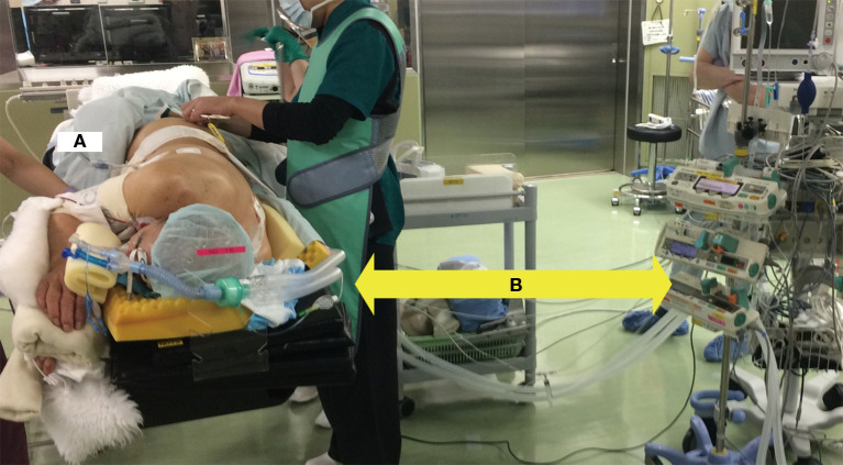
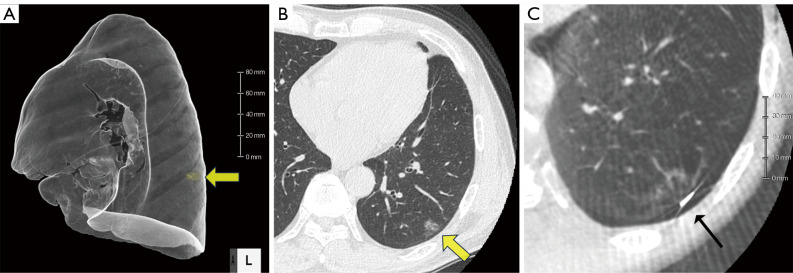
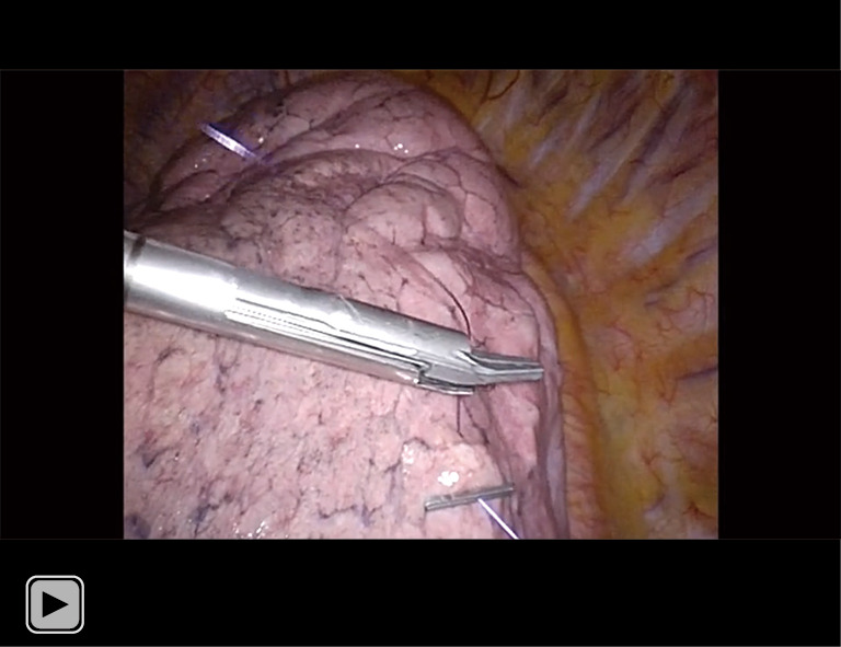
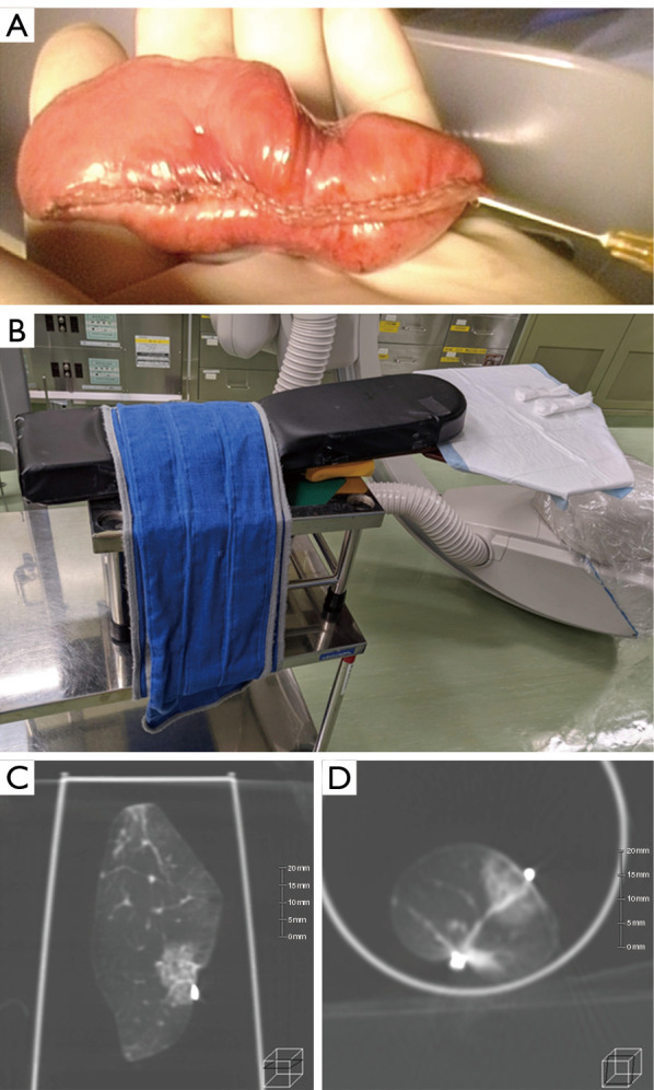
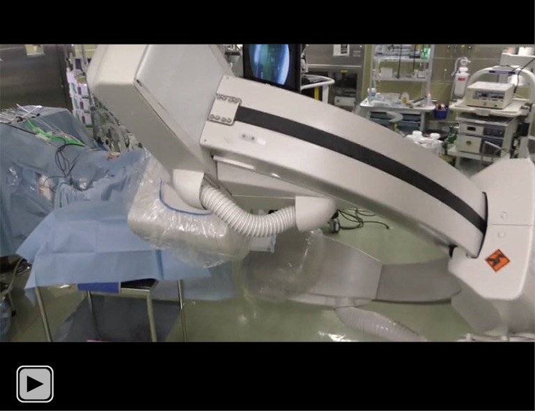
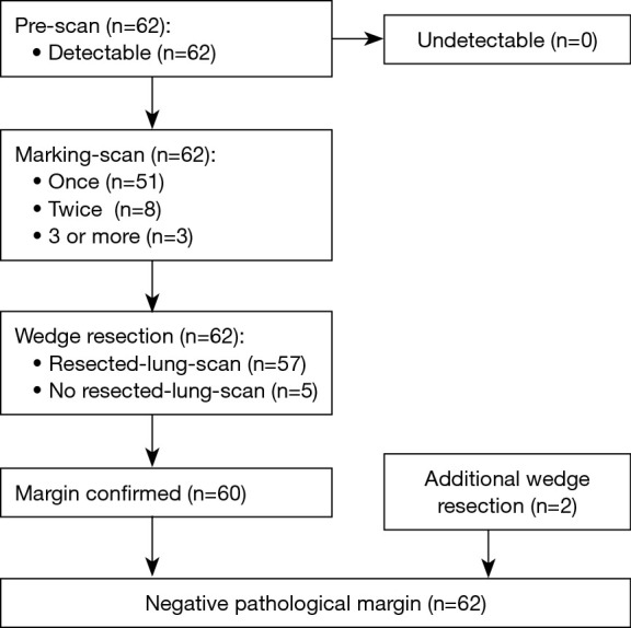
When performing thoracoscopic partial resections of nonpalpable lung tumors such as ground-glass opacities (GGOs) and small tumors, detecting the location of the lesion and assessing the resection margins can be challenging. We have developed a novel method to ease this difficulty, the One-stop Solution for a nonpalpable lung tumor, Marking, Resection, and Confirmation of the surgical margin in a Hybrid operating room (OS-MRCH), which uses a hybrid operating room wherein the operating table is seamlessly integrated with cone-beam computed tomography (CBCT). We performed the OS-MRCH method on 62 nodules including primary lung cancer presenting with GGO. Identification of the lesion and confirmation of the margin were performed in 58 of the cases, while nodules were detected in all. The frequency of computed tomography (CT) scans performed prior to resection was one time in 51 cases, two times in eight cases, and ≥3 times in three cases. Additional resection was performed in two cases. The median operative time was 85.0 minutes, and the median pathological margin was 11.0 mm. The key advantages of this method are that all surgical processes can be completed in a single session, specialized skill sets are not required, and it is feasible to perform in any facility equipped with a hybrid operating room. To overcome its disadvantages, such as longer operating time and limited patient positioning, we devised various methods for positioning patients and for CT imaging of the resected specimens. OS-MRCH is a simple, useful, and practical method for performing thoracoscopic partial resection of nonpalpable lung tumors.

Keywords: Nonpalpable lung tumor; ground-glass opacity (GGO); hybrid operating room; surgical margin; video-assisted thoracoscopic surgery (VATS).


Conflict of interest statement

Conflicts of Interest: All authors have completed the ICMJE uniform disclosure form (available at https://tlcr.amegroups.com/article/view/10.21037/tlcr-24-25/coif). The authors have no conflicts of interest to declare.

Figures

Figure 1
Figure 1
Position of the patient and operating table. This image is published with the patient’s consent. (A) The patient needs to be positioned in a lateral position with both elbows folded in order to stay within the rotation range of the C-arm of the hybrid surgical system; (B) an arrow indicates a longer distance from the anesthesia machine is necessary to prevent the C-arm from interfering with the rotation. Therefore, longer-than usual anesthesia tubes and venous/arterial lines are needed.
Figure 2
Figure 2
Preoperative CT-scan and Marking-scan images. Clip marking is performed based on preoperative 3D-CT (A) or thin-slice HRCT (B) performed prior to the day of surgery. Yellow arrows indicate GGO tumors. Intraoperative Marking-scan (C) indicating that the clip is placed on the lung surface directly above the GGO (line arrow). CT, computed tomography; HRCT, high-resolution computed tomography; GGO, ground-glass opacity.
Video 1
Video 1
How to make a clip marking on the lung surface. A 5-mm clip is attached to the end of the thread sutured to the target lung surface. After drawing the thread to the end, a second 5-mm clip is applied to include the lesion, and the thread is cut. This method shortens the time required for marking, without requiring intrathoracic ligation.
Figure 3
Figure 3
Resected-lung-scan images. (A) Air injection is performed using a low-gauge needle before the Resected-lung-scan. Note that rapid air injection causes emphysema in the resected lung. (B) A platform for Resected-lung-scan assembly using the arm board of an operating table. A plastic cup containing the specimen is placed on this platform for Resected-lung-scan imaging. (C,D) A Resected-lung-scan indicates the relationship of the lesion to the clips and staple line, and confirms that the resection margin is adequate.
Video 2
Video 2
Video of a resected-lung scan. Immediately following air injection, the resected lung is scanned using CBCT imaging in the same operating room. Prompt CBCT imaging has several advantages: it prevents lung collapse, improves image reproducibility, and allows for the confirmation of surgical margins immediately following resection. CBCT, cone-beam computed tomography.
Figure 4
Figure 4
Flowchart of 62 cases. In all 62 cases, tumors were identified by Pre-scan; Marking-scan was performed once in 51 cases (82.3%); Resected-lung-scan was performed in 57 cases (91.9%).

References

    1. Altorki N, Wang X, Kozono D, et al. Lobar or Sublobar Resection for Peripheral Stage IA Non-Small-Cell Lung Cancer. N Engl J Med 2023;388:489-98. 10.1056/NEJMoa2212083 - DOI - PMC - PubMed
    1. Kanazawa S, Ando A, Yasui K, et al. Localization of pulmonary nodules for thoracoscopic resection: experience with a system using a short hookwire and suture. AJR Am J Roentgenol 1998;170:332-4. 10.2214/ajr.170.2.9456940 - DOI - PubMed
    1. Yoshida Y, Inoh S, Murakawa T, et al. Preoperative localization of small peripheral pulmonary nodules by percutaneous marking under computed tomography guidance. Interact Cardiovasc Thorac Surg 2011;13:25-8. 10.1510/icvts.2011.266932 - DOI - PubMed
    1. Yi JH, Choi PJ, Bang JH, et al. Systemic air embolism after computed tomography-guided hook wire localization: two case reports and literature review. J Thorac Dis 2018;10:E59-64. 10.21037/jtd.2017.12.04 - DOI - PMC - PubMed
    1. Oikawa T, Nomoto Y, Kinoshita K. A case of left-sided hemiplegia due to cerebral air embolism caused by CT-guided marking for small peripheral lung cancer. The Journal of the Japanese Association for Chest Surgery 2008;22:914-9.