. 2024 May;32(5):102058.
doi: 10.1016/j.jsps.2024.102058. Epub 2024 Apr 1.

Fabrication and evaluation of ribavirin-loaded electrospun nanofibers as an antimicrobial wound dressing

Affiliations

Fabrication and evaluation of ribavirin-loaded electrospun nanofibers as an antimicrobial wound dressing

Khulud A Alsulami et al. Saudi Pharm J. 2024 May.

Abstract

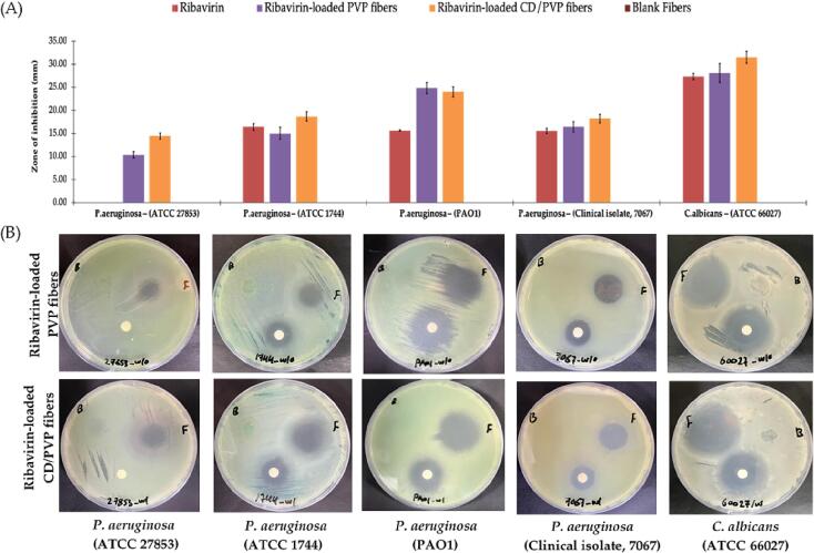
Background: Skin is regarded as an essential first line of defense against harmful pathogens and it hosts an ecosystem of microorganisms that create a widely diverse skin microbiome. In chronic wounds, alterations in the host-microbe interactions occur forming polymicrobial biofilms that hinder the process of wound healing. Ribavirin, an antiviral drug, possesses antimicrobial activity, especially against Pseudomonas aeruginosa and Candida albicans, which are known as the main opportunistic pathogens in chronic wounds.

Rationale: In this study, electrospun nanofiber systems loaded with ribavirin were developed as a potential wound dressing for topical application in chronic wounds. Ribavirin was chosen in this study owing to the emerging cases of antimicrobial (antibiotics and antifungal) resistance and the low attempts to discover new antimicrobial agents, which encouraged the repurposing use of current medication as an alternative solution in case of resistance to the available agents. Additionally, the unique mechanism of action of ribavirin, i.e., perturbing the bacterial virulence system without killing or stopping their growth and rendering the pathogens disarmed, might be a promising choice to prevent drug resistance. Cyclodextrin (CD) was utilized to formulate ribavirin as an electrospun nanofibers delivery system to enhance the absorption and accelerate the release of ribavirin for topical use.

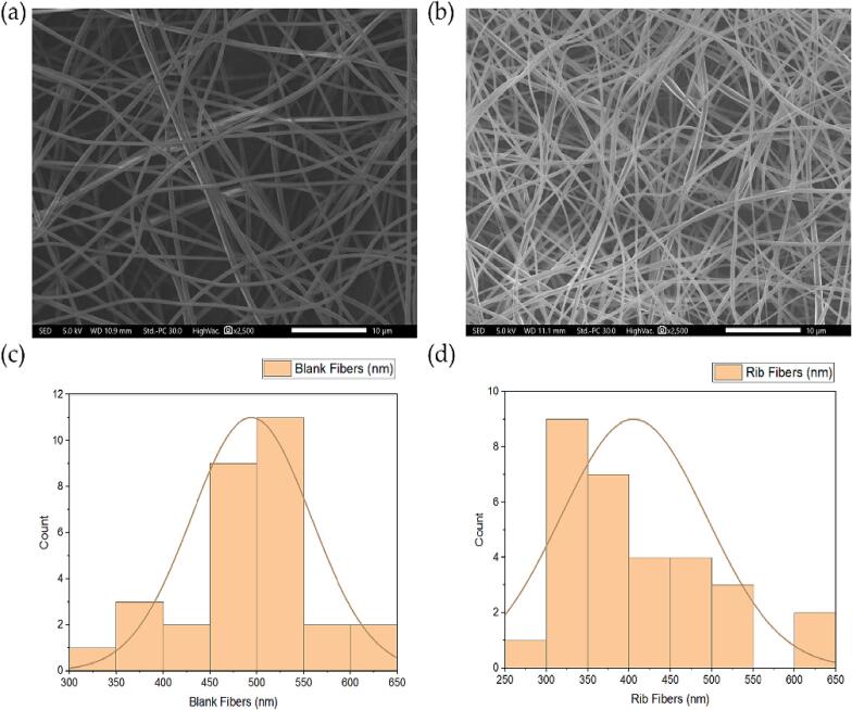
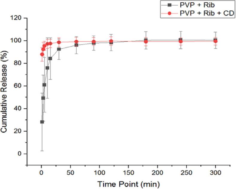
Results: The results demonstrated a successful ribavirin nanofibers fabrication that lacked beads and pores on the nanofibrous surfaces. Ribavirin underwent a physical transformation from crystalline to amorphous form, as confirmed by X-ray diffraction analysis. This change occurred due to the molecular dispersion after the electrospinning process. Additionally, the CD enhanced the encapsulation efficiency of ribavirin in the nanofibers as observed from the drug-loading results. Polyvinylpyrrolidone (PVP) and CD increased ribavirin released into the solution and the disintegration of fibrous mats which shrank and eventually dissolved into a gel-like substance as the ribavirin-loaded fibers began to break down from their border toward the midpoint. Cytotoxicity of ribavirin and CD was evaluated against human dermal fibroblasts (HFF-1) and the results showed a relatively safe profile of ribavirin upon 24-hour cell exposure, while CD was safe within 24- and 48-hour.

Conclusion: This study provides valuable insights into the potential application of our nanofibrous system for treating chronic wounds; however, further antimicrobial and in-vivo studies are required to confirm its safety and effectiveness.

Keywords: Candida albicans; Chronic wounds; Cyclodextrin; Electrospinning; Nanofibers; Pseudomonas aeruginosa; Ribavirin.


Conflict of interest statement

The authors declare that they have no known competing financial interests or personal relationships that could have appeared to influence the work reported in this paper.

Figures

None
Graphical abstract
Fig. 1
Fig. 1
SEM images that represent (a) PVP fibers and (b) ribavirin-loaded PVP fibers. The diameter distribution of (c) PVP fibers and (d) ribavirin-loaded PVP fibers showed their average diameters of 494 ± 64 nm and 406 ± 88 nm, respectively (n = 30).
Fig. 2
Fig. 2
SEM images that represent (a) blank PVP/CD fibers and (b) ribavirin-loaded PVP/CD fibers. The distribution of diameter of (c) blank PVP/CD fibers and (d) ribavirin-loaded PVP/CD fibers showed their average fiber diameters of 790 ± 431 nm and 1,133 ± 234 nm, respectively (n = 30).
Fig. 3
Fig. 3
XRD patterns of PVP, CD, ribavirin, blank PM (PVP + CD), ribavirin PM (PVP + Rib + CD), blank and ribavirin-loaded PVP/CD nanofibers demonstrating the molecular dispersion of ribavirin in the ribavirin-loaded fibers. Grey arrows represent the distinctive peaks. PM, physical mixture; PVP, polyvinylpyrrolidone; CD, cyclodextrin; Rib, ribavirin.
Fig. 4
Fig. 4
FTIR transmissions at a wavenumber range 4000 to 400 cm−1 of (A) PVP, CD, ribavirin, PVP/CD PM, ribavirin/PVP PM, ribavirin/PVP/CD PM, and (B) blank PVP nanofibers, PVP/CD nanofibers, ribavirin-loaded PVP nanofibers and ribavirin-loaded PVP/CD nanofibers. PM, physical mixture; PVP, polyvinylpyrrolidone; CD, cyclodextrin; Rib, ribavirin.
Fig. 5
Fig. 5
HPLC calibration curve of ribavirin-loaded nanofibers showed excellent linearity (R2 = 0.9991).
Fig. 6
Fig. 6
The disintegration of PVP/CD fibers (PVP + CD), ribavirin-loaded fibers (PVP + Rib + CD), blank PVP fibers (blank fibers) and ribavirin-loaded fibers (Rib fibers) showing an ultra-rapid disintegration of all systems in ≤ 3 s. PVP, polyvinylpyrrolidone; CD, cyclodextrin; Rib, ribavirin.
Fig. 7
Fig. 7
The release profile of the ribavirin demonstrated a rapid release in the initial 10 min and at 1 min from ribavirin-loaded fibers (PVP + Rib) and ribavirin-loaded PVP/CD fibers (PVP + Rib + CD), respectively. Results are shown as the average ± SD (n = 3). PVP, polyvinylpyrrolidone; CD, cyclodextrin; Rib, ribavirin.
Fig. 8
Fig. 8
Cell viability of (A) ribavirin, (B) CD, and (C) ribavirin and CD combined in a ratio of 1:20, after being applied at different concentrations upon 24- and 48-hour exposure to HFF-1 cells. Results are shown as average ± SD (n = 3).
Fig. 9
Fig. 9
The zone of inhibition assay showing (A) the antimicrobial activity of ribavirin, ribavirin-loaded PVP nanofibers, ribavirin-loaded PVP/CD nanofibers and blank nanofibers at dose equivalent to 625 μg against P. aeruginosa (ATCC 27853, ATCC 1744, PAO1, and the clinical isolate 7067) and C. albicans, and (B) MHA plate images that were taken after an overnight incubation against all test microbes. Data shown are the diameters of the zones of inhibition measured in millimeters (mm). The results are shown as average ± SD (n = 3). PVP, polyvinylpyrrolidone; CD, cyclodextrin; Rib, ribavirin.

References

    1. Aburayan W.S., Booq R.Y., BinSaleh N.S., et al. The delivery of the novel drug ‘halicin’ using electrospun fibers for the treatment of pressure ulcer against pathogenic bacteria. Pharmaceutics. 2020;12:1189. - PMC - PubMed
    1. Ahmed T.A., Almehmady A.M., Alharbi W.S., et al. Incorporation of perillyl alcohol into lipid-based nanocarriers enhances the antiproliferative activity in malignant glioma cells. Biomedicines. 2023;11:2771. - PMC - PubMed
    1. Alamer A.A., Alsaleh N.B., Aodah A.H., et al. Development of imeglimin electrospun nanofibers as a potential buccal antidiabetic therapeutic approach. Pharmaceutics. 2023;15:1208. - PMC - PubMed
    1. Alkahtani M.E., Aodah A.H., Abu Asab O.A., et al. Fabrication and characterization of fast-dissolving films containing escitalopram/quetiapine for the treatment of major depressive disorder. Pharmaceutics. 2021;13:891. - PMC - PubMed
    1. Alshaya H.A., Alfahad A.J., Alsulaihem F.M., et al. Fast-dissolving nifedipine and atorvastatin calcium electrospun nanofibers as a potential buccal delivery system. Pharmaceutics. 2022;14:358. - PMC - PubMed

LinkOut - more resources