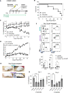
. 2024 Apr 11;9(9):e165936.
doi: 10.1172/jci.insight.165936.

Minnelide suppresses GVHD and enhances survival while maintaining GVT responses

Affiliations

Minnelide suppresses GVHD and enhances survival while maintaining GVT responses

Sabrina N Copsel et al. JCI Insight. .

Abstract

Allogeneic hematopoietic stem cell transplantation (aHSCT) can cure patients with otherwise fatal leukemias and lymphomas. However, the benefits of aHSCT are limited by graft-versus-host disease (GVHD). Minnelide, a water-soluble analog of triptolide, has demonstrated potent antiinflammatory and antitumor activity in several preclinical models and has proven both safe and efficacious in clinical trials for advanced gastrointestinal malignancies. Here, we tested the effectiveness of Minnelide in preventing acute GVHD as compared with posttransplant cyclophosphamide (PTCy). Strikingly, we found Minnelide improved survival, weight loss, and clinical scores in an MHC-mismatched model of aHSCT. These benefits were also apparent in minor MHC-matched aHSCT and xenogeneic HSCT models. Minnelide was comparable to PTCy in terms of survival, GVHD clinical score, and colonic length. Notably, in addition to decreased donor T cell infiltration early after aHSCT, several regulatory cell populations, including Tregs, ILC2s, and myeloid-derived stem cells in the colon were increased, which together may account for Minnelide's GVHD suppression after aHSCT. Importantly, Minnelide's GVHD prevention was accompanied by preservation of graft-versus-tumor activity. As Minnelide possesses anti-acute myeloid leukemia (anti-AML) activity and is being applied in clinical trials, together with the present findings, we conclude that this compound might provide a new approach for patients with AML undergoing aHSCT.

Keywords: Cellular immune response; Immunology; Stem cell transplantation; Transplantation.


Figures
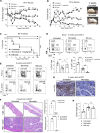
Figure 1
Figure 1. Recipients treated with Minnelide exhibited diminished acute GVHD after MHC-mismatched aHSCT.
(AC) An aHSCT was performed utilizing a B6→BALB/c donor and recipient mouse model (day –1: 7.5–8.5 Gy; day 0: 5.5 × 10⁶ TCD BM cells and pooled splenocytes containing 0.8 × 10⁶ T cells) and recipients were treated with 0.1 mg/kg Minnelide from day −2 to 28 after transplantation. Weight loss (A), clinical scores (B), and GVHD survival (C) are presented (n = 8 untreated, n = 16 Minnelide-treated, and n = 4 BM-only mice). (D) Representative flow cytometry contour plots and frequency of CD4+ and CD8+ cells in the blood on day 14 after aHSCT. (E) Eight weeks after aHSCT, thymic and splenic T cell populations were evaluated and representative flow cytometry plots are shown. (F) Representative H&E staining and pathology scores (on the right) (n = 2–6) from the skin and colon 8 weeks after aHSCT. Original magnification, ×200 (top) and ×100 (bottom), respectively. (G) Representative photographs of colon anti-CD3 staining 8 weeks after transplantation. Original magnification, ×100. (H) Increased colon length in recipients of Minnelide treatment 8 weeks after aHSCT. These data are representative of 4 independent aHSCT experiments in this model. Clinical scores were compared using Benjamini-Krieger-Yekutieli–corrected multiple t tests over time. Groups were compared using 1-way ANOVA with Tukey’s multiple-comparison test and a log-rank test was used for survival analyses. *P < 0.05; **P < 0.01; ***P < 0.001; ****P < 0.0001. Data are presented as mean ± SEM.
Figure 2
Figure 2. Minnelide treatment promotes lymphoid engraftment after MHC-mismatched aHSCT.
(AH) An aHSCT was performed utilizing the same model as in Figure 1 (B6→BALB/c donor and recipient strain combination), but transplanted BM was derived from congenic B6-CD45.1 donors and T cells from B6-CD45.2 donors. Recipients were treated with 0.1 mg/kg Minnelide from day −2 to 28 after transplantation. Seven weeks after aHSCT, splenic and thymic tissues were analyzed for total cell numbers (A and C), splenic CD4+/CD8+ ratio (B), and CD4+CD8+ DP thymocytes (D). (E) Representative flow cytometry contour plots and frequency of donor (transplanted hematopoietic progenitors) BM–derived (Kb+CD45.1+) CD4+ and CD8+ cells in the spleen seven weeks after aHSCT are shown. Kb, MHC H2Kb. Frequency of splenic CD19+ (F), CD11b+ (G), and NK1.1+ (H) cells derived from donor BM 7 weeks after transplantation. GVHD, n = 3; Minnelide, n = 3; BM only + Minnelide, n = 3. Groups compared using 1-way ANOVA with Dunnett’s multiple-comparison test. *P < 0.05; **P < 0.01; ***P < 0.001. Data are presented as mean ± SEM.
Figure 3
Figure 3. Minnelide can inhibit T cell proliferation and decrease T cell proinflammatory cytokines early after MHC-mismatched aHSCT.
Recipients were treated with 0.1 mg/kg Minnelide from day −2 to 7 after transplantation, and on day 7 colons and spleens were evaluated. (A) Representative photographs of colon anti-CD3 staining show a marked decrease in CD3+ cell infiltrate. Original magnification, ×100. (B) Purified T cells were seeded in 96-well plates and treated with different doses of triptolide for 120 hours. A dose-dependent decrease in proliferation was observed in response to 0.5–50 nM triptolide. A significant and lasting reduction in proliferation was observed in response to treatment. (CG) On day 7 after aHSCT, spleens were analyzed for frequency of donor (C) Th1 cells producing IFN-γ, (D) CD8+ cells producing IFN-γ, (E) Th22 cells producing IL-13, and (F) CD4+FoxP3+ Tregs. (G) Summary data of the frequency of the indicated donor (MHC H2Kb–positive, Kb+) cell populations. These data were pooled from 2 independent aHSCT experiments (untreated, n = 5; Minnelide, n = 5). Groups were compared using a 2-tailed, unpaired t test. *P < 0.05, **P < 0.01, ***P < 0.001, ****P < 0.0001. Data are presented as mean ± SEM.
Figure 4
Figure 4. Minnelide decreases cytokine storm in recipient mice following MHC-mismatched aHSCT.
(AK) Using the major BM transplantation model described in Figure 1, recipients were treated with 0.1 mg/kg Minnelide from day −2 to 7 after transplantation, and on days 4 (AG) and 7 (HK) serum was collected via cardiac puncture for cytokine quantification using the LEGENDplex Mouse Inflammation Panel (see Supplemental Methods). The specific cytokines evaluated are indicated on the y axis of each graph. Groups were compared using 1-way ANOVA with Tukey’s multiple-comparison test. *P < 0.05; **P < 0.01; ***P < 0.001. Data are presented as mean ± SEM.
Figure 5
Figure 5. Minnelide treatment increases innate and adaptive regulatory cell populations that promote immunosuppressive activity in GVHD target tissues following MHC-mismatched aHSCT.
Using the major BM transplantation model, B6→BALB/c (described in Figure 1), recipients were treated with 0.1 mg/kg Minnelide from day −2 to day +20 after transplantation. On day 20 after aHSCT, lamina propria (LP) from colon (AJ) as well as lung lymphocytes (K) was evaluated. Data are presented as frequency of colonic CD11b+ cells (A), frequency of MDSCs (Ly6G+Ly6CCD11b+) (B), frequency of ILC2s (GATA3+CD90.2+Lin) (C), frequency of KLRG1+cells within the ILC2 population (D), and ICOS+ cells (E). (F) Representative flow cytometry contour plots and frequency of Tregs (CD4+FoxP3+) in colonic LP from BM-only, untreated (received BM + T cells), and Minnelide-treated (received BM + T cells) mice (n = 3). Frequency of LP T cell subsets, CD4+FoxP3+KLRG1+ (G), total CD4+ (H), total CD8+ (I), and Th2 (CD4+FoxP3GATA3+) (J). (K) Representative flow cytometric contour plots of Treg cells in the lung (CD4+FoxP3+CD4+). (L) Frequency of CD4+ and CD8+ cells in the spleen 3 weeks after aHSCT. Groups were compared using 1-way ANOVA with Tukey’s multiple-comparison test. *P < 0.05; **P < 0.01; ***P < 0.001.
Figure 6
Figure 6. Recipients treated with Minnelide exhibited maintained GVT, while GVHD was ameliorated after MHC-mismatched aHSCT.
(AC) aHSCT was performed utilizing a B6→BALB/c donor and recipient mouse model and recipients were treated with 0.1 mg/kg Minnelide from day −2 to 28 after transplantation. GVHD survival (A), clinical scores (B), and weight loss (C) are presented (n = 8 GVHD, n = 16 Minnelide, and n = 4 BM-only mice). (D) Tumor burden in recipient blood on day 22 after BM transplantation. The data are from 2 independent experiments. Clinical scores were compared using Benjamini-Krieger-Yekutieli–corrected multiple t tests over time. Groups were compared using 1-way ANOVA with Tukey’s multiple-comparison test or log-rank test for survival analyses. *P < 0.05 for B6 BM + T cells + MLL-AF9 vs. B6 BM + T cells + MLL-AF9 + Minnelide; #P < 0.05 for B6 BM + MLL-AF9 vs. B6 BM + MLL-AF9 + Minnelide; **P < 0.01. Data are presented as mean ± SEM.
Figure 7
Figure 7. Functional immunity is intact in Minnelide-treated recipients.
Two months after aHSCT, recipients received 2 skin grafts, applied on the trunk of each mouse, 1 from B6 × BALB/c F1 (H2b/d) mice and 1 from C3H/HeJ third-party (H2k) donors. Grafts were assessed and scored on the indicated days (Minnelide n= 5; BM only n = 2). (A) Allograft score. Graft scoring was performed as follows: 0, intact graft and healthy appearance; 1, inflamed graft, but without signs of necrosis observed; 2, inflamed graft and less than 25% necrosis observed; 3, inflamed graft and between 25% and 75% necrosis observed; and 4, greater than 75% necrosis detected or loss of graft. (B) Allograft survival. All mice accepted the F1 (B6 × BALB/c) skin grafts, whereas all C3H/HeJ grafts were rejected in both Minnelide and BM-only transplant recipients by day 21. (C) Representative photographs of skin grafts present on recipient mice on days 8 and 31 from both groups. Groups were compared using log-rank for survival analyses. Data are presented as mean ± SEM.
Figure 8
Figure 8. Treatment with Minnelide reduced xGVHD.
(AF) NSG recipient mice were irradiated (2 Gy) and the following day underwent transplantation with human mobilized PBMCs (6 × 10⁶). Recipients were treated with 0.1 mg/kg Minnelide from day −2 to 28 after transplantation. Schematic of experimental outline (A), overall survival (B), weight loss (C), and xGVHD clinical scores (D) are presented (n = 6 xGVHD or Minnelide and n = 3 control [no xGVHD] mice). (E) Representative photographs of xGVHD and Minnelide-treated recipients 4 weeks after transplantation. (F) Flow cytometry contour plots and frequency of human CD4+, CD8+, and CD14+ cells in the blood 2 weeks after transplantation. These data are representative of 2 independent (n = 6/group) human-to-mouse HSCTs. Clinical scores were compared using Benjamini-Krieger-Yekutieli–corrected multiple t tests over time. (G) Proliferation assays of CellTrace Violet–labeled human PBMCs stimulated by either anti-CD3 (OKT3 mAb) in vitro for 4 days and a 1-way mixed lymphocyte reaction for 6 days. Groups compared using 2-way ANOVA with Dunnett’s multiple-comparison test or log-rank for survival analyses. *P < 0.05; **P < 0.01; **P < 0.001; ****P < 0.0001. Data are presented as mean ± SEM.

Similar articles

Cited by

References

    1. Appelbaum FR. Haematopoietic cell transplantation as immunotherapy. Nature. 2001;411(6835):385–389. doi: 10.1038/35077251. - DOI - PubMed
    1. Zeiser R, Blazar BR. Acute graft-versus-host disease - biologic process, prevention, and therapy. N Engl J Med. 2017;377(22):2167–2179. doi: 10.1056/NEJMra1609337. - DOI - PMC - PubMed
    1. Horowitz MM, et al. Graft-versus-leukemia reactions after bone marrow transplantation. Blood. 1990;75(3):555–562. doi: 10.1182/blood.V75.3.555.555. - DOI - PubMed
    1. Ferrara JL, Reddy P. Pathophysiology of graft-versus-host disease. Semin Hematol. 2006;43(1):3–10. doi: 10.1053/j.seminhematol.2005.09.001. - DOI - PubMed
    1. Ferrara JL, et al. Pathophysiologic mechanisms of acute graft-vs.-host disease. Biol Blood Marrow Transplant. 1999;5(6):347–356. doi: 10.1016/S1083-8791(99)70011-X. - DOI - PubMed

Publication types

MeSH terms