. 2024 Apr 23;121(17):e2317083121.
doi: 10.1073/pnas.2317083121. Epub 2024 Apr 11.

Hox gene-specific cellular targeting using split intein Trojan exons

Affiliations

Hox gene-specific cellular targeting using split intein Trojan exons

Fengqiu Diao et al. Proc Natl Acad Sci U S A. .

Abstract

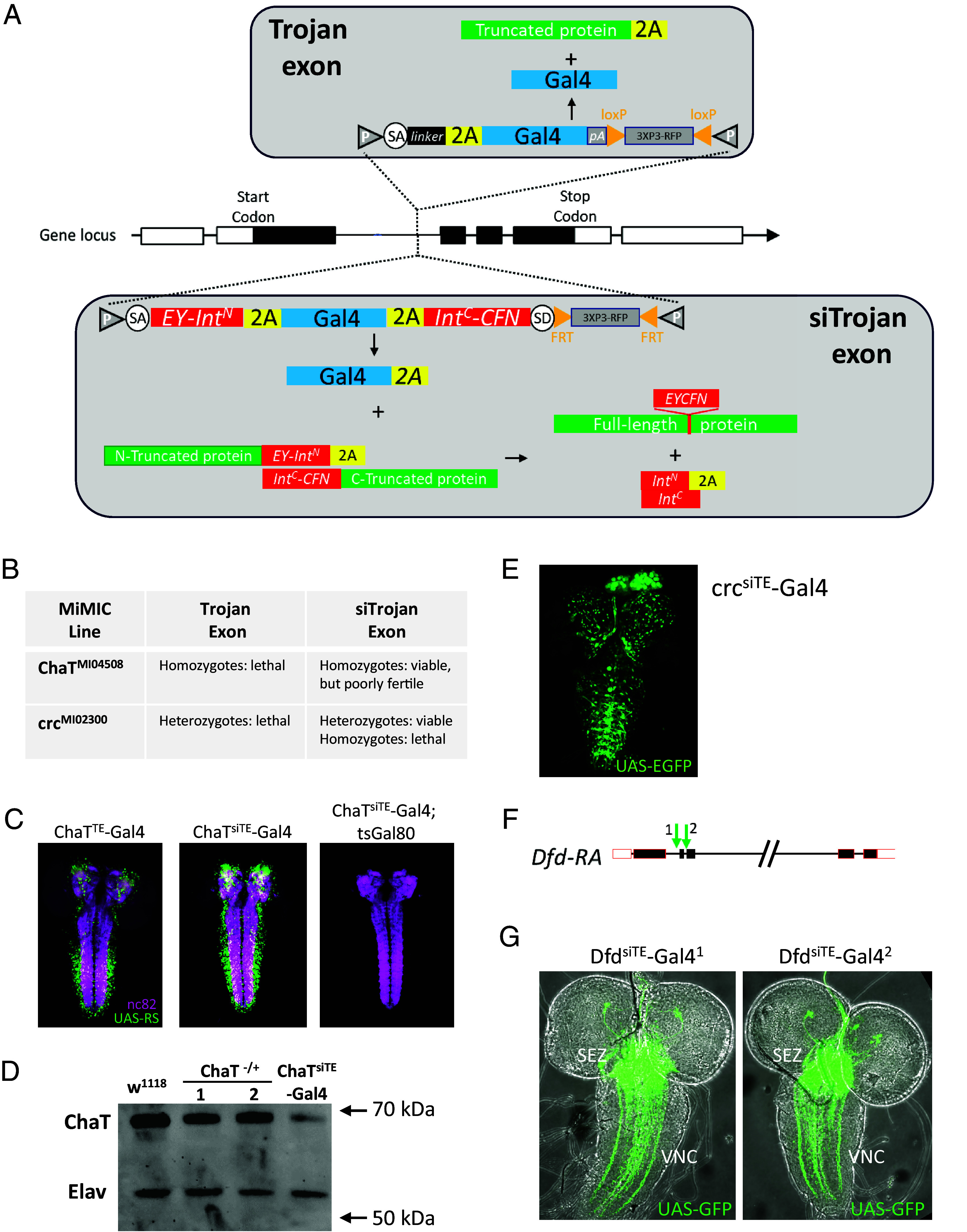
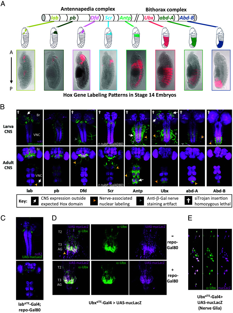
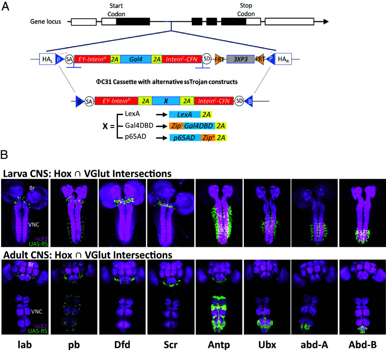
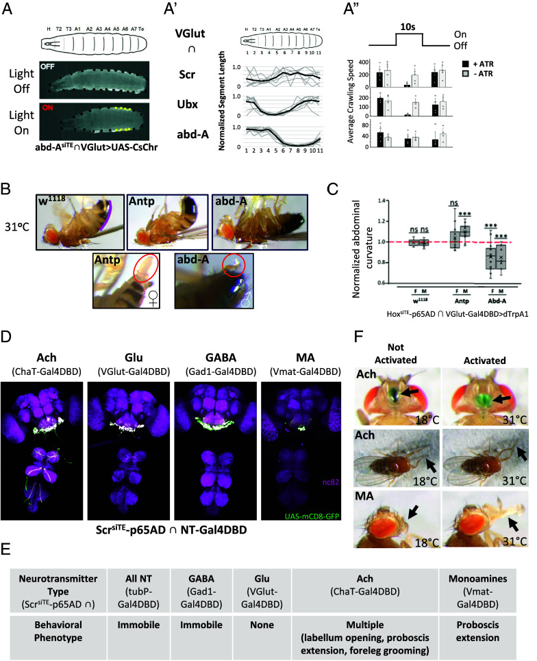
The Trojan exon method, which makes use of intronically inserted T2A-Gal4 cassettes, has been widely used in Drosophila to create thousands of gene-specific Gal4 driver lines. These dual-purpose lines provide genetic access to specific cell types based on their expression of a native gene while simultaneously mutating one allele of the gene to enable loss-of-function analysis in homozygous animals. While this dual use is often an advantage, the truncation mutations produced by Trojan exons are sometimes deleterious in heterozygotes, perhaps by creating translation products with dominant negative effects. Such mutagenic effects can cause developmental lethality as has been observed with genes encoding essential transcription factors. Given the importance of transcription factors in specifying cell type, alternative techniques for generating specific Gal4 lines that target them are required. Here, we introduce a modified Trojan exon method that retains the targeting fidelity and plug-and-play modularity of the original method but mitigates its mutagenic effects by exploiting the self-splicing capabilities of split inteins. "Split Intein Trojan exons" (siTrojans) ensure that the two truncation products generated from the interrupted allele of the native gene are trans-spliced to create a full-length native protein. We demonstrate the efficacy of siTrojans by generating a comprehensive toolkit of Gal4 and Split Gal4 lines for the segmentally expressed Hox transcription factors and illustrate their use in neural circuit mapping by targeting neurons according to their position along the anterior-posterior axis. Both the method and the Hox gene-specific toolkit introduced here should be broadly useful.

Keywords: Drosophila; cell type; development; genetic access; neural circuit-mapping.


Conflict of interest statement

Competing interests statement:The authors declare no competing interest.

Figures

Fig. 1.
Fig. 1.
The architecture and application of split intein Trojan exons. (A) Schematic illustrating the original Trojan exon strategy (Top) and the new split intein Trojan exon strategy (Bottom). In both cases, the synthetic exon is inserted into an intron separating two coding exons of a native gene. The original Trojan exon produces the Gal4 protein and a truncation product of the native protein. The siTrojan exon technique produces three translation products: the Gal4 molecule fused at its C terminus to a T2A peptide and two truncated fragments of the native protein. The N-terminal fragment is fused at its C terminus to the CfaN split intein moiety (EY-IntN) and a T2A peptide; the C-terminal fragment is fused at its N terminus to the CfaC split intein moiety (IntC-CFN). These two truncated products of the native protein are ligated by split intein-mediated trans-splicing. (B) Driver lines made by inserting siTrojan-Gal4 constructs into the ChaT and crc genes by ΦC31-mediated MiMIC cassette exchange have reduced lethality compared to classical Trojan-Gal4 lines made via insertions into the same MiMIC sites. (C) Larval CNS expression of ChaT-Gal4 lines made as in (B) using classical Trojan exon technology (Left, ChaTTE-Gal4) and siTrojan exon technology (Middle, ChaTsiTE-Gal4). The ChaTsiTE-Gal4 line remains sensitive to tsGal80 inhibition at the restrictive temperature of 31 °C (Right) despite the T2A tail on Gal4. Green, UAS-RS (RedStinger) expression; magenta, nc82 neuropil staining. (D) Western blot showing full-length ChaT protein in CNS of flies homozygous for the ChaTsiTE-Gal4 insertion. 2X CNS equivalents were loaded in all lanes for flies of either the control (w1118) or experimental (ChaTsiTE-Gal4/ChaTsiTE-Gal4) genotypes and immunostained for ChaT protein. For comparison, results are shown for hemizygous flies with exonic insertions into the same ChaT intron (1: MiMIC line, ChaTMI04508/TM3, Ser; 2: the original Trojan exon line, ChaTTE-Gal4/TM6B). MW markers are as indicated and immunostaining of the panneuronal protein Elav is included as a loading control. (E) L3 larval CNS expression of the crcsiTE-Gal4 line created by conversion of MiMIC line MI02300 using a siTrojan-Gal4 construct. (F) Intronic sites of CRISPR/Cas9-mediated insertion (green arrows) of Trojan-Gal4 and siTrojan-Gal4 constructs into the Deformed gene. Insertions of the original Trojan exon produced a viable line only at site 1, siTrojan exon insertions at both sites 1 and 2 produced viable lines with robust Gal4 expression in the expected patterns of the Dfd gene: (G) Larval CNS expression of DfdsiTE-Gal4 lines produced by siTrojan exon insertion as in (F). Strong labeling is seen in cell bodies in the SEZ and their axons projecting to the ventral nerve cord (VNC).
Fig. 2.
Fig. 2.
Embryonic and nervous system expression of Hox Gene–specific siTrojan Gal4 drivers. (A) Expression of the HoxsiTE-Gal4 drivers (p65AD∩tubP-Gal4DBD for Scr) varies across the A-P axis in stage 14 embryos (Bottom images). The patterns are consistent with those observed in previous studies (schematics from Hughes and Kaufman (13) used with permission; License 5645480954289) and change progressively with Hox gene position along the 3R chromosome. The two complexes of Hox genes on 3R are indicated. Red, UAS-6XmCherry. “Saturation” and image “Exposure” in the red channel were adjusted in Photoshop (to settings of 25 and 2.0, respectively) to enhance visualization. (B) CNS expression of the Hox-specific drivers in third instar larvae (Top) and adults (Bottom). Br, brain; VNC, ventral nerve cord. Here and subsequent panels: green, UAS-nucLacZ visualized with anti-beta-galactosidase antibody; magenta, nc82 neuropil labeling. Arrows and arrowheads, unexpected labeling; crosses, patterns of drivers that are homozygous lethal, as indicated in key. (C) CNS expression of the labsiTE-Gal4 driver in third instar larvae (Top) and adults (Bottom) is limited to the tritocerebral region of the brain when repo-Gal80 is used to block Gal4 activity in glia. (D) Cells in the abdominal nerves of the VNC (yellow arrowhead) are labeled by the UbxsiTE-Gal4 driver in the absence (Top), but not in the presence (Bottom) of repo-Gal80, indicating that they are glia. Thoracic neuromeres T2-T3 and abdominal ganglia (AG) are indicated. (E) Glia cells of the abdominal nerves labeled by UbxsiTE-Gal4 are immunopositive for anti-Ubx antibodies, indicating that the driver expression in these cells is not ectopic.
Fig. 3.
Fig. 3.
Targeting glutamatergic subsets of Hox gene–expressing neurons using siTrojan Split Gal4 cassettes. (A) siTrojan technology retains the modularity of the original Trojan exon system: An siTrojan-Gal4 insertion can easily be substituted with Split Gal4 hemidrivers, LexA::GAD drivers, or any other desired construct by ΦC31 integrase-mediated cassette exchange. (B) CNS expression of the Hox-specific hemidrivers in glutamatergic neurons of third instar larvae (Top) and adults (Bottom). For all Hox genes except Dfd, a p65AD hemidriver was used; for Dfd a Gal4DBD hemidriver was used. Br, brain; VNC, ventral nerve cord. Green, UAS-RS (RedStinger); magenta, nc82 neuropil labeling.
Fig. 4.
Fig. 4.
Activating subsets of Hox-gene expressing neurons has diverse behavioral effects. (AA”) Activation of glutamatergic motor neurons in third instar larvae resulted in selective contraction of body wall segments, consistent with the pattern of Hox gene expression. (A) Stimulation of motor neurons in the abd-A expression pattern using the light-activated channel UAS-Cs.Chrimson.mVenus results in contraction of posterior segments when light is ON (Bottom, yellow dots) compared to when lights are OFF (Middle). In larval body wall schematic (Top): H, head; T2-T3, thoracic segments; A1-A7, abdominal segments; Te, fused terminal abdominal segments. (A’) Length changes in individual body wall segments (relative to lights off) during motor neuron activation for five animals of each indicated genotype/Hox gene. Gray values, segment lengths for each animal; dark lines, mean segment lengths. (A”) Crawling speed typically decreased during motor neuron activation (i.e., during 10 s lights-on stimulus vs. 10 s prior to or after the stimulus) in experimental animals of the same three genotypes indicated in (A’) that were fed all-trans-retinal (+ATR), a cofactor for Cs.Chrimson. Control animals of the same genotypes, but not fed ATR (-ATR), showed little change in locomotion with light. (B) In adult animals, dTrpA1-mediated stimulation of glutamatergic motor neurons expressing the Hox genes Antp and abd-A resulted in diverse behaviors that were sexually dimorphic. In males, these included persistent abdominal flexion (Top Middle, Antp) or extension (Top Right, abd-A) relative to the abdominal posture of w1118 control animals (Top Left) at the restricted temperature of 31 °C. Stimulation of abd-A-expressing motor neurons in males also robustly induced extension of the aedeagus and secretion of seminal fluid (Bottom Right, oval), while stimulation of Antp-expressing motor neurons in females induced egg extrusion in 5/6 animals (Bottom Left, oval). (C) Abdominal curvature was measured as described in Materials and Methods from the contours of the abdomen from segments A2 or A3 to the tip [see the magenta line for the w1118 animal in (B), Top Left]. Normalized abdominal curvature is the ratio of the curvature measured during motor neuron stimulation by dTrpA1 to the curvature measured prior to stimulation and is shown for both males and females of the indicated genotypes. N ≥ 10 for all conditions, significance determined by the t test. The dashed red line indicates no change in curvature with stimulation. (D) CNS expression pattern of the Scr gene subdivided according to the type of neurotransmitter used by component neurons. Four major subtypes were isolated by intersectional labeling using the ScrsiTE-p65AD hemidriver and Gal4DBD hemidrivers with traditional Trojan exon insertions into the indicated genes. Green, UAS-mCD8-GFP; magenta, nc82 neuropil staining. (E) The behavioral phenotypes resulting from dTrpA1-mediated stimulation of either all Scr-expressing neurons, or those subsets shown in (D) that use a particular neurotransmitter. (F) Behavioral changes observed upon activation of cholinergic (Ach) and monoaminergic (MA) subsets of Scr-expressing neurons. Left and Right panels show individual flies at 18 °C and 31 °C, i.e., prior to and during dTrpA1-mediated stimulation, respectively. Top panels, dye placed on the labellum of a fly emphasizes its robust opening (arrows) upon stimulation of the cholinergic subset of Scr-expressing neurons. Middle panels, headless flies groom their forelegs (arrows) after the onset of stimulation of the same neuronal subset. Bottom panels, extension of the proboscis (arrows) accompanies activation of the monoaminergic subset of Scr-expressing neurons.

Similar articles

Cited by

References

    1. Nagarkar-Jaiswal S., et al. , A library of MiMICs allows tagging of genes and reversible, spatial and temporal knockdown of proteins in Drosophila. Elife 4, e05338 (2015). - PMC - PubMed
    1. Fisher Y. E., et al. , FlpStop, a tool for conditional gene control in. Elife 6, e22279 (2017). - PMC - PubMed
    1. Venken K. J., et al. , MiMIC: A highly versatile transposon insertion resource for engineering Drosophila melanogaster genes. Nat. Methods 8, 737–743 (2011). - PMC - PubMed
    1. Diao F., et al. , Plug-and-play genetic access to drosophila cell types using exchangeable exon cassettes. Cell Rep. 10, 1410–1421 (2015). - PMC - PubMed
    1. Gnerer J. P., Venken K. J. T., Dierick H. A., Gene-specific cell labeling using MiMIC transposons. Nucleic Acids Res. 43, e56 (2015). - PMC - PubMed

Substances

LinkOut - more resources