. 2024 Jul 1;73(7):1127-1139.
doi: 10.2337/db23-0123.

Efficient Vascular and Neural Engraftment of Stem Cell-Derived Islets

Affiliations

Efficient Vascular and Neural Engraftment of Stem Cell-Derived Islets

Julia Thorngren et al. Diabetes. .

Abstract

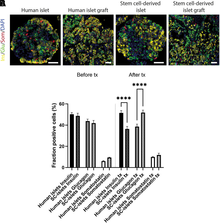
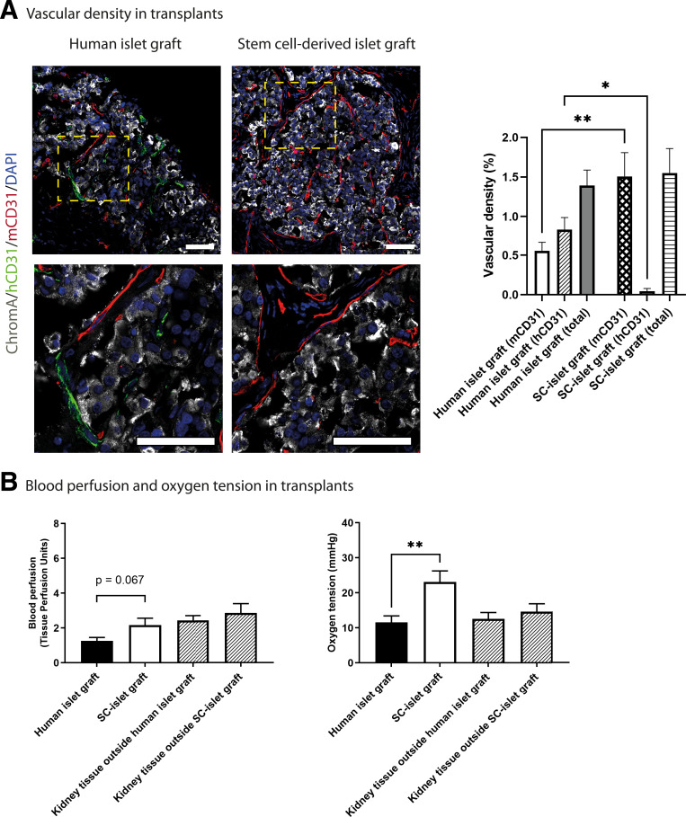
Pluripotent stem cell-derived islets (SC-islets) have emerged as a new source for β-cell replacement therapy. The function of human islet transplants is hampered by excessive cell death posttransplantation; contributing factors include inflammatory reactions, insufficient revascularization, and islet amyloid formation. However, there is a gap in knowledge of the engraftment process of SC-islets. In this experimental study, we investigated the engraftment capability of SC-islets at 3 months posttransplantation and observed that cell apoptosis rates were lower but vascular density was similar in SC-islets compared with human islets. Whereas the human islet transplant vascular structures were a mixture of remnant donor endothelium and ingrowing blood vessels, the SC-islets contained ingrowing blood vessels only. Oxygenation in the SC-islet grafts was twice as high as that in the corresponding grafts of human islets, suggesting better vascular functionality. Similar to the blood vessel ingrowth, reinnervation of the SC-islets was four- to fivefold higher than that of the human islets. Both SC-islets and human islets contained amyloid at 1 and 3 months posttransplantation. We conclude that the vascular and neural engraftment of SC-islets are superior to those of human islets, but grafts of both origins develop amyloid, with potential long-term consequences.


Conflict of interest statement

Duality of Interest. No potential conflicts of interest relevant to this article were reported.

Figures

Figure 1
Figure 1
Endocrine composition of human islets and SC-islets before and after transplantation. A: Images of human islets and SC-islets immunostained for insulin (ins; yellow), glucagon (glu; green), somatostatin (som; red), and nuclei (blue) before and after transplantation. B: Comparison of percentages of endocrine cells in human islets and SC-islets before and after transplantation. All values are given as mean ± SEM (n = 4–10). One biological replicate corresponded to one mouse receiving a transplant of SC-islets from one differentiation or islets from one human donor. Scale bar, 50 μm. ****P < 0.0001 by one-way ANOVA and Šídák multiple comparisons test.
Figure 2
Figure 2
Maturation markers of human islets and SC-islets before and after transplantation. Images of human islets and SC-islets immunostained for MAFA, NEUROD1, NKX6.1, NKX2.2, PCNA, and PDX1 (green), insulin (ins; red), and nuclei (blue) before and after transplantation. Scale bar, 50 μm.
Figure 3
Figure 3
Amyloid detected in SC-islets transplanted beneath the renal capsule. AD: One month posttransplantation, retrieved human islet grafts (A) (C is higher magnification) and SC-islet grafts (B) (D is higher magnification) were immunostained for the endocrine marker chromogranin A (CgA; gray), mouse endothelial marker CD31 (red), and amyloid marker pentameric formylthiophene acetic acid (pFTAA; green). EH: Three months posttransplantation, human islet grafts (E) (G is higher magnification) and SC-islet grafts (F) (H is higher magnification) were also immunostained for amyloid. I: Quantification of amyloid was performed in both human islet grafts and SC-islet grafts. J: Measurement of the distance from amyloid to blood vessels. All values are given as mean ± SEM (n = 4–6). One biological replicate corresponded to one mouse receiving a transplant of SC-islets from one differentiation or islets from one human donor. Scale bar, 100 μm. *P < 0.05 by two-tailed Student t test.
Figure 4
Figure 4
Characterization of blood vessels in transplanted human islets or SC-islets. A: Three months posttransplantation, retrieved human islet grafts and SC-islet grafts were immunostained for the endocrine marker chromogranin A (gray), human endothelial marker CD31 (green), mouse endothelial marker CD31 (red), and nuclei (blue). Bottom row shows grafts above in higher magnification. Vascular density in these grafts was evaluated. All values are given as mean ± SEM (n = 6). B: Tendency toward increased blood flow in SC-islet grafts compared with human islet grafts (P = 0.067 by two-tailed Student t test; n = 8–9). Oxygen tension was superior in SC-islet grafts compared with human islet grafts (n = 7–8). All values are given as mean ± SEM. Measurements of blood flow and oxygen tension in the superficial kidney cortex (1–2 mm) in the immediate vicinity of the graft is included for reference. One biological replicate corresponded to one mouse receiving a transplant of SC-islets from one differentiation or islets from one human donor, but exceptions were made for blood flow and oxygen tension measurements where two mice received transplants with human islets from same donor and four mice received transplants with SC-islets from two differentiations (i.e., two mice per differentiation). Scale bar, 50 μm. *P < 0.05, **P < 0.01 by one-way ANOVA and Šídák multiple comparisons test (A) or two-tailed Student t test (B).
Figure 5
Figure 5
Nerve density in transplanted human islets or SC-islets. Three months posttransplantation, retrieved human islet grafts and SC-islet grafts were immunostained for the endocrine marker chromogranin A (gray), nerve marker NF-L (green), mouse endothelial marker CD31 (red), and nuclei (blue). Bottom row shows grafts above in higher magnification. Nerve density in these grafts was evaluated. All values are given as mean ± SEM (n = 6). One biological replicate corresponded to one mouse transplanted with SC-islets from one differentiation or islets from one human donor. Scale bar, 50 μm. ***P < 0.001 by two-tailed Student t test.
Figure 6
Figure 6
β-Cell apoptosis of human islets and SC-islets 3 months after transplantation. AD: Three months posttransplantation, retrieved human islet grafts (A) (C is higher magnification) and SC-islet grafts (B) (D is higher magnification) were immunostained for insulin (gray), apoptosis marker, active cleaved caspase-3 (green), and nuclei (blue). E: Caspase-3 reactivity was evaluated in both human islet grafts and SC-islet grafts. All values are given as mean ± SEM (n = 6). One biological replicate corresponded to one mouse receiving a transplant of SC-islets from one differentiation or islets from one human donor. Scale bar, 100 μm. *P < 0.05 by Mann-Whitney test.
Figure 7
Figure 7
RNA sequencing (RNA seq) of human islets and SC-islets before transplantation. A: Principal component analysis. Samples group together by sample type (human islets vs. SC-islets). Majority of the expression variation among samples (PC1) is explained by sample type. B: Volcano plot showing expression differences between human islets and SC-islets. Human islets are treated as reference for fold change (FC). DE genes belonging to gene ontology (GO) term positive regulation of angiogenesis (GO:0045766) are highlighted; colors correspond to sample types in which the expression is higher. C: Heat map of selected genes with known functions in angiogenesis and neurogenesis. D: Gene expression analysis of human islets or SC-islets before transplantation. Log2 transformation of FC of the relative mRNA expression levels in the 11 DEGs (genes involved in angiogenesis and neurogenesis) selected for validation with quantitative RT-PCR (qRT-PCR; black bars) parallels the direction of change in RNA seq (blue bars).

References

    1. Maffi P, Secchi A. Clinical results of islet transplantation. Pharmacol Res 2015;98:86–91 - PubMed
    1. Eriksson O, Eich T, Sundin A, et al. . Positron emission tomography in clinical islet transplantation. Am J Transplant 2009;9:2816–2824 - PubMed
    1. Moberg L, Johansson H, Lukinius A, et al. . Production of tissue factor by pancreatic islet cells as a trigger of detrimental thrombotic reactions in clinical islet transplantation. Lancet 2002;360:2039–2045 - PubMed
    1. Lee Y, Ravazzola M, Park BH, Bashmakov YK, Orci L, Unger RH. Metabolic mechanisms of failure of intraportally transplanted pancreatic beta-cells in rats: role of lipotoxicity and prevention by leptin. Diabetes 2007;56:2295–2301 - PubMed
    1. Mattsson G, Jansson L, Carlsson PO. Decreased vascular density in mouse pancreatic islets after transplantation. Diabetes 2002;51:1362–1366 - PubMed